Introduction
Adenocarcinoma of the rete testis is a rare, highly
aggressive tumor originating from the nonspermatogenic epithelium
of the intratesticular excretory ducts. Only ~60 cases have been
reported in the literature to date. Rete testis adenocarcinoma
occurs most frequently in elderly males and is usually associated
with a poor prognosis (1). The
majority of cases present as a scrotal mass with diffuse
enlargement of the testis. However, it is difficult to make a
differential diagnosis with other testicular lesions, as rete
testis adenocarcinoma also invariably presents with epididymitis,
hydrocele, inflammatory lumps or inguinal hernia (2–4). The
delayed diagnosis of right testis adenocarcinoma is often made by
the pathologist following surgery, due to non-specific clinical
presentation and symptoms. Ultrasound is a proven, safe diagnostic
procedure with a high degree of sensitivity and specificity for
testicular tumors (5). Due to the
low prevalence of adenocarcinoma of the rete testis, the
sonographic characteristics of this highly malignant tumor type
have not been studied in sufficient depth. This report presents a
case of primary adenocarcinoma of the rete testis, as confirmed
pathologically. The diagnosis and differential diagnosis of this
neoplasm, with regard to sonographic characteristics, are reviewed
and discussed.
Case report
Patient presentation
A 46-year-old male was admitted with complaints of
swelling and pain in the right side of the scrotum for a 1-year
period. No associated symptoms were observed. Anti-tuberculosis
chemotherapy was performed in Sichuan Provincial People’s Hospital
(Chengdu, China), which yielded no response. Physical examination
revealed a swelling of the right scrotum and hard nodules at the
head of the right epididymis exhibiting severe tenderness. Written
informed consent was obtained from the patient.
Ultrasonography
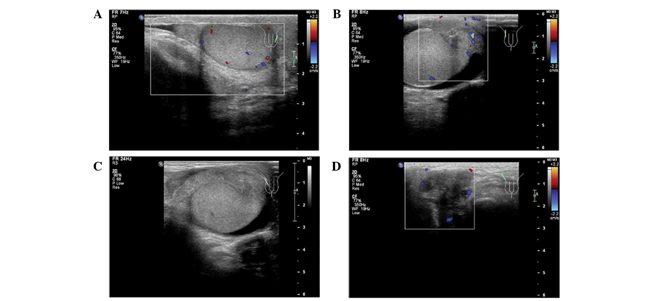
Ultrasonography of the scrotum revealed a 1.0×2.1-cm
hypoechoic nodule at the right epididymis with a poorly-defined
border. In addition, a relatively abundant blood flow was detected
by color Doppler ultrasound and a weak echo was recorded in the
right side of the tunica vaginalis area (Fig. 1B and C). A 5.0×1.9-cm irregular mass
with an unclear boundary and low mixed echo structure was localized
in the right inguinal region, which also showed increased
vascularization (Fig. 1D).
Diagnosis
The levels of lactate dehydrogenase (LDH),
α-fetoprotein (AFP) and β-human chorionic gonadotropin (β-HCG) were
normal in the serum. Chest radiographs, abdominal ultrasonography
and computed tomography (CT) of the abdomen revealed no remarkable
results. Considering the aforementioned results, a diagnosis of
primary testicular tumor was proposed.
Surgical procedures
Based on this provisional diagnosis, scrotal
incision was performed for testicular exploration. This revealed
blood-mixed fluid in the scrotal skin and an enlarged right
epididymis with multiple yellow-grey, rough nodules, forming
adhesions with the scrotal skin. Intraoperative frozen sections
revealed adenocarcinoma in the specimen and the scrotal incision
was extended and a high right inguinal orchidectomy was
performed.
Microscopy results
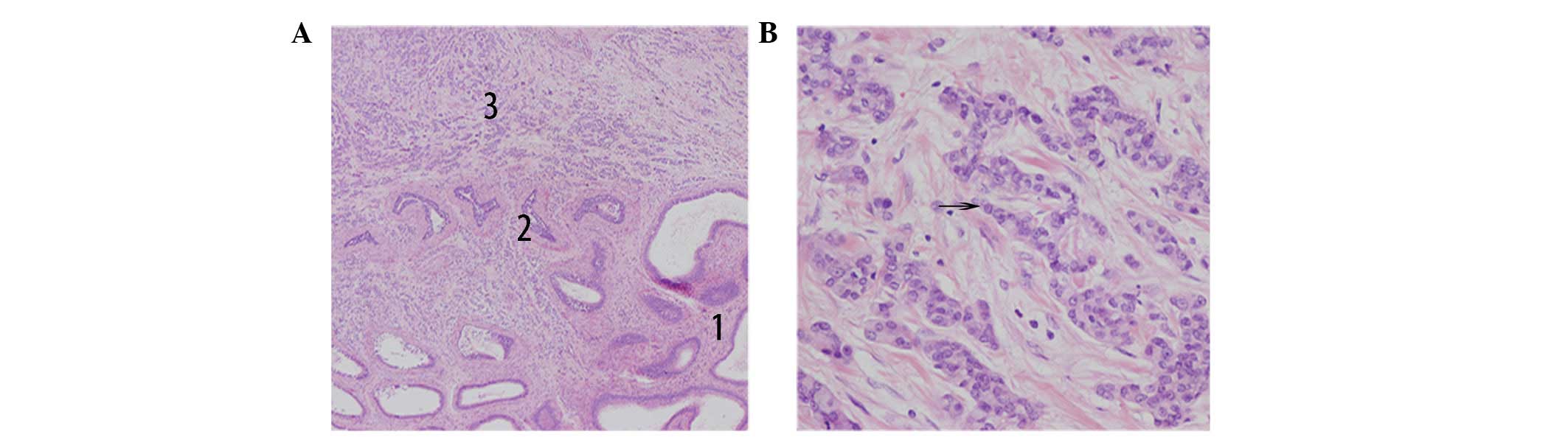
Light microscopic examination revealed an absence of
normal testicular microstructure, with nests of cells separated by
fibrovascular stroma. In addition, a transition from normal to
tumorous epithelium was detected. A small portion of lumens of the
rete testis were packed with cuboidal tumorous cells. Cells
exhibited heterotypic hyperplasia and were partially arranged in a
glandular pattern, with hyperchromatic nuclei, an increase in the
nuclear/cytoplasmic ratio and a visible nucleolus (Fig. 2). No involvement of the tumor to the
testicular parenchyma or tunica was detected. A diagnosis of
poorly-differentiated adenocarcinoma of the rete testis was
established based on the diagnostic criteria of Nochomovitz and
Orenstein (1).
Patient outcome
Taking into account that adenocarcinoma of the rete
testis is highly resistant to adjuvant radiotherapy and any known
chemotherapeutic regimens (4,6), no
further treatment was administered. Follow-up was carried out by
serum LDH, AFP and β-HCG testing and abdominal CT every three
months. The patient subsequently underwent metastasis at multiple
sites and succumbed to adenocarcinoma 11 months following
surgery.
Discussion
Primary adenocarcinoma of the rete testis is
resistant to adjuvant therapy and is associated with a poor
prognosis. As many as 40% of patients succumb to this condition
within one year of diagnosis. Survival rates for 3- and 5-years are
49 and 13%, respectively (4). Early
diagnosis with surgical management is recommended by the majority
of urologists (4,7–9). There
are no specific clinical manifestations but tumor markers,
including AFP and β-HCG, may help to detect the tumor earlier.
CT-positron emission tomography may provide improved diagnostic
sensitivity but is considered expensive and is not cost-effective
(10). Ultrasound has been shown to
be a reliable and valuable tool in the diagnosis of scrotal
abnormalities. This procedure is relatively cheap and noninvasive.
In addition, it provides real-time imaging, reveals internal blood
flow properties, causes little discomfort and is easily repeatable,
as well as being suitable for X-ray-sensitive organs as an ionizing
radiation-free test. Ultrasound diagnostics are therefore
recommended for confirming the presence of testicular masses
(11,12).
Ultrasonography provides information regarding
composition of the lesion which may facilitate diagnosis and
differentiation from other pathological tumor types. Adenocarcinoma
of the rete testis is typically located in the region of the
epididymis or testicular hilum, rather than the intratesticular
region, as reported in the majority of the current literature
(3,8,13,14).
The majority of patients exhibit hydrocele, and echoic
paratesticular regions are observed (3,15,16).
Nodular septations with cystic solid components have also been
reported as an unusual observation which varies from other
pathological tumor types (17). In
the majority of cases, the lesions present as hypoechoic masses
with poorly-defined borders, although an uneven echo pattern is
detected on occasion (3,9). Increased vascularization of the tumor
is another ultrasonographic feature which is helpful in
differential diagnosis (8). The
case presented in the current report is in accordance with the
majority of these features.
In a number of cases, other imaging methods,
including CT, magnetic resonance imaging and nuclear medicine, may
be necessary to complete the imaging work-up of patients with
testicular tumors. However, pathological examination is still the
gold standard for diagnosis confirmation.
In conclusion, adenocarcinoma of the rete testis is
an extremely rare tumor type with a poor prognosis. Sonography is
the most promising tool for early diagnosis and increased case
examples providing sonographic tumor observations must be presented
to achieve an improved rate of diagnosis.
References
|
1
|
Nochomovitz LE and Orenstein JM:
Adenocarcinoma of the rete testis. Case report, ultrastructural
observations, and clinicopathologic correlates. Am J Surg Pathol.
8:625–634. 1984. View Article : Google Scholar : PubMed/NCBI
|
|
2
|
Gruber H, Ratschek M, Pummer K, et al:
Adenocarcinoma of the rete testis: report of a case with surgical
history of adenomatous hyperplasia of the rete testis. J Urol.
158:1525–1526. 1997. View Article : Google Scholar : PubMed/NCBI
|
|
3
|
Wu CA, Chen YH, Man KM, et al: Papillary
adenocarcinoma of rete testis mimics inflammatory lump: a case
report. Case Rep Urol. 2011:8578122011.PubMed/NCBI
|
|
4
|
Sánchez-Chapado M, Angulo JC and Haas GP:
Adenocarcinoma of the rete testis. Urology. 46:468–475.
1995.PubMed/NCBI
|
|
5
|
London NJ, Smart JG, Kinder RB, et al:
Prospective study of routine scrotal ultrasonography in urological
practice. Br J Urol. 63:416–419. 1989. View Article : Google Scholar : PubMed/NCBI
|
|
6
|
Jones EC, Murray SK and Young RH: Cysts
and epithelial proliferations of the testicular collecting system
(including rete testis). Semin Diagn Pathol. 17:270–293.
2000.PubMed/NCBI
|
|
7
|
Fukunaga M, Aizawa S, Furusato M, et al:
Papillary adenocarcinoma of the rete testis: a case report. Cancer.
50:134–138. 1982. View Article : Google Scholar : PubMed/NCBI
|
|
8
|
Mermershtain W, Vardi N, Gusakova I and
Klein J: Serous papillary adenocarcinoma of the rete testis:
unusual ultrasonography and pathological findings. J Cancer Res
Ther. 3:37–39. 2007. View Article : Google Scholar : PubMed/NCBI
|
|
9
|
Perimenis P, Athanasopoulos A and Speakman
M: Primary adenocarcinoma of the rete testis. Int Urol Nephrol.
35:373–374. 2003. View Article : Google Scholar : PubMed/NCBI
|
|
10
|
Musser JE, Ernest AJ, Thibault GP and
McMann LP: Primary adenocarcinoma of the rete testis: improved
staging accuracy with CT-PET. Urology. 77:3342011. View Article : Google Scholar : PubMed/NCBI
|
|
11
|
Motzer RJ, Agarwal N, Beard C, et al: NCCN
clinical practice guidelines in oncology: testicular cancer. J Natl
Compr Canc Netw. 7:672–693. 2009.PubMed/NCBI
|
|
12
|
Albers P, Albrecht W, Algaba F, et al:
Guidelines on testicular cancer. Eur Urol. 48:885–894. 2005.
View Article : Google Scholar : PubMed/NCBI
|
|
13
|
Mehra BR, Thawait AP, Narang RR, Gangane
NM and Vyas VJ: Adenocarcinoma of the rete testis with uncommon
presentation as haematocele. Singapore Med J. 48:e311–e313.
2007.PubMed/NCBI
|
|
14
|
Amin MB: Selected other problematic
testicular and paratesticular lesions: rete testis neoplasms and
pseudotumors, mesothelial lesions and secondary tumors. Mod Pathol.
18(Suppl 2): S131–S145. 2005. View Article : Google Scholar
|
|
15
|
Nakagawa T, Hiraoka N, Ihara F, Komiyama
M, Kanai Y and Matsuno Y: Primary adenocarcinoma of the rete testis
with preceding diagnosis of pulmonary metastases. Int J Urol.
13:1532–1535. 2006. View Article : Google Scholar : PubMed/NCBI
|
|
16
|
Hagiuda J, Matsumoto M, Hanawa Y, Ishikawa
H and Marumo K: Adenocarcinoma of the rete testis. A case report.
Nihon Hinyokika Gakkai Zasshi. 101:749–753. 2010.(In Japanese).
|
|
17
|
Glazier DB, Vates TS, Cummings KB and
Antoun S: Adenocarcinoma of the rete testis. World J Urol.
14:397–400. 1996. View Article : Google Scholar : PubMed/NCBI
|