Introduction
Oral leukoplakia is the most common type of
premalignant lesion affecting the oral mucosa (1,2).
Occasionally, types of oral cancer are preceded by clinically
evident potential malignant oral disorders. Leukoplakia is the most
common of these disorders and exhibits a malignant transformation
rate ranging between 0.6 and 18% (3). Its malignant transformation rate may
be directly associated with the severity of epithelial dysplasia,
as it ranges between 5% for leukoplakia with mild epithelial
dysplasia and 43% for leukoplakia with severe epithelial dysplasia
(4). Thus, the risk of the
malignant transformation of leukoplakia may be evaluated based on
microscopic assessments of epithelial dysplasia (5–7). Oral
squamous cell carcinoma (OSCC) is considered to develop from
precancerous dysplastic lesions via multi-step carcinogenic
processes, in which oncogene activation and the loss of tumor
suppressor gene expression are the key features (8).
As a family of transcription factors, homeobox genes
are not only important in embryonic development and
differentiation, but also control the differentiation and
proliferation of mature tissues (9). Paired-like homeodomain 1 (PITX1) was
originally described as a bicoid-related homeobox transcription
factor that is involved in the transcription of the
proopiomelanocortin gene in the adult pituitary. In addition, PITX1
is involved in the differentiation of pituitary cells and the
formation of the pituitary gland (10). PITX1 is exclusively expressed
throughout the developing hindlimb, but not the forelimb bud; it
determines the morphology of the muscles, tendons and bones of the
hindlimbs (11,12). The development of the oral
epithelium, the first branchial arch and its derivatives, are also
known to require PITX1 (13,14).
In previous studies, the downregulation of PITX1
expression has been consistently associated with human OSCC
(15), esophageal (16), gastric (17,18),
bronchial (19,20), hepatic (21), colorectal (22), pancreatic (23) and prostatic (24) malignancies, as well as malignant
melanoma (18,25). However, the clinical significance of
PITX1 expression in the development of OSCC remains unclear. In the
present study, PITX1 expression was examined in oral epithelial
dysplasia, which is considered to be a precancerous lesion of
OSCC.
Materials and methods
Tissue samples
Tissue samples were analyzed from 26 individuals
with normal oral mucosa, 106 cases of oral epithelial dysplasia and
97 OSCC patients. All the specimens were obtained from the Division
of Oral and Maxillofacial Biopathological Surgery, Tottori
University Faculty of Medicine (Yanago, Japan). Approval for the
study was obtained from the institutional review board of Tottori
University Faculty of Medicine (no. 1558). All specimens were fixed
in 10% buffered formalin and embedded in paraffin wax. The
resultant paraffin blocks were sectioned into 4-μm slices. All
patients provided written informed consent.
The histological diagnosis of OSCC and oral
epithelial dysplasia was performed according to the World Health
Organization criteria for the histological typing of cancer and
precancer of the oral mucosa (26).
Immunohistochemistry
All the specimens were fixed with 10% formalin and
embedded in paraffin. Histofine SAB-PO®
immunohistochemical staining kit (Nichirei Corporation, Tokyo,
Japan) and 4-μm-thick sections were used for the
immunohistochemical analysis. As primary antibodies, a rabbit
polyclonal antibody raised against PITX1 (1:800; Abcam, Cambridge,
UK) and a mouse monoclonal antibody raised against Ki-67 (1:50;
MIB-1; DakoCytomation, Glostrup, Denmark) were used. Briefly,
paraffin-embedded sections were dewaxed with xylene and gradually
hydrated. Antigen retrieval was performed by autoclaving in 10 mM
citrate buffer (pH 6.0) for 10 min after endogenous peroxidase
activity had been blocked by immersing the sections in 0.3%
hydrogen peroxide in methanol for 30 min. The sections were then
reacted with each primary antibody overnight at 4°C, prior to being
treated with secondary antibody and biotin-streptavidin complex
(Nichirei Corporation, Tokyo, Japan) for 30 min each at 37°C. The
resultant immunoreactions were visualized with diaminobenzidine
(DakoCytomation, Glostrup, Denmark) and the sections were
counterstained with hematoxylin (Wako Pure Chemical, Osaka,
Japan).
Evaluation of immunohistochemical
observations
To evaluate PITX1 and Ki-67 expression, images of
positive tumor cell nuclei were captured using a charge-coupled
device camera (Nikon, Tokyo, Japan) in the most strongly labeled
region. Subsequently, the number of positive cells was counted in
high-magnification fields using the FLVFS Image Filing Software
(Flovel, Co., Ltd., Tachikawa, Japan) and the percentage of
positive cells was determined for each antibody. The numbers of
normal squamous, dysplastic and OSCC cells were counted among
>1,000 cells and the percentage of positively stained cells was
designated as the labeling index (LI; %).
Statistical analysis
The correlations between the PITX1 or Ki-67 LIs and
malignant transformation were calculated using Pearson’s
correlation test. Student’s t-test or Mann-Whitney U test were used
for comparisons between two categorical variables. P<0.05 was
considered to indicate a statistically significant difference.
Results
PITX1 expression levels in the normal
oral mucosa, oral epithelial dysplasia and OSCC
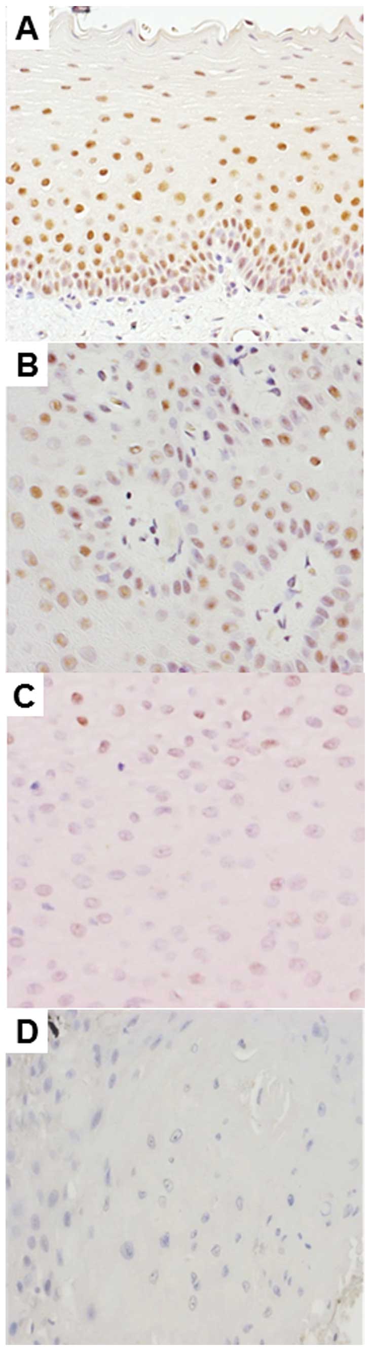
Immunohistochemical analysis showed that
PITX1-positive cells were distributed in the basal cell layer of
the normal oral mucosa (Fig. 1A)
and in numerous oral epithelial dysplasia samples (Fig. 1B). By contrast, only weak nuclear
staining was detected in the OSCC (Fig.
1D) and sections of oral epithelial dysplasia samples (Fig. 1C). The PITX1 LIs of the normal oral
mucosa, oral epithelial dysplasia and OSCC specimens are summarized
in Fig. 2. The PITX1 LI was
72.8±6.5, 52.3±9.24 and 4.8±4.25 [mean ± standard deviation (SD)]
in the normal oral mucosa, oral epithelial dysplasia and OSCC
samples, respectively. The mean PITX1 LI of the oral epithelial
dysplasia samples was significantly lower than that of the normal
oral mucosa specimens, but significantly higher than that of the
OSCC samples (P<0.001).
Downregulation of PITX1 expression in
oral epithelial dysplasia is a predictive marker of malignant
transformation in OSCC
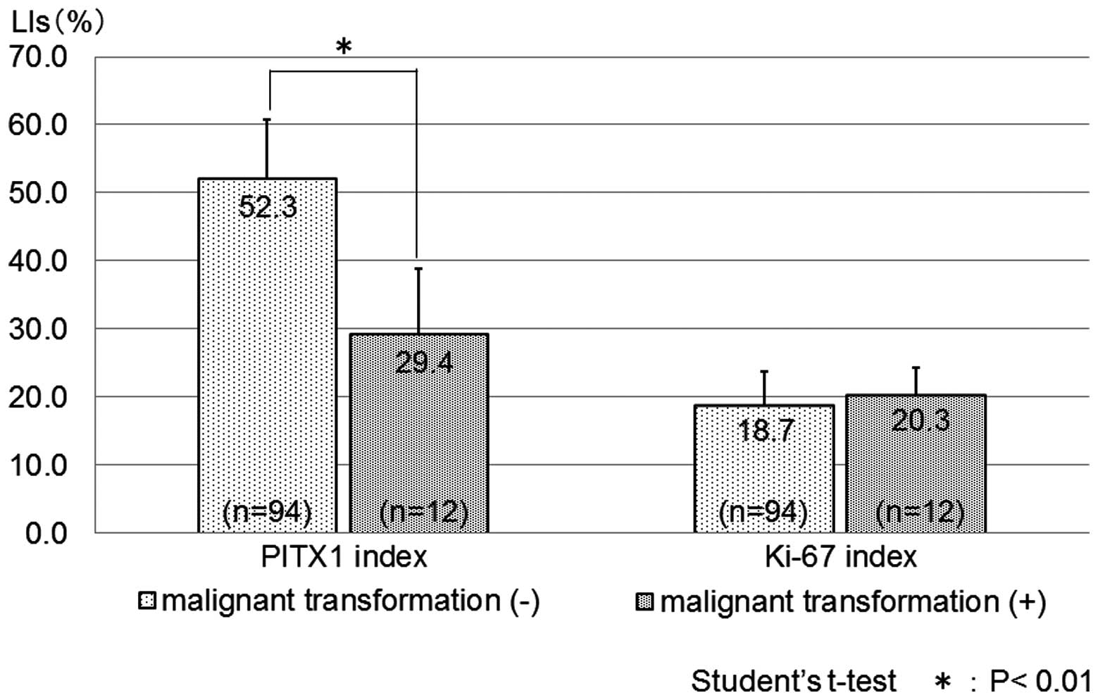
The malignant transformation of oral epithelial
dysplasia into OSCC occurred in 12 (11.3%) of the 106 patients with
oral epithelial dysplasia. Immunohistochemical analysis showed that
the malignant transformation-negative oral epithelial dysplasia
cases were detected for PITX1 nuclear staining (Fig. 1B). By contrast, only weak nuclear
staining was detected in the malignant transformation-positive oral
epithelial dysplasia cases (Fig.
1C). The PITX1 LI was 52.3±8.40 (mean ± SD) in the cases of
oral epithelial dysplasia that did not undergo malignant
transformation (P<0.01) and 29.4±9.40 in those that did
(Fig. 3), and the difference was
significant (P<0.01). In addition, low PITX1 expression was
found to significantly correlate with malignant transformation.
By contrast, the Ki-67 LIs were 18.7±5.07 (mean ±
SD) in the malignant transformation-negative oral epithelial
dysplasia cases (P<0.01) and 20.3±3.99 in the malignant
transformation-positive cases (Fig.
3). The difference between the two groups was not
significant.
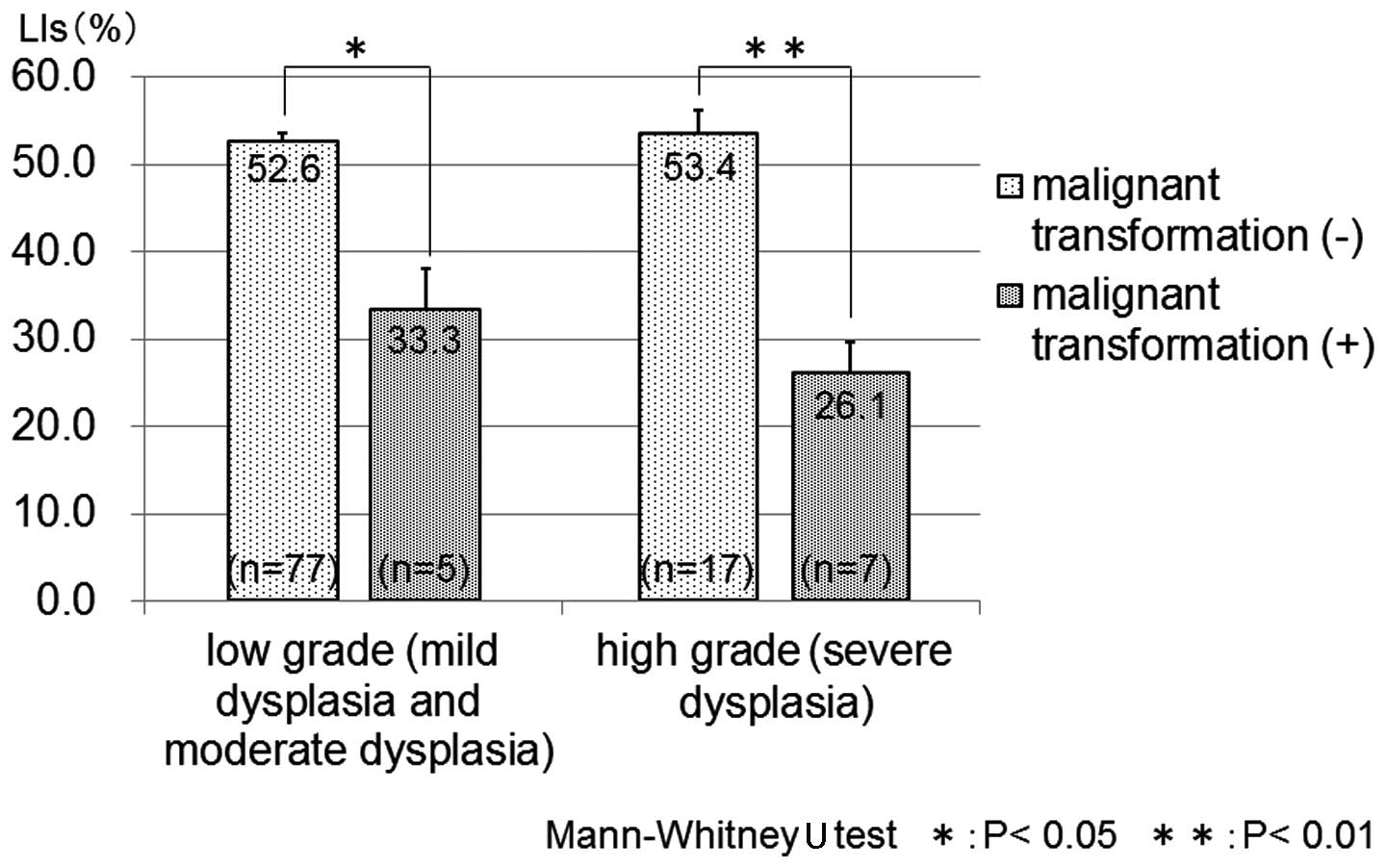
Next, the oral dysplasia cases were separated into
low-grade (mild or moderate dysplasia) and high-grade (severe
dysplasia) groups. In the low-grade group, the PITX1 LI was
52.6±0.94 [median ± standard error (SE)] for malignant
transformation-negative oral epithelial dysplasia and 33.3±4.85 for
malignant transformation-positive oral epithelial dysplasia. In the
high-grade group, the PITX1 LI was 53.4±2.75 (median ± SE) for
malignant transformation-negative oral epithelial dysplasia and
26.1±3.53 for malignant transformation-positive oral epithelial
dysplasia. The median PITX1 LI was significantly decreased in
malignant transformation-positive oral epithelial dysplasia,
regardless of the histological grade of the dysplasia (Fig. 4).
Discussion
The activation of oncogenes, inactivation of tumor
suppressor genes and increased cellular proliferation are key
events in the multi-step carcinogenesis process (27). The presence of oral epithelial
dysplasia is generally accepted as one of the most important
predictors of malignant development in premalignant oral lesions
(1,4). In addition, the presence of certain
genetic abnormalities in carcinoma-carrying patients may be an
important prognostic indicator of patient survival. Such indicators
may be useful in the clinical setting for selecting the initial
treatment or developing tumor-specific therapies. Accordingly, a
number of previous molecular studies have attempted to detect
prognostic indicators for oral cancer. Several proteins, including
Mcm2 (27,28), p53 (29), Bcl-2 family (30), ΔNp63 (31) and Ki-67 (29,32–34),
have been identified as prognostic markers in oral epithelial
dysplasia. In particular, a number of previous studies have
examined Ki-67 expression in the transformation of oral epithelial
dysplasia. The Ki-67 antigen was first reported by Gerdes et
al, which is a nuclear protein associated with cellular
proliferation (35). Ki-67 has
since become one of the biomarkers frequently used as a prognostic
indicator and proliferation marker (36). Ki-67 is a nuclear non-histone
protein that is maximally expressed in cells in the G2 and M phases
of the cell cycle, but is absent from resting cells. Therefore,
Ki-67 may be employed to evaluate the percentage of proliferating
cells in normal tissues, as well as premalignant and malignant
lesions (34). Previous studies
have suggested a strong correlation between Ki-67 expression and
the degree of dysplasia, indicating that Ki-67 may be involved in
proliferative events as well as neoplastic transformation (33). However, in the present study, the
Ki-67 LI did not differ between the malignant
transformation-positive and -negative cases of oral epithelial
dysplasia, indicating that the Ki-67 LI is not useful as a
prognostic marker in oral epithelial dysplasia. By contrast, it was
found that PITX1 downregulation was significantly more common in
malignant transformation-positive oral epithelial dysplasia cases
than in malignant transformation-negative cases, independent of the
histological grade of dysplasia.
The reduced PITX1 expression observed in the
malignant cases suggested that PITX1 may have a tumor-suppressing
function. This encouraged the current study to examine its
potential role in the carcinogenesis of oral epithelium.
Previously, Kolfschoten et al reported that PITX1 suppresses
tumorigenicity by downregulating the Ras pathway through RASAL1, a
member of the Ras-GTPase activating protein family of genes
(GTP-activating negative regulators of Ras expression) (37). Activation of the
Ras/mitogen-activated protein kinase (MAPK) pathway contributes to
the tumorigenesis and progression of OSCC and inhibition of the
pathway was found to suppress the proliferation of human OSCC
(15). Therefore, it has been
suggested that the loss of PITX1 expression results in the
activation of the Ras/MAPK pathway via the downregulation of
RASAL1, leading to the hyperproliferation of oral epithelia.
The present study was the first to report a
correlation between PITX1 and oral epithelial dysplasia. The
downregulation of PITX1 in oral epithelial cells may be involved in
the carcinogenesis of OSCC. Thus, PITX1 is considered to be a
candidate tumor suppressor gene. In addition, PITX1 may serve as a
novel biomarker for predicting prognosis in oral epithelial
dysplasia. Further studies must examine the mechanisms that
modulated PITX1 expression during the progression of OSCC.
Acknowledgements
The authors would like to thank Mr. N. Itaki for
providing excellent technical assistance. The present study was
supported by a Grant-in-Aid for Scientific Research (C; no.
22501012) from the Ministry of Education, Culture, Sports, Science
and Technology of Japan.
References
|
1
|
Bánóczy J and Csiba A: Occurrence of
epithelial dysplasia in oral leukoplakia. Analysis and follow-up
study of 12 cases. Oral Surg Oral Med Oral Pathol. 42:766–774.
1976.
|
|
2
|
van der Waal I: Potentially malignant
disorders of the oral and oropharyngeal mucosa; terminology,
classification and present concepts of management. Oral Oncol.
45:317–323. 2009.
|
|
3
|
Gupta PC, Mehta FS, Daftary DK, et al:
Incidence rates of oral cancer and natural history of oral
precancerous lesions in a 10-year follow-up study of Indian
villagers. Community Dent Oral Epidemiol. 8:283–333.
1980.PubMed/NCBI
|
|
4
|
Silverman S Jr, Gorsky M and Lozada F:
Oral leukoplakia and malignant transformation. A follow-up study of
257 patients. Cancer. 53:563–568. 1984. View Article : Google Scholar : PubMed/NCBI
|
|
5
|
Bouquot JE and Whitaker SB: Oral
leukoplakia - rationale for diagnosis and prognosis of its clinical
subtypes or ‘phases’. Quintessence Int. 25:133–140. 1994.PubMed/NCBI
|
|
6
|
Napier SS and Speight PM: Natural history
of potentially malignant oral lesions and conditions: an overview
of the literature. J Oral Pathol Med. 37:1–10. 2008. View Article : Google Scholar : PubMed/NCBI
|
|
7
|
Piemonte ED, Lazos JP and Brunotto M:
Relationship between chronic trauma of the oral mucosa, oral
potentially malignant disorders and oral cancer. J Oral Pathol Med.
39:513–517. 2010.PubMed/NCBI
|
|
8
|
Reibel J: Prognosis of oral pre-malignant
lesions: significance of clinical, histopathological, and molecular
biological characteristics. Crit Rev Oral Biol Med. 14:47–62. 2003.
View Article : Google Scholar
|
|
9
|
Imoto I, Pimkhaokham A, Watanabe T, et al:
Amplification and overexpression of TGIF2, a novel homeobox gene of
the TALE superclass, in ovarian cancer cell lines. Biochem Biophys
Res Commun. 276:264–270. 2000. View Article : Google Scholar : PubMed/NCBI
|
|
10
|
Lamonerie T, Tremblay JJ, Lanctôt C, et
al: Ptx1, a bicoid-related homeo box transcription factor involved
in transcription of the pro-opiomelanocortin gene. Genes Dev.
10:1284–1295. 1996. View Article : Google Scholar : PubMed/NCBI
|
|
11
|
DeLaurier A, Schweitzer R and Logan M:
Pitx1 determines the morphology of muscle, tendon, and bones of the
hindlimb. Dev Biol. 299:22–34. 2006. View Article : Google Scholar : PubMed/NCBI
|
|
12
|
Shang J, Li X, Ring HZ, et al: Backfoot, a
novel homeobox gene, maps to human chromosome 5 (BFT) and mouse
chromosome 13 (Bft). Genomics. 40:108–113. 1997. View Article : Google Scholar : PubMed/NCBI
|
|
13
|
Lanctôt C, Moreau A, Chamberland M, et al:
Hindlimb patterning and mandible development require the Ptx1 gene.
Development. 126:1805–1810. 1999.PubMed/NCBI
|
|
14
|
Shang J, Luo Y and Clayton DA: Backfoot is
a novel homeobox gene expressed in the mesenchyme of developing
hindlimb. Dev Dyn. 209:242–253. 1997. View Article : Google Scholar : PubMed/NCBI
|
|
15
|
Libório TN, Acquafreda T,
Matizonkas-Antonio LF, et al: In situ hybridization detection of
homeobox genes reveals distinct expression patterns in oral
squamous cell carcinomas. Histopathology. 58:225–233.
2011.PubMed/NCBI
|
|
16
|
Lord RV, Brabender J, Wickramasinghe K, et
al: Increased CDX2 and decreased PITX1 homeobox gene expression in
Barrett’s esophagus and Barrett’s-associated adenocarcinoma.
Surgery. 138:924–931. 2005.PubMed/NCBI
|
|
17
|
Chen YN, Chen H, Xu Y, et al: Expression
of pituitary homeobox 1 gene in human gastric carcinogenesis and
its clinicopathological significance. World J Gastroenterol.
14:292–297. 2008. View Article : Google Scholar : PubMed/NCBI
|
|
18
|
Qi DL, Ohhira T, Fujisaki C, et al:
Identification of PITX1 as a TERT suppressor gene located on human
chromosome 5. Mol Cell Biol. 31:1624–1636. 2011. View Article : Google Scholar : PubMed/NCBI
|
|
19
|
Chen Y, Knösel T, Ye F, et al: Decreased
PITX1 homeobox gene expression in human lung cancer. Lung Cancer.
55:287–294. 2007. View Article : Google Scholar : PubMed/NCBI
|
|
20
|
Stender JD, Stossi F, Funk CC, et al: The
estrogen-regulated transcription factor PITX1 coordinates
gene-specific regulation by estrogen receptor-alpha in breast
cancer cells. Mol Endocrinol. 25:1699–1709. 2011. View Article : Google Scholar
|
|
21
|
Calvisi DF, Ladu S, Conner EA, et al:
Inactivation of Ras GTPase-activating proteins promotes
unrestrained activity of wild-type Ras in human liver cancer. J
Hepatol. 54:311–319. 2011. View Article : Google Scholar
|
|
22
|
Knösel T, Chen Y, Hotovy S, et al: Loss of
desmocollin 1–3 and homeobox genes PITX1 and CDX2 are associated
with tumor progression and survival in colorectal carcinoma. Int J
Colorectal Dis. 27:1391–1399. 2012.
|
|
23
|
Hamidov Z, Altendorf-Hofmann A, Chen Y, et
al: Reduced expression of desmocollin 2 is an independent
prognostic biomarker for shorter patients survival in pancreatic
ductal adenocarcinoma. J Clin Pathol. 64:990–994. 2011. View Article : Google Scholar
|
|
24
|
Kwok SC, Liu X, Mangel P and Daskal I:
PTX1(ERGIC2)-VP22 fusion protein upregulates interferon-beta in
prostate cancer cell line PC-3. DNA Cell Biol. 25:523–529. 2006.
View Article : Google Scholar : PubMed/NCBI
|
|
25
|
Osaki M, Chinen H, Yoshida Y, et al:
Decreased PITX1 gene expression in human cutaneous malignant
melanoma and its clinicopathological significance. Eur J Dermatol.
Jun 28–2013.(Epub ahead of print).
|
|
26
|
Pindborg JJ, Smith CJ and van der Waal I:
Histological typing of cancer and precancer of the oral mucosa (2nd
edition), World Health Organization. Springer-Verlag; Berlin: pp.
24–40. 1997
|
|
27
|
Kodani I, Shomori K, Osaki M, et al:
Expression of minichromosome maintenance 2 (MCM2), Ki-67, and
cell-cycle-related molecules, and apoptosis in the
normal-dysplasia-carcinoma sequence of the oral mucosa.
Pathobiology. 69:150–158. 2001. View Article : Google Scholar
|
|
28
|
Gouvêa AF, Vargas PA, Coletta RD, et al:
Clinicopathological features and immunohistochemical expression of
p53, Ki-67, Mcm-2 and Mcm-5 in proliferative verrucous leukoplakia.
J Oral Pathol Med. 39:447–452. 2010.PubMed/NCBI
|
|
29
|
Nasser W, Flechtenmacher C, Holzinger D,
et al: Aberrant expression of p53, p16INK4a and Ki-67 as basic
biomarker for malignant progression of oral leukoplakias. J Oral
Pathol Med. 40:629–635. 2011. View Article : Google Scholar : PubMed/NCBI
|
|
30
|
Nogami T, Kuyama K and Yamamoto H:
Histopathological and immunohistochemical study of malignant
transformation of oral leukoplakia, with special reference to
apoptosis-related gene products and proliferative activity. Acta
Otolaryngol. 123:767–775. 2003.
|
|
31
|
Matsubara R, Kuwano S, Kiyosue T, et al:
Increased ΔNp63 expression is predictive of malignant
transformation in oral epithelial dysplasia and poor prognosis in
oral squamous cell carcinoma. Int J Oncol. 39:1391–1399. 2011.
|
|
32
|
Thomson PJ, Soames JV, Booth C and O’Shea
JA: Epithelial cell proliferative activity and oral cancer
progression. Cell Prolif. 35(Suppl 1): S110–S120. 2002. View Article : Google Scholar
|
|
33
|
Kumar P, Kane S and Rathod GP:
Coexpression of p53 and Ki 67 and lack of c-erbB2 expression in
oral leukoplakias in India. Braz Oral Res. 26:228–234. 2012.
View Article : Google Scholar : PubMed/NCBI
|
|
34
|
Iamaroon A, Khemaleelakul U, Pongsiriwet S
and Pintong J: Co-expression of p53 and Ki67 and lack of EBV
expression in oral squamous cell carcinoma. J Oral Pathol Med.
33:30–36. 2004. View Article : Google Scholar : PubMed/NCBI
|
|
35
|
Gerdes J, Lemke H, Baisch H, et al: Cell
cycle analysis of a cell proliferation-associated human nuclear
antigen defined by the monoclonal antibody Ki-67. J Immunol.
133:1710–1715. 1984.PubMed/NCBI
|
|
36
|
Oliveira LR and Ribeiro-Silva A:
Prognostic significance of immunohistochemical biomarkers in oral
squamous cell carcinoma. Int J Oral Maxillofac Surg. 40:298–307.
2011. View Article : Google Scholar : PubMed/NCBI
|
|
37
|
Kolfschoten IG, van Leeuwen B, Berns K, et
al: A genetic screen identifies PITX1 as a suppressor of RAS
activity and tumorigenicity. Cell. 121:849–858. 2005. View Article : Google Scholar : PubMed/NCBI
|