Introduction
Esophageal carcinoma is one of the most common types
of malignancy in China, and squamous cell carcinoma (SCC) is the
main histological type. Concurrent chemoradiotherapy (CCRT) is an
accepted standard treatment for patients with locally advanced
esophageal squamous cell carcinoma (ESCC). However, prognosis for
these patients remains poor (1–3), with
a five-year overall survival (OS) rate of ~20% (4). The median survival time (MST) of all
patients with T3–4M0 esophageal cancer who received CCRT was
demonstrated to be only 16 months (5). Local recurrence and distant metastasis
following definitive chemoradiation are the primary patterns of
failure (6). Therefore, predicting
the failure patterns and OS following CCRT is important. However,
identification of reliable markers predicting treatment outcome
following CCRT remains limited in the previous literature (7). In the surgical groups of previous
studies, epidermal growth factor receptor (EGFR) overexpression has
been found in ESCC and may predict the postoperative recurrence and
OS (6,8–10). The
clinical importance of EGFR overexpression remains unsettled in
ESCC patients undergoing CCRT (11). In the present study, the prognostic
relevance of EGFR was studied in locally advanced ESCC.
Materials and methods
Patients
In total, 47 locally advanced ESCC patients with a
median age of 63 years (range, 45–72 years) were admitted to the
Shandong Cancer Hospital and Institute, University of Jinan (Jinan,
China). All patients fulfilled the following criteria: i)
histologically confirmed ESCC; ii) no previous treatment; iii)
endoscopically evaluable primary lesions; iv) Karnofsky Performance
Status scale of 70–100; v) retained function of the major organs
(bone marrow, heart, liver and kidneys); vi) no significant medical
disease (such as myocardial infarction and pneumonectasis); vii)
clinically diagnosed T2–4, Nany and Many
(Union for International Cancer Control, 6th edition; 2002); viii)
physical examination and computed tomography (CT) performed prior
to and following treatment; ix) received 5-fluorouracil and
cisplatin (CF scheme); and x) written informed consent obtained
prior to treatment. All patients were administered the same regimen
of CCRT.
Immunohistochemical staining methods
Histological analysis confirmed that all esophageal
tumors were squamous cell carcinoma. All pretherapeutic endoscopic
biopsy specimens were examined for EGFR expression.
Immunohistochemical staining was performed with labeled
streptavidin biotin (LSAB) method using a Dako LSAB kit (Dako,
Carpinteria, CA, USA). Primary antibody used for the
immunohistochemical staining was anti-EGFR monoclonal antibody
(dilution of 1:60; clone 31G7; Cytomed GmbH, Baden-Baden,
Germany).
Formalin-fixed, paraffin-embedded biopsy samples
were cut into 4 μm sections. Following deparaffinization, the
sections were incubated three times in a microwave oven for 10 min
and incubated with 0.3% H2O2 for 30 min.
Next, these sections were incubated with the primary antibody.
Following six washes in phosphate-buffered saline, sections were
incubated with rabbit monoclonal antibodies against EGFR for 20 min
at room temperature. The primary antibodies were localized by the
sequential application of biotinylated goat polyclonal anti-rabbit
IgG gout immunoglobulins and streptavidin-peroxide conjugate
(Dako). Immunostaining was visualized by developing the slides in
diaminobenzidine (Dako) and counterstaining with Mayer’s
hematoxylin (Abcam, Cambridge, UK). Finally, the slides underwent
alcohol and xylene immersion and were mounted for examination. For
the negative controls, the primary antibody solutions were replaced
with blocking buffer.
Staining evaluation
The sections were evaluated by two pathologists who
were not informed of the results of chemoradiotherapy and the
patients’ follow-up. The immunoreactivity of EGFR was characterized
into the following five grades according to the percentage of
immunoreactive tumor cells: 0, 0–4% positive tumor cells; 1, 5–24%;
2, 25–49%; 3, 50–74%; and 4, 75–100%. Staining grades of 3 and 4
were defined as positive for EGFR expression, while staining grades
of 0, 1 and 2 were defined as negative, consistent with previous
interpretations of EGFR in ESCC (12).
Treatment schedule
Chemotherapy and radiotherapy were initiated on the
same day. Patients received a total radiation dose of ~60 Gy,
administered in 30 fractions (1.8–2 Gy per fraction; five times per
week). Radiation was delivered by high-energy (≥6 MV) linear
accelerators as a requirement. Three-dimensional treatment planning
was used to ensure adequate dose delivery to the target while
simultaneously limiting the total dose to normal structures. All
fields were treated each day (five times per week). The gross tumor
volume was defined as any evidence of disease as documented by
pretreatment staging procedures, including CT, positron emission
tomography or endoscopic ultrasonography. The clinical target
volume was defined as the gross tumor volume plus inclusion of the
regional draining lymphatics based on the primary tumor location.
The planning target volume was based on tumor size as assessed by
CT or endoscopy (whichever was larger), with superior and inferior
borders extending 5 cm beyond the tumor and lateral borders
extending 1.5 cm beyond the tumor. A barium swallow radiograph was
also obtained at the time of treatment simulation to confirm the
location of the tumor and esophagus. The spinal cord dose did not
exceed 45.0 Gy. Doses to normal lung tissue were calculated by
dose-volume histograms. The maximum dose to the entire heart was
limited to 40.0 Gy, but a dose as high as 45.0 Gy could be
administered to <50% of the heart. Chemotherapeutics consisted
of the protracted infusion of 5-fluorouracil (750–1,000
mg/m2/day) on days one to five in combination with
cisplatin (30 mg/m2/day) with adequate hydration and
antiemetics continuous intravenous drip coverage between days one
and three A total of two cycles of chemotherapeutics were performed
during radiotherapy at four-week intervals. This was followed by
two more periods of chemotherapeutics with the same doses performed
at three-week intervals, three weeks following the completion of
radiotherapy (13).
Follow-up and observational indices
Patients were followed up at regular intervals
(every three to six months) after CCRT. The follow-up included CT
and barium swallow radiograph. Endoscopic ultrasonography was
adopted when abnormal esophagus was found by the abovementioned
examinations. In addition, the time of local recurrence, distant
metastasis and OS were documented. OS was defined as the interval
between the date of CCRT initiation and the date of mortality or
final follow-up. The deadline of the follow-up was December 20,
2012.
Statistical analysis
The SPSS software package, version 13.0 (SPSS, Inc.,
Chicago, IL, USA) was used for statistical analysis. A logistic
regression analysis was applied to evaluate the association between
the expression of EGFR and clinicopathological features. Survival
curves of the patients were calculated by the Kaplan-Meier method
and analyzed by the log-rank test. The prognostic significance of
clinicopathological factors was assessed using the Cox
proportional-hazards regression model. Two-sided significance
levels of P<0.05 were considered to indicate a statistically
significant difference.
Results
Patient characteristics
All patients with locally advanced ESCC treated with
CCRT at the Shandong Cancer Hospital and Institute, Jinan
University (Jinan, China) between December 2008 and November 2011
were candidates for the present study. In total, 47 patients with a
median age of 63 years (range, 45–72 years) fulfilled the inclusion
criteria and their clinicopathological features are presented in
Table I. All patients belonged to
TNM stage II/III.
 | Table IPatient characteristics. |
Table I
Patient characteristics.
| Patients |
|---|
|
|
|---|
| Characteristic | n | % |
|---|
| Age, years |
| <60 | 20 | 42.6 |
| ≥60 | 27 | 57.4 |
| Gender |
| Male | 30 | 63.8 |
| Female | 17 | 36.2 |
| Location |
| Cervical | 4 | 8.5 |
| Upper | 25 | 53.2 |
| Middle | 14 | 29.8 |
| Lower | 4 | 8.5 |
| Histological
grade |
| G1 | 13 | 27.7 |
| G2 | 21 | 44.6 |
| G3 | 13 | 27.7 |
| T stage |
| ≤T2a | 8 | 17 |
| T3 | 30 | 63.8 |
| T4 | 9 | 19.2 |
| N stage |
| N0 | 10 | 21.3 |
| N1 | 37 | 78.7 |
| M stage |
| M0 | 41 | 87.2 |
| M1a | 6 | 12.8 |
| TNM stage |
| II | 14 | 29.8 |
| III | 33 | 70.2 |
Immunoreactivity
All 47 samples were immunohistochemically detected
for EGFR. EGFR expression was observed in the cell membrane and
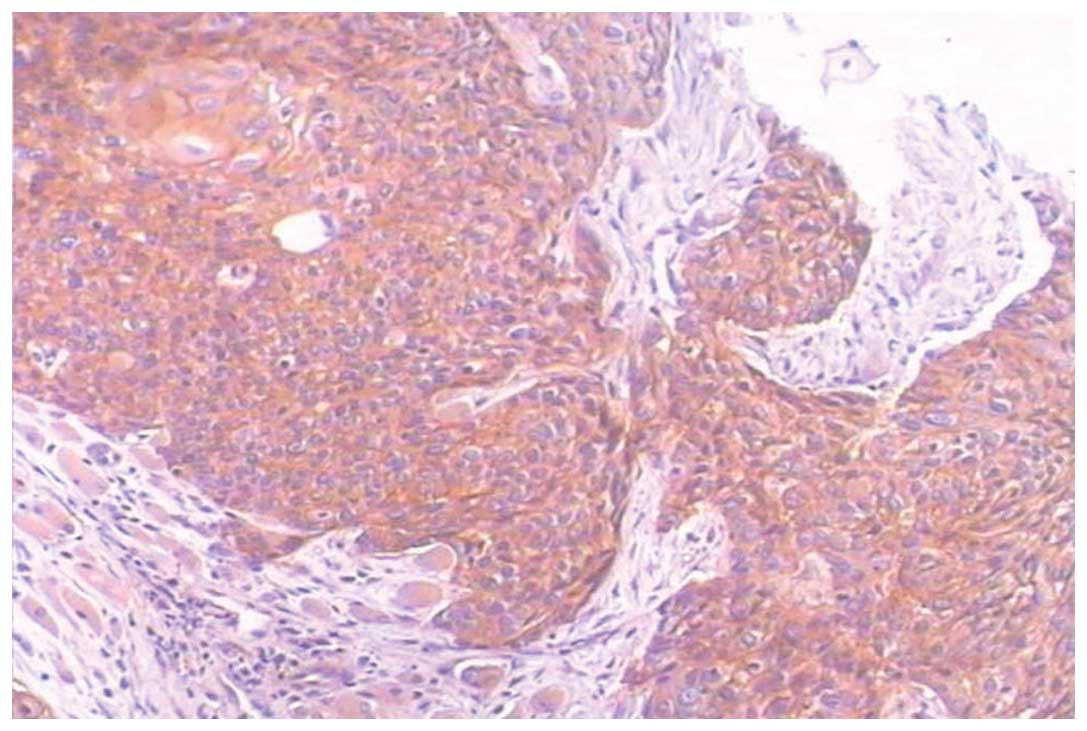
cytoplasm. Positive expression of EGFR (Fig. 1) in ESCC cells was observed in 28
(59.6%) cases; grade 3 in 11 (23.4%) cases and grade 4 in 17
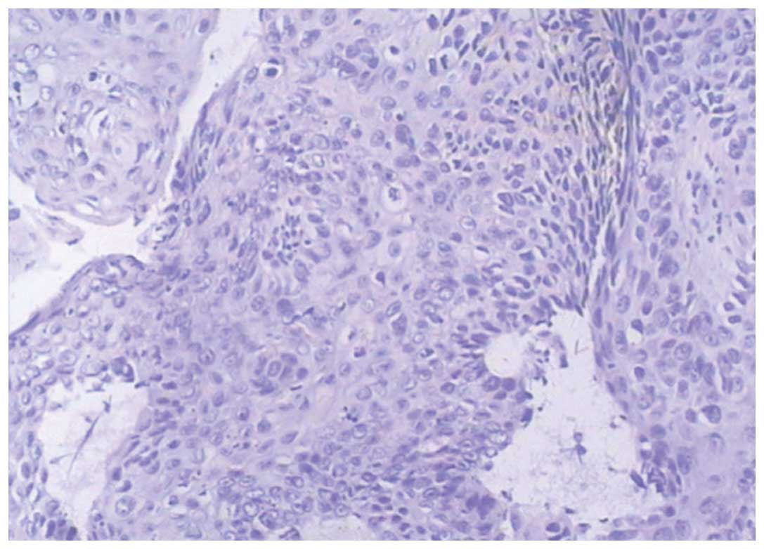
(36.2%) cases. In total, 19 (40.4%) cases were EGFR-negative
(Fig. 2); grade 0 in two (4.2%)
cases, grade 1 in seven (14.9%) cases and grade 2 in 10 (21.3%)
cases. EGFR overexpression was found to correlate with the presence
of lymph node metastasis (P=0.011; Table II). By contrast, no correlation was
detectable between EGFR overexpression and gender (P=0.120), age
(P=0.882), tumor differentiation grade (P=0.582), tumor location
(P=0.314), depth of invasion (T; P=0.593) and distant metastasis
(M; P=0.051).
| Table IIResults of the logistic regression
analysis between the expression of EGFR and clinicopathological
features. |
Table II
Results of the logistic regression
analysis between the expression of EGFR and clinicopathological
features.
| Features | B | SE | Wald | df | P-value | Exp (B) |
|---|
| Gender | −2.130 | 1.372 | 2.411 | 1 | 0.120 | 0.119 |
| Age, years | −0.180 | 1.214 | 0.022 | 1 | 0.882 | 0.836 |
| G | 0.648 | 1.175 | 0.304 | 1 | 0.582 | 1.911 |
| T | −1.092 | 2.045 | 0.285 | 1 | 0.593 | 0.335 |
| N | −6.445 | 2.520 | 6.541 | 1 | 0.011a | 0.002 |
| M | −4.121 | 2.115 | 3.798 | 1 | 0.051 | 0.016 |
| Location | −1.531 | 1.521 | 1.014 | 1 | 0.314 | 0.216 |
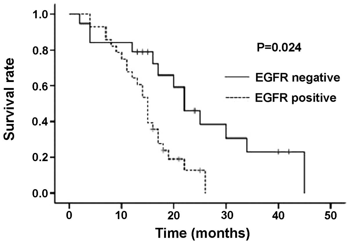
Survival analyses
The median duration of follow-up was 15 months and
only three patients were lost to follow-up. A difference in OS was
identified between patients with and without EGFR overexpression.
Fig. 3 shows the survival curves
according to EGFR expression using Kaplan-Meier analysis. The MST
of all 47 patients in the present study who received CCRT was 16.5
months. The MST of the EGFR-positive group was 15 months and the
MST of the EGFR-negative group was 23.5 months. A significant
difference was identified between the groups in terms of EGFR
expression (P=0.024). In total, 10 patients survived (four
EGFR-positive and six EGFR-negative cases), and of the 37 deceased
patients, 24 were EGFR-positive and 13 were EGFR-negative. In
addition, correlations between OS and gender (P=0.021), age
(P=0.018), T stage (P=0.035) and tumor location (P=0.023) were
detected in the Cox proportional hazard model. Local recurrence was
found to correlate with T (P=0.015) and M (P=0.026) stage, while
distant metastasis was found to correlate with age (P=0.048).
Discussion
In the present study, the expression of EGFR in a
series of 47 locally advanced ESCC patients was studied. In total,
59.6% of the biopsy specimens exhibited overexpression of EGFR on
immunohistochemical analysis. EGFR overexpression was revealed to
correlate with the presence of lymph node metastasis and poor
survival.
Previous studies have assessed the correlations
between EGFR overexpression and clinicopathological features in
ESCC. Firstly, certain studies have suggested that the expression
of EGFR significantly correlates with depth of invasion in ESCC
(14,15). The authors considered EGFR
overexpression to be a predictor of T stage. However, in the
current study, no correlation was observed between the expression
of EGFR and T stage (P=0.593). This result may be explained by the
fact that the T stage was determined by imaging and not by
pathology. Secondly, the expression of EGFR in lymph node-positive
groups was higher than in the negative groups (P=0.011). It must be
noted that the majority of the enrolled patients were locally
advanced and exhibited positive lymph nodes. Therefore, the results
may correlate with the deviation of the sample size. However, other
previous studies (14–17) have also demonstrated that EGFR
amplification or overexpression significantly correlates with lymph
node metastasis. Thirdly, the correlation between EGFR expression
and the differentiation degree of ESCC remains unclear. In a study
by Sunpaweravong et al (16), high-level protein expression of EGFR
was found to correlate with well-differentiated tumors (P=0.02),
while a correlation (P=0.032) was found between EGFR overexpression
and poorly differentiated histology in a study by Zhang et
al (18). However, in the
present study, no significant correlation was found between the
expression of EGFR and the differentiation degree of ESCC. This may
be the result of a small sample size. Finally, no significant
correlations were detected between the expression of EGFR and other
parameters.
Previously, hyperexpression of HER-2 in the tumor
has been found to correlate with ESCC progression and is
significantly more common in patients developing early local
relapses or distant metastases following surgery, however, this
correlation has not been found in EGFR (19), as shown in the current study. This
suggests that EGFR may not be a predictive factor for local
relapses or distant metastases in ESCC. Although, in a study by
Yamamoto et al (6), EGFR in
the surgical group of patients was found to independently correlate
with postoperative recurrence (P=0.036). In the current study, the
survival rate of EGFR-positive patients appeared worse than that
for EGFR-negative patients following CCRT. However, a prospective
study (12) reported no correlation
between EGFR expression and the OS in ESCC patients who underwent
neoadjuvant chemoradiotherapy and subsequent esophagectomy. In
addition, a certain study (22)
found no correlation between EGFR overexpression and ESCC. In the
chemotherapy group of a previous study (6), EGFR-positive patients showed an
improved prognosis (P=0.022). We conclude that EGFR expression may
have a predictive value in patients with ESCC treated with CCRT.
However, the number of samples analyzed in the current study was
small and the results require confirmation in a greater number of
patients. In addition, the median follow-up time was only 15
months; therefore, the follow-up of these patients must be
continued in the future. The results of a study by Gotoh et
al (5) suggested that EGFR may
aid in predicting the response of primary sites to definitive CRT
in esophageal SCC, and that EGFR is not predictive of the response
to concurrent CRT. With regard to the retrospective nature of the
current study, inadequate information was available with regard to
the patients details.
In the present study, 38 patients did not reach T4
stage and did not receive resection of the esophageal carcinoma.
This was due to intolerability and unwillingness. In addition,
concerning the curability of treatment for advanced localized
esophageal cancer, no clear difference has previously been
identified between surgery and radical CRT (1–3), and
even local advanced esophageal cancer impossible to curatively
resect has been reported to be cured by CRT alone in specific
patients (23). In the present
study, the tumor tissue of 10 patients was investigated for
mutation status, but no mutations were found and the incidence of
EGFR mutations in patients with ESCC was extremely low. Therefore,
the correlation between the presence of EGFR mutations and
clinicopathological features and outcomes was not studied following
CCRT.
In conclusion, EGFR overexpression may be observed
as a potentially useful biomarker, clinically; however, further
larger and more homogeneous prospective studies are required to
demonstrate the predictive value of EGFR for ESCC patients who have
received CCRT.
Acknowledgements
The current study was supported by the National
Nature Science Foundation (grant no. 81201827).
References
|
1
|
Stahl M, Stuschke M, Lehmann N, et al:
Chemoradiation with and without surgery in patients with locally
advanced squamous cell carcinoma of the esophagus. J Clin Oncol.
23:2310–2317. 2005. View Article : Google Scholar : PubMed/NCBI
|
|
2
|
Bedenne L, Michel P, Bouché O, et al:
Chemoradiation followed by surgery compared with chemoradiation
alone in squamous cancer of the esophagus: FFCD 9102. J Clin Oncol.
25:1160–1168. 2007. View Article : Google Scholar
|
|
3
|
Bonnetain F, Bouché O, Michel P, et al: A
comparative longitudinal quality of life study using the Spitzer
quality of life index in a randomized multicenter phase III trial
(FFCD 9102): chemoradiation followed by surgery compared with
chemoradiation alone in locally advanced squamous resectable
thoracic esophageal cancer. Ann Oncol. 17:827–834. 2006.
|
|
4
|
McNamara MJ and Adelstein DJ: Current
Developments in the management of locally advanced esophageal
cancer. Curr Oncol Rep. 14:342–349. 2012. View Article : Google Scholar : PubMed/NCBI
|
|
5
|
Gotoh M, Takiuchi H, Kawabe S, et al:
Epidermal growth factor receptor is a possible predictor of
sensitivity to chemoradiotherapy in the primary lesion of
esophageal squamous cell carcinoma. Jpn J Clin Oncol. 37:652–657.
2007. View Article : Google Scholar
|
|
6
|
Yamamoto Y, Yamai H, Seike J, et al:
Prognosis of esophageal squamous cell carcinoma in patients
positive for human epidermal growth factor receptor family can be
improved by initial chemotherapy with docetaxel, fluorouracil, and
cisplatin. Ann Surg Oncol. 19:757–765. 2012. View Article : Google Scholar
|
|
7
|
Lee JM, Yang SY, Yang PW, et al:
Polymorphism in epidermal growth factor receptor intron 1 predicts
prognosis of patients with esophageal cancer after chemoradiation
and surgery. Ann Surg Oncol. 18:2066–2073. 2011. View Article : Google Scholar
|
|
8
|
Kawaguchi Y, Kono K, Mimura K, et al:
Targeting EGFR and HER-2 with cetuximab- and trastuzumab-mediated
immunotherapy in oesophageal squamous cell carcinoma. Br J Cancer.
97:494–501. 2007. View Article : Google Scholar : PubMed/NCBI
|
|
9
|
Itakura Y, Sasano H, Shiga C, et al:
Epidermal growth factor receptor overexpression in esophageal
carcinoma. An immunohistochemical study correlated with
clinicopathologic findings and DNA amplification. Cancer.
74:795–804. 1994. View Article : Google Scholar
|
|
10
|
Gibault L, Metges JP, Conan-Charlet V, et
al: Diffuse EGFR staining is associated with reduced overall
survival in locally advanced oesophageal squamous cell cancer. Br J
Cancer. 93:107–115. 2005. View Article : Google Scholar : PubMed/NCBI
|
|
11
|
Yu WW, Guo YM, Zhu M, et al:
Clinicopathological and prognostic significance of EGFR
over-expression in esophageal squamous cell carcinoma: a
meta-analysis. Hepatogastroenterology. 58:426–431. 2011.PubMed/NCBI
|
|
12
|
Sarbia M, Ott N, Pühringer-Oppermann F and
Brücher BL: The predictive value of molecular markers (p53, EGFR,
ATM, CHK2) in multimodally treated squamous cell carcinoma of the
oesophagus. Br J Cancer. 97:1404–1408. 2007. View Article : Google Scholar : PubMed/NCBI
|
|
13
|
Minsky BD, Pajak TF, Ginsberg RJ, et al:
INT 0123 (Radiation Therapy Oncology Group 94-05) phase III trial
of combined-modality therapy for esophageal cancer: high-dose
versus standard-dose radiation therapy. J Clin Oncol. 20:1167–1174.
2002. View Article : Google Scholar
|
|
14
|
Yang YL, Xu KL, Zhou Y, et al: Correlation
of epidermal growth factor receptor overexpression with increased
epidermal growth factor receptor gene copy number in esophageal
squamous cell carcinomas. Chin Med J (Engl). 125:450–454.
2012.PubMed/NCBI
|
|
15
|
Hanawa M, Suzuki S, Dobashi Y, et al: EGFR
protein overexpression and gene amplification in squamous cell
carcinomas of the esophagus. Int J Cancer. 118:1173–1180. 2006.
View Article : Google Scholar : PubMed/NCBI
|
|
16
|
Sunpaweravong P, Sunpaweravong S,
Puttawibul P, et al: Epidermal growth factor receptor and cyclin D1
are independently amplified and overexpressed in esophageal
squamous cell carcinoma. J Cancer Res Clin Oncol. 131:111–119.
2005. View Article : Google Scholar : PubMed/NCBI
|
|
17
|
Marx AH, Zielinski M, Kowitz CM, et al:
Homogeneous EGFR amplification defines a subset of aggressive
Barrett’s adenocarcinomas with poor prognosis. Histopathology.
57:418–426. 2010.PubMed/NCBI
|
|
18
|
Zhang G, Zhang Q, Zhang Q, et al:
Expression of nucleostemin, epidermal growth factor and epidermal
growth factor receptor in human esophageal squamous cell carcinoma
tissues. J Cancer Res Clin Oncol. 136:587–594. 2010. View Article : Google Scholar
|
|
19
|
Delektorskaya VV, Chemeris YG, Zavalishina
LE, et al: Squamous cell carcinoma of the esophagus: evaluation of
the status of epidermal growth factor receptors (EGFR and HER-2) by
immunohistochemistry and in situ hybridization. Bull Exp Biol Med.
149:615–620. 2010. View Article : Google Scholar
|
|
20
|
Fukai Y, Masuda N, Kato H, et al:
Correlation between laminin-5gamma2 chain and epidermal growth
factor receptor expression in esophageal squamous cell carcinomas.
Oncology. 69:71–80. 2005. View Article : Google Scholar
|
|
21
|
Wang KL, Wu TT, Choi IS, et al: Expression
of epidermal growth factor receptor in esophageal and
esophagogastric junction adenocarcinomas: association with poor
outcome. Cancer. 109:658–667. 2007. View Article : Google Scholar : PubMed/NCBI
|
|
22
|
Kii T, Takiuchi H, Kawabe S, et al:
Evaluation of prognostic factors of esophageal squamous cell
carcinoma (stage II–III) after concurrent chemoradiotherapy using
biopsy specimens. Jpn J Clin Oncol. 37:583–589. 2007.
|
|
23
|
Hironaka S, Ohtsu A, Boku N, et al:
Nonrandomized comparison between definitive chemoradiotherapy and
radical surgery in patients with T(2–3)N(any) M(0) squamous cell
carcinoma of the esophagus. Int J Radiat Oncol Biol Phys.
57:425–433. 2003.PubMed/NCBI
|