Introduction
Chordoma is a rare tumor that constitutes only 3–4%
of all primary bone tumors (1) and
is considered to arise from the remnants of a primitive notochord.
Chordoma arise from the sacrococcygeal region, base of the skull
and vertebral column, with a common onset age of 60 years (1,2). The
tumor usually develops in adults and grows slowly, however,
repeated local recurrences occasionally occur (3,4).
Previous studies have demonstrated that chordoma in
children has a greater tendency to metastasize than those in adults
(5,6). Furthermore, the clinical course of
chordoma in children is different compared with that in adults. The
reason for this difference is that chordoma in children frequently
exhibits an atypical histology with a diffuse and solid growth
pattern, an increased degree of nuclear atypia and high mitotic
activity. In adult cases, the clinical course generally shows
indolent progression (1,2,7).
Certain studies have reported that the local recurrence rates in
adult cases were 33–71% (8,9) and Hulen et al (10) identified that following a surgical
procedure the mean time of first recurrence was 29 months (range,
12–66 months) and the mean time of metastases was 50 months (range,
16–122 months). In addition, previous studies demonstrated that the
five- and 10-year overall survival rates of chordoma patients were
74–83 and 50–56%, respectively (8,11,12).
The current study encountered a rare case of sacral
chordoma in an adult who exhibited multiple metastases nine months
following surgery and subsequently succumbed to the disease six
months later. This case is presented along with the autopsy
observations, which demonstrated an aggressive clinical course,
although the tumor did not result in a sarcomatoid change. The
family of the patient provided written informed consent.
Case report
A 78-year-old male was admitted to an orthopedic
clinic with a slow-growing, hard mass of the sacral region, which
had developed over approximately two years, as well as chronic
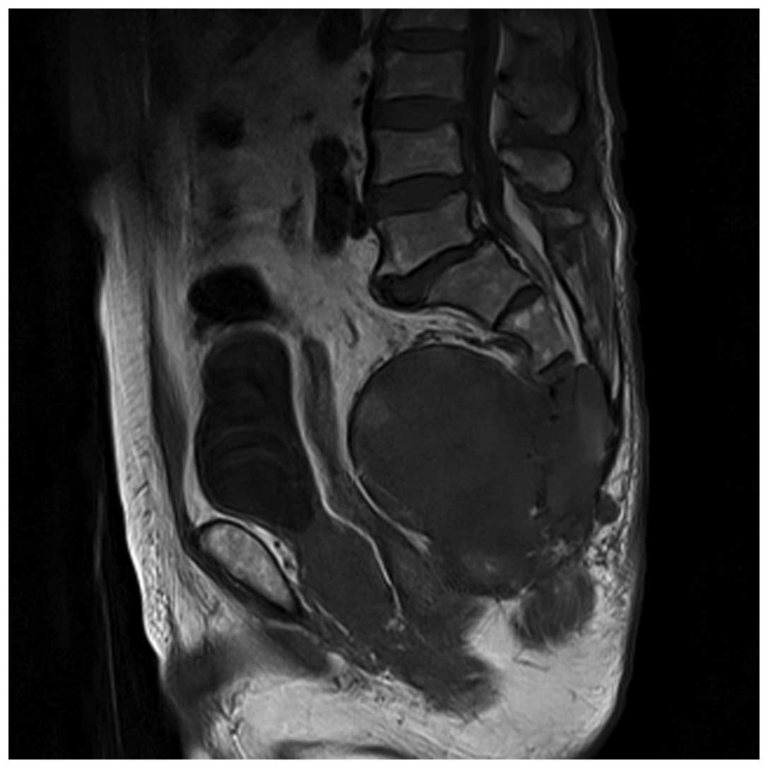
constipation. A large sacral mass was detected by magnetic
resonance imaging (MRI) and the patient was referred to the
Department of Orthopedic Surgery, Faculty of Medicine, Tottori
University (Yonago, Japan) for evaluation and treatment. A physical
examination revealed a diphasic elastic hard mass, measuring 8 × 3
cm in diameter with a smooth surface, in the sacral and gluteal
regions. The mass was fixed to the sacrum and not adhered to the
skin. The results of the neurological assessment were normal, with
the exception of bowel dysfunction. Radiographs showed an
osteolytic lesion in the sacrum, and MRI revealed a large tumor and
compression of the rectum (Fig. 1).
An open biopsy was conducted and histology of the specimen
confirmed the diagnosis of a chordoma. Sacral amputation at S2 was
performed along with resection of the soft component of the tumor.
Adhesion to the presacral membrane was not prominent. The surgical
margin was minor, although microscopically the margin was
identified as R0. Adjuvant radiotherapy was initiated two months
postoperatively due to an infection that was associated with
catheter use and surgical wound dehiscence. An infection of the
sacral region was revealed following radiotherapy (60 Gy), which
was treated via curettage and administration of an antimicrobial
agent. The patient was subsequently discharged.
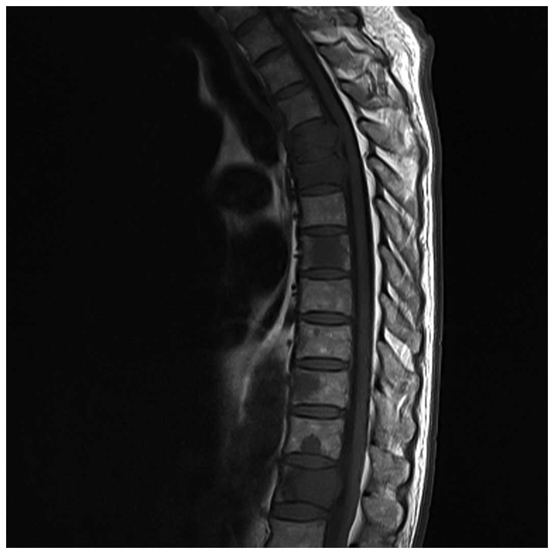
Nine months postoperatively, the patient complained
of back and left shoulder girdle pain. MRI revealed multiple low
intensity areas in the thoracic spine (4th, 5th, 7th, 10th and 12th
vertebrae), and the spinal canal narrowed at the 4th and 5th
thoracic vertebrae (Fig. 2). A
needle biopsy and percutaneous vertebroplasty were performed on
these lesions and a pathological examination demonstrated that
these lesions were metastases of chordoma. Thereafter, metastatic
lesions of the spine rapidly increased in size and number. In
addition, a recurrent tumor was detected in the sacral region. Two
months later, computed tomography and MRI detected metastases to
the liver, cervical spine and right scapula. Tetraplegia
subsequently occurred and gradually advanced, and four months later
the patient succumbed to respiratory dysfunction. A subsequent
autopsy demonstrated multiple metastases to the liver, vertebrae,
kidneys, heart, pancreas and cervical lymph nodes. Pathological
observations revealed apparent tumor emboli of the lungs. The
causes of mortality were, therefore, identified to be pulmonary
tumor emboli and respiratory dysfunction resulting from congested
lungs.
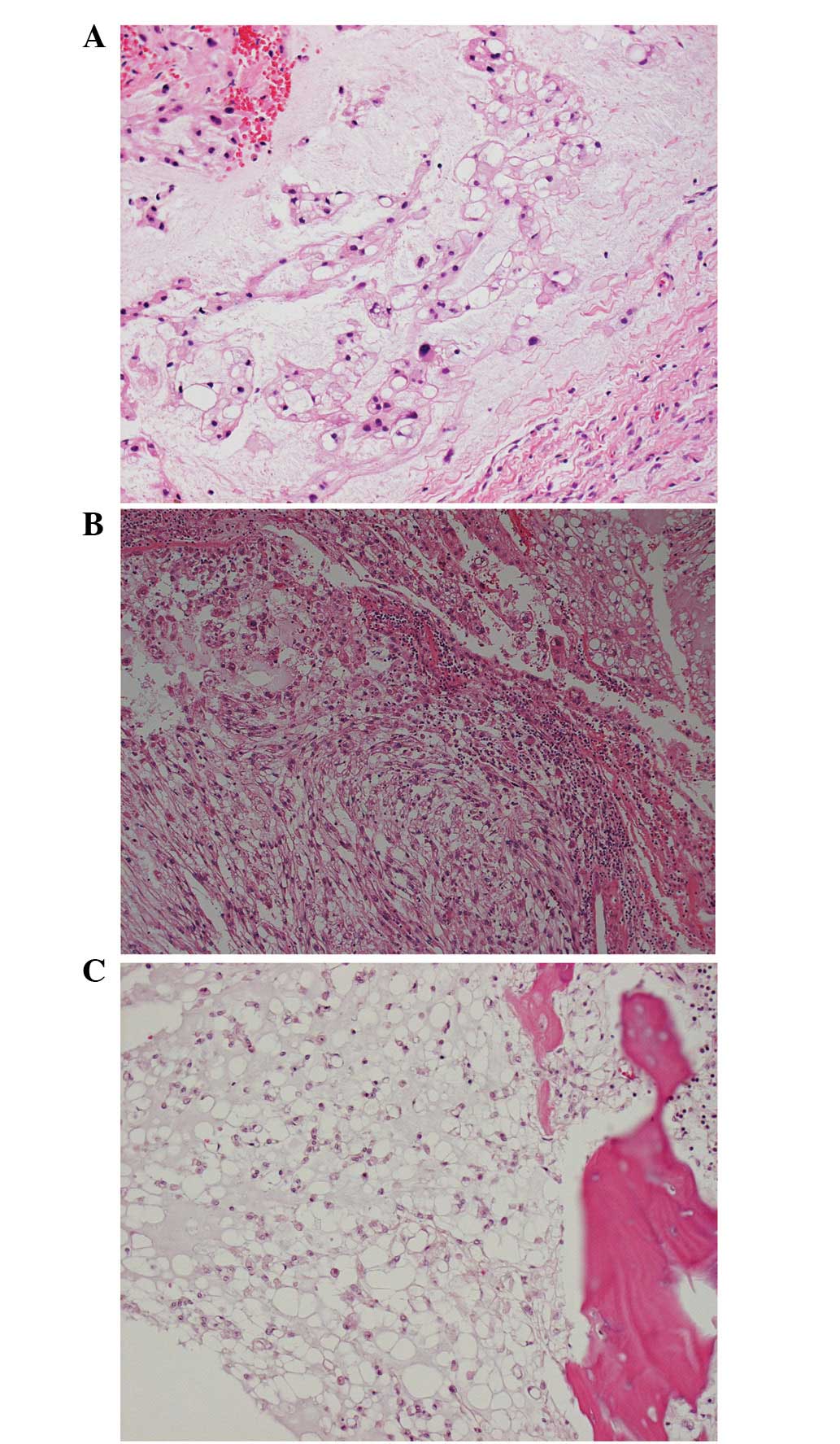
Histological examination of the primary tumor
demonstrated that physaliferous cells were embedded in a myxoid
matrix; spindle cells were also observed in other areas. The
spindle cells did not exhibit nuclear atypia (Fig. 3A and B). In one of the metastatic
tumors, the tumor cells were smaller than those in the primary
region (Fig. 3C). These cells were
termed stellate cells and no proliferation of anaplastic cells was
identified in the primary or metastatic tumor.
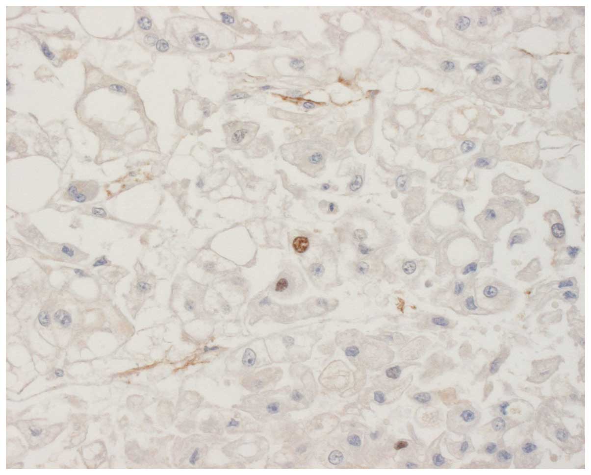
Upon immunohistochemical staining of the primary and
metastatic tumors, the tumor cells were found to be positive for
epithelial membrane antigen (EMA), cytokeratin and vimentin.
However, the metastatic tumor cells were only moderately stained
for vimentin and the Ki-67 labeling indices were <5% in the two
tumors (Fig. 4). Therefore, the
capacity for cellular proliferation of the tumors was considered to
be low.
Discussion
Chordomas are regarded as a low-grade malignancy,
however, these lesions tend to recur locally and to metastasize to
distant sites due to the specificity of their localization. Sites
of chordoma involvement are the axial spine (sacral, 60%;
spheno-occipital/nasal, 25%; cervical, 10%; and thoracolumbar, 5%)
(13).
Sacrococcygeal chordomas in adults are generally
considered to be slow-growing (2).
However, previous studies of cases in infants described sacral
chordomas with an aggressive clinical course (14,15).
Shinmura et al (14)
reported an autopsy case of a three-year-old male showing a tumor
composed of ‘pink’ cells, which were hypothesized to reflect the
earliest embryonic organs of the notochord, along with scattered
physaliferous cells within a myxoid matrix. This case showed
occasional mitotic figures with mild nuclear atypia. The tumor was
not termed dedifferentiated chordoma, but atypical chordoma, since
the tumor cells were positive for epithelial markers, such as
cytokeratin and EMA, and did not demonstrate bizarre nuclei or
sarcomatous features. In adiditon, Iwasa et al (15) reported two atypical chordomas in
infancy and described that the tumors did not show proliferation of
anaplastic cells or features of dedifferentiation. Adult cases of
aggressive sacrococcygeal chordomas have previously been reported;
these cases showed dedifferentiation to the fibrosarcoma,
osteosarcoma and malignant fibrous histiocytoma (3,4,16). The
present case did not demonstrate such findings, although the tumor
cells exhibited mild nuclear atypia, for example, the metastatic
cells were smaller than the tumor cells from the first biopsy.
These stellate cells are responsible for tumor progression and it
is hypothesized that physaliferous cells are degenerated stellate
cells. If stellate cells are predominant in a tumor, this indicates
an aggressive clinical behavior (17,18).
The present tumor did not show any sarcomatoid features and the
tumor cells were positive for epithelial markers; therefore, the
tumor was diagnosed as comprising of features of conventional and
atypical chordoma.
Ki-67 protein is a cellular marker of proliferation
(19) and the fraction of
Ki-67-positive tumor cells (the Ki-67 labeling index) often
correlates with the clinical course of cancer. Previously, Holton
et al (20) and Bergh et
al (21) reported that the
presence of mitotic figures and/or a Ki-67 labeling index >5–6%
were associated with faster growing tumors and earlier metastases.
In the current study, the Ki-67 labeling indices in the resected
and recurrent tumors were <5%. Therefore, these observations did
not explain the aggressive clinical behavior of this case.
Previously, Klingler et al (22) investigated microsatellite
instability in sacral chordoma. The study demonstrated that a
patient, who manifested no microsatellite instability, but a loss
of heterozygosity (LOH) at 9p and 18q, exhibited an
aggressive clinical cancer course, presenting with lymph node
metastasis and succumbing to widespread metastatic disease. In
addition, Horbinski et al (23) indicated that chordoma with 9p
LOH and/or 9p21 homozygous deletion may deomonstrate a risk
for a more aggressive clinical course and shorter survival. This
observation was of interest to the present study, however, our
patient was not investigated for chromosomal anomalies. It is
possible that this chromosomal anomaly was present and should have
been investigated during the open biopsy.
The effects of adjuvant therapies, such as
chemotherapy and radiation therapy, for chordoma are not as
apparent as the response to surgery. Shinmura et al
(14) and Iwasa et al
(15) indicated that chemotherapy
did not appear to benefit the control of recurrent and metastatic
tumors in the cases of infants; furthermore, radiation therapy was
not identified to be effective. Conversely, York et al
(24) demonstrated that
conventional radiation therapy extended the disease-free interval
for patients that received subtotal resection. The current case
showed low proliferative activity, however, the progression of the
recurrent or metastatic tumor was not controlled by radiation
therapy. Therefore, the complete resection of chordoma was
considered to be the most important approach in the present study,
as has been indicated in previous reports (24,25).
In conclusion, orthopedic surgeons must be aware
that sacral chordoma may become aggressive, even in adults. In
addition, due to its potential prognostic relevance, chromosomal
anomalies in chordoma must be investigated during open
biopsies.
Acknowledgements
Aspects of the present study were presented in
poster form at a conference of the International Society of
Orthopaedic Surgery and Traumatology (Prague, Czech Republic) in
2011.
References
|
1
|
Huvos AG: Bone Tumors: Diagnosis,
Treatment and Prognosis. WB Saunders; Philadelphia, PA: pp.
373–391. 1979
|
|
2
|
Unni KK: Dahlin’s Bone Tumors: General
Aspects and Data on 11,087 cases. 5th edition. Lippincott-Raven;
Philadelphia, PA: pp. 291–305. 1996
|
|
3
|
Chambers PW and Schwinn CP: Chordoma: A
clinicopathologic study of metastasis. Am J Clin Pathol.
72:765–776. 1979.PubMed/NCBI
|
|
4
|
Hruban RH, May M, Marcove RC and Huvos AG:
Lumbo-sacral chordoma with high-grade malignant cartilaginous and
spindle cell components. Am J Surg Pathol. 14:384–389. 1990.
View Article : Google Scholar
|
|
5
|
Coffin CM, Swanson PE, Wick MR and Dehner
LP: Chordoma in childhood and adolescence: A clinicopathologic
analysis of 12 cases. Arch Pathol Lab Med. 117:927–933.
1993.PubMed/NCBI
|
|
6
|
Kaneko Y, Sato Y, Iwaki T, Shin RW,
Tateishi J and Fukui M: Chordoma in early childhood: a
clincopathological study. Neurosurgery. 29:442–446. 1991.
View Article : Google Scholar : PubMed/NCBI
|
|
7
|
Volpe R and Mazabraud A: A
clinicopathologic review of 25 cases of chordoma (a pleomorphic and
metastasizing neoplasm). Am J Surg Pathol. 7:161–170. 1983.
View Article : Google Scholar : PubMed/NCBI
|
|
8
|
Osaka S, Kodoh O, Sugita H, Osaka E,
Yoshida Y and Ryu J: Clinical significance of a wide excision
policy for sacrococcygeal chordoma. J Cancer Res Clin Oncol.
132:213–218. 2006. View Article : Google Scholar : PubMed/NCBI
|
|
9
|
Gennari L, Azzarelli A and Quagliuolo V: A
posterior approach for the excision of sacral chordoma. J Bone
Joint Surg Br. 69:565–568. 1987.PubMed/NCBI
|
|
10
|
Hulen CA, Temple HT, Fox WP, Sama AA,
Green BA and Eismont FJ: Oncologic and functional outcome following
sacrectomy for sacral chordoma. J Bone Joint Surg Am. 88:1532–1539.
2006. View Article : Google Scholar : PubMed/NCBI
|
|
11
|
Samson IR, Springfield DS, Suit HD and
Mankin HJ: Operative treatment of sacrococcygeal chordoma. A review
of twenty-one cases. J Bone Joint Surg Am. 75:1476–1484. 1993.
|
|
12
|
Fuchs B, Dickey ID, Yaszemski MJ, Inwards
CY and Sim FH: Operative management of sacral chordoma. J Bone
Joint Surg Am. 87:2211–2216. 2005. View Article : Google Scholar : PubMed/NCBI
|
|
13
|
Mirra JM, Rocca CD, Nelson SD and Mertens
F: Chordoma. World Health Organization Classification of Tumors.
Pathology and Genetics of Tumours of Soft Tissue and Bone. Fletcher
CDM: IARC Press; Lyon: pp. 316–317. 2002
|
|
14
|
Shinmura Y, Miura K, Yajima S and Tsutsui
Y: Sacrococcygeal chordoma in infancy showing an aggressive
clinical course: an autopsy case report. Pathol Int. 53:473–477.
2003. View Article : Google Scholar : PubMed/NCBI
|
|
15
|
Iwasa Y, Nakashima Y, Okajima H and
Morishita S: Sacral chordoma in early childhood:
clinicopathological and immunohistochemical study. Pediatr Dev
Pathol. 1:420–426. 1998. View Article : Google Scholar : PubMed/NCBI
|
|
16
|
Hanna SA, Tirabosco R, Amin A, et al:
Dedifferentiated chordoma: a report of four cases arising ‘de
novo’. J Bone Joint Surg Br. 90:652–656. 2008.PubMed/NCBI
|
|
17
|
Murad TM and Murthy MS: Ultrastructure of
a chordoma. Cancer. 25:1204–1215. 1970. View Article : Google Scholar : PubMed/NCBI
|
|
18
|
Makek M and Leu HJ: Malignant fibrous
histiocytoma arising in a recurrent chordoma. Case report and
electron microscopic findings. Virchows Arch A Pathol Anat Histol.
397:241–250. 1982. View Article : Google Scholar
|
|
19
|
Scholzen T and Gerdes J: The Ki-67
protein: from the known and the unknown. J Cell Physiol.
182:311–322. 2000. View Article : Google Scholar : PubMed/NCBI
|
|
20
|
Holton JL, Steel T, Luxsuwong M, Crockard
HA and Revesz T: Skull base chordomas: correlation of the tumour
doubling time with age, mitosis and Ki67 proliferation index.
Neuropathol Appl Neurobiol. 26:497–503. 2000. View Article : Google Scholar : PubMed/NCBI
|
|
21
|
Bergh P, Kindblom LG, Gunterberg B,
Remotti F, Ryd W and Meis-Kindblom JM: Prognostic factors in
chordoma of the sacrum and mobile spine: a study of 39 patients.
Cancer. 88:2122–2134. 2000. View Article : Google Scholar : PubMed/NCBI
|
|
22
|
Klingler L, Shooks J, Fiedler PN, Marney
A, Butler MG and Schwartz HS: Microsatellite instability in sacral
chordoma. J Surg Oncol. 73:100–103. 2000. View Article : Google Scholar : PubMed/NCBI
|
|
23
|
Horbinski C, Oakley GJ, Cieply K, et al:
The prognostic value of Ki-67, p53, epidermal growth factor
receptor, 1p36, 9p21, 10q23, and 17p13 in skull base chordomas.
Arch Pathol Lab Med. 134:1170–1176. 2010.PubMed/NCBI
|
|
24
|
York JE, Kaczaraj A, Abi-Said D, et al:
Sacral chordoma: 40-year experience at a major cancer center.
Neurosurgery. 44:74–80. 1999.PubMed/NCBI
|
|
25
|
Boriani S, Bandiera S, Biagini R, et al:
Chordoma of the mobile spine: fifty years of experience. Spine
(Phila Pa 1976). 31:493–503. 2006.PubMed/NCBI
|