Introduction
Colorectal carcinoma (CRC) is one of the leading
causes of cancer-associated mortality worldwide. Five-year survival
is 90% if the disease is diagnosed while still localized (i.e.,
confined to the wall of the bowel), but only 68% for regional
disease (i.e., disease with lymph node involvement) and just 10% if
distant metastases are present (1).
Detection of early-stage cancer and precancerous lesions is a key
measure in reducing the mortality rate. Fecal occult blood tests
(FOBT) have reduced the mortality and morbidity of CRC. However,
these have a significant false result rate. Alternative screening
tests, including flexible sigmoidoscopy and colonoscopy, have been
investigated and used as alternatives, but none have been shown to
be as cost effective as FOBT in reducing mortality rates. DNA
mutation detection is a potentially beneficial test, but has yet to
demonstrate superior sensitivity and specificity in a clinical
setting (2). Optimized screening
methods and markers must be established with high sensitivity and
specificity for early-stage cancers and precancerous lesions.
Silencing of tumor suppressor genes by promoter
methylation is a common feature of cancer and is recognized to play
a crucial role in human tumors. DNA methylation in CRCs has been
studied extensively and numerous genes specifically affected by CpG
methylation have been identified, including death-associated
protein kinase (DAPK) (3),
adenomatous polyposis coli, Ras-associated family 2A, Wnt
inhibitory factor 1 (4), secreted
frizzled-related protein 2 (5),
P16, DAPK1, hypermethylated in cancer 1,
O-6-methylguanine-DNA transferase (6), GATA4, GATA5 (7), thrombospondin 1 (8), A-kinase anchor protein 12 (9) and WNT5A (10). These investigations demonstrated
that promoter CpG methylation is a frequent event and often occurs
early during CRC carcinogenesis. Aberrantly methylated DNA has also
been proposed as a potential tumor marker (11).
Additionally, it has been demonstrated that DNA
hypermethylation can be detected in DNA from the feces of CRC
patients, suggesting that fecal DNA methylation analysis is a
promising approach to non-invasive screening for early colorectal
lesions (12–15). Although these reports have confirmed
the potential for early detection of colon cancer-derived
aberrantly methylated DNA in feces, the majority of these
investigations analyzed only a small number of samples. Current
detection methods and their sensitivity for known markers are not
optimal (16,17). Thus, the development of improved
screening methods and identification of new cancer markers is
highly desirable.
The p33 inhibitor of growth 1b (p33ING1b)
gene has been identified as a novel growth inhibitor and tumor
suppressor gene, with reduced expression in ovarian, esophageal,
gastric, brain and breast cancers. However, mutation of the
p33ING1b gene is rare (18–22).
The p33ING1b gene promoter region is rich in CpG
dinucleotides (23), suggesting
that other mechanisms, including DNA methylation, may contribute to
the reduced expression of this gene. In the present study, the
methylation of p33ING1b in feces from patients with CRCs
and precancerous lesions was analyzed and compared with normal
controls. In addition, the sensitivity and specificity of this test
were compared with those of the immunochemical FOBT (IFOBT), and
the feasibility of detecting hypermethylation in feces as a
non-invasive screening tool for CRCs and precancerous lesions was
evaluated.
Materials and methods
Patients and specimens
In total, 61 CRC patients, 27 advanced adenoma (AA)
patients and 20 patients with endoscopically normal colons
undergoing colonoscopy for routine clinical examination at the
First Affiliated Hospital of Guangxi Medical University (Nanning,
China) were enrolled in this study. Fecal specimens were collected
by patients, sent to the laboratory within 2 h after defecation and
stored at −80°C until analyzed. In addition, 37 cancer tissue
samples resected by surgery were obtained from the 61 CRC patients.
This study was approved by the ethical committee of Guangxi Medical
University (Nanning, China). Written informed consent was obtained
from the families of the patients.
IFOBT
All stool samples were examined using a single IFOBT
with MagStream HemSp (Fujirebio, Tokyo, Japan), an immunochemical
test for human hemoglobin. The FOBT was performed at the laboratory
of the First Affiliated Hospital of Guangxi Medical University.
Fecal DNA isolation
Fecal DNA was isolated by the QIAamp DNA Stool mini
kit (Qiagen, Hilden, Germany). The extracted DNA was quantitated by
ultraviolet detection and stored at −20°C.
CRC tissue DNA isolation
Tissue uvDNA isolation was performed by the
conventional method of phenol/chloroform treatment followed by
proteinase K digestion. The extracted DNA was stored at −20°C.
Bisulfite modification
The bisulfite modification procedure was performed
according to Olek et al (24) and Zhang et al (25) with minor modifications. In brief,
500 ng genomic DNA was denatured by incubation in 0.3 M NaOH for 15
min at 37°C, then mixed with 2 volumes 2% low melting point
agarose. Agarose/DNA mixtures were pipetted into chilled mineral
oil to form agarose beads. Each bead was placed in an individual
tube to which 200 μl aliquots of 5 M bisulfate solution were
added [2.5 M sodium metabisulfite (Sigma, St. Louis, MO, USA) and
100 mM hydroquinone (Sigma); pH 5.0]. The reaction mixture was then
incubated in the dark for 16 h at 50°C. Treatment was completed by
equilibration against 1 ml Tris-EDTA buffer followed by
desulfonation in 500 μl NaOH (0.2 M). Finally, the beads
were washed with 1 ml H2O and then used directly in
polymerase chain reaction (PCR).
Nested methylation-specific PCR
(nMSP)
Blood DNA treated in vitro with CpG
methyltransferase (M.SssI) (Fermentas, Hanover, MD, USA) was used
as a positive control for methylated alleles, and placental DNA was
used as a negative control for the nMSP assay. The
bisulfite-modified DNA underwent nMSP in a blind manner using
primer pairs designed to specifically amplify the methylated or
unmethylated alleles of p33ING1b. The sequences of PCR
primers specific for methylated and unmethylated alleles of
p33ING1b and the sizes of the expected PCR products are
summarized in Table I (22).
 | Table IPrimers for nested
methylation-specific polymerase chain reaction. |
Table I
Primers for nested
methylation-specific polymerase chain reaction.
| Primer names | Sequences, 5′-3′ | Size, bp |
|---|
| Nested primer | | 44 |
| Forward |
AGATAAGGTTTAGGGAAGGYGTT | |
| Reverse |
AACAACCRCAATAACCAATCTACT | |
| Methylated
p33ING1b primer | | 151 |
| Forward |
CGGATGGCGTAGGCGCGGGAGTC | |
| Reverse |
CCGAACACGAACGAAAATAACGACGC | |
| Unmethylated
p33ING1b primer | | 151 |
| Forward |
TGGATGGTGTAGGTGTGGGAGTT | |
| Reverse |
CCAAACACAAACAAAAATAACAACACA | |
Briefly, 3 μl bisulfite-modified DNA was
added in a final volume of 25 μl PCR mixture containing 2.5
μl 10X PCR buffer, 2 μl deoxynucleotide triphosphates
(2.5 mmol/l), 0.5 μl each nested primer (10 μmol/l)
and 1 unit Long Taq polymerase (Tiangen Biotech (Beijing)
Co., Ltd., Beijing, China). The PCR amplification protocol for
stage 1 was as follows: 94°C for 4 min, followed by 35 cycles at
94°C for 30 sec, specific annealing temperature 53°C for 30 sec and
72°C for 30 sec, and finally 72°C for 5 min. For stage 2
amplifications [PCR with the methylation-specific primers (Sangon
Biotech (Shanghai) Co. Ltd., Shanghai, China)] 1 μl
first-step PCR products was added in a final volume of 25 μl
PCR mixture: 2.5 μl 10X PCR buffer, 2 μl
deoxynucleotide triphosphates (2.5 mmol/l), 0.5 μl each MSP
primer (10 μmol/l) and 1 unit Taq polymerase (Takara,
Dalian, China). The PCR amplifications were performed at 95°C for 4
min, followed by 35 cycles at 94°C for 30 sec, specific annealing
temperature (62.5°C for the methylated-p33ING1b primer
and 58°C for the unmethylated-p33ING1b primer) for 30
sec and 72°C for 25 sec, and finally at 72°C for 5 min. nMSP
products were analyzed by 2% agarose gel electrophoresis stained
with ethidium bromide.
Statistical analysis
Statistical probabilities were analyzed by 2×2
contingency tables using binomial distribution of differences and
Pearson’s χ2 test. Calculations were performed with SPSS
13.0 software (SPSS Inc., Chicago, IL, USA). P<0.05 was
considered to indicate a statistically significant difference.
Results
Detection of p33ING1b
methylation in fecal DNA
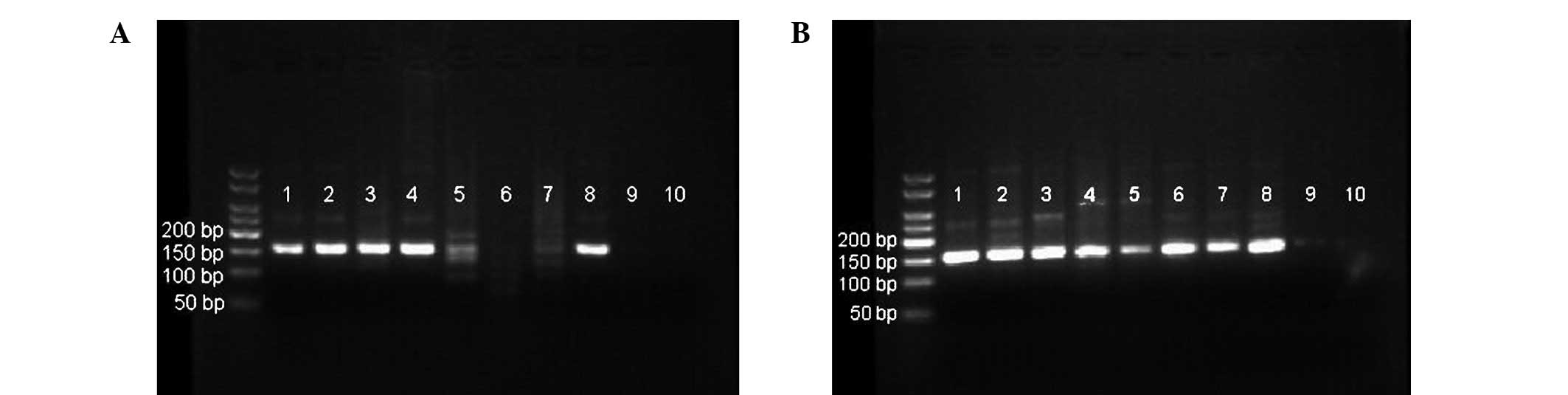
nMSP was performed on all 108 samples, including 37
CRC tissues with matched fecal samples. The correlation of
p33ING1b methylation between the DNA from CRC tissues
and fecal DNA was examined first (Fig.
1). Of 37 cases, 31 (84%) and 29 (78%) exhibited
p33ING1b methylation in the tissue and matched fecal
samples, respectively. The coincident rate of p33ING1b
methylation status in tissues and the matched feces was 94.6%, with
significant correlation (R=0.838; P<0.01; Table II).
| Table IIConsistency analysis of p33 inhibitor
of growth 1b methylation in tissues and matched fecal samples of
colorectal cancer. |
Table II
Consistency analysis of p33 inhibitor
of growth 1b methylation in tissues and matched fecal samples of
colorectal cancer.
| Cancerous
tissues | |
|---|
|
| |
|---|
| Fecal DNA
samples | Methylated | Unmethylated | Total |
|---|
| Methylated | 29 | 0 | 29 |
| Unmethylated | 2 | 6 | 8 |
| Total | 31 | 6 | 37 |
An overview of the frequency of p33ING1b
methylation in the investigated fecal DNA is provided in Table III. The positive rates of
p33ING1b promoter methylation in fecal DNA from 61
sporadic CRC patients and 27 AA patients were 73.77 and 62.96%,
respectively. When compared with the control group (5%), there were
statistically significant differences (P<0.01) (Table III). The methylation of
p33ING1b in the 61 CRC patients was analyzed for
association with clinicopathological data. No correlations were
identified between the methylation of p33ING1b in feces
and gender, age, tumor location, pathological differentiation,
lymph node metastasis, distant metastasis and Dukes’ stage
(Table IV).
| Table IIIp33ING1b promoter
methylation in fecal DNA of CRC and AA patients and the control
group. |
Table III
p33ING1b promoter
methylation in fecal DNA of CRC and AA patients and the control
group.
| | p33ING1b
methylation, n (%) |
|---|
| |
|
|---|
| Fecal DNA | Samples, n | Methylated | Unmethylated |
|---|
| CRCa | 61 | 45 (73.77) | 16 (26.23) |
| AAa | 27 | 17 (62.96) | 10 (37.04) |
| Control | 20 | 1 (5.00) | 19 (95.00) |
| Table IVCorrelations between clinical
parameters and the methylation of p33ING1b promoter in
fecal DNA from colorectal cancer patients. |
Table IV
Correlations between clinical
parameters and the methylation of p33ING1b promoter in
fecal DNA from colorectal cancer patients.
| | p33ING1b
methylation, n (%) | | |
|---|
| |
| | |
|---|
| Group | Patients, n | Methylated | Unmethylated | χ2 | P-value |
|---|
| Gender | | | | 0.176 | 0.674 |
| Male | 37 | 28 (75.68) | 9 (24.32) | | |
| Female | 24 | 17 (70.83) | 7 (29.17) | | |
| Age, years | | | | 0.434 | 0.510 |
| ≤58 | 30 | 21 (70.00) | 9 (30.00) | | |
| >58 | 31 | 24 (77.42) | 7 (22.58) | | |
| Tumor location | | | | 79.31 | 0.349 |
| Colon | 29 | 23 (79.31) | 6 (20.69) | | |
| Rectum | 32 | 22 (68.75) | 10 (31.25) | | |
| Pathological
differentiation | | | | 1.490 | 0.222 |
| High or
middle | 38 | 26 (68.42) | 12 (31.58) | | |
| Low | 23 | 19 (82.61) | 14 (17.39) | | |
| Lymph node
metastasis | | | | 0.218 | 0.640 |
| Presence | 22 | 17 (77.27) | 5 (22.73) | | |
| Absence | 39 | 28 (71.79) | 11 (28.21) | | |
| Distant
metastasis | | | | 0.085 | 0.771 |
| Presence | 9 | 9 (81.82) | 2 (18.18) | | |
| Absence | 52 | 36 (72.00) | 14 (28.00) | | |
| Dukes’ stage | | | | 0.617 | 0.432 |
| A/B | 33 | 23 (69.70) | 10 (30.30) | | |
| C/D | 28 | 22 (78.57) | 6 (21.43) | | |
The positive detection rates of FOBT in 61 CRCs, 27
AA cases and 20 control patients were 49.18, 33.33 and 10%,
respectively (Table V). The
positive rate of FOBT in cases of CRC was significantly higher than
in the control group (P<0.01). However, there was no
statistically significant difference between FOBT results in cases
of AA and the control group (P>0.05).
| Table VFOBT detection rates in CRC, AA and
the control group. |
Table V
FOBT detection rates in CRC, AA and
the control group.
| | FOBT, n (%) |
|---|
| |
|
|---|
| Group | Samples, n | Positive | Negative |
|---|
| CRCa | 61 | 30 (49.18) | 31 (50.82) |
| AA | 27 | 9 (33.33) | 18 (66.67) |
| Control group | 20 | 2 (10.00) | 18 (90.00) |
The sensitivity, specificity, crude accuracy and
Youden’s index of the test for p33ING1b methylation in
fecal DNA were compared with those of the FOBT (Table VI) (26). Youden’s index was used to measure
the effectiveness of the diagnostic marker and is as follows:
Youden’s index (J) = Se(c) + Sp(c) - 1 (where Se is senstivity and
Sp is specificity). The p33ING1b methylation test had a
sensitivity of 73.77% for CRCs and 62.96% for detecting AAs, and a
specificity, crude accuracy and Youden’s index of 95%, 84.26% and
0.75, respectively. The FOBT had a sensitivity of 49.18% for CRCs
and 33.33% for AAs, and a specificity, crude accuracy and Youden’s
index of 90%, 52.78% and 0.44, respectively (Table VI). The sensitivities of the
p33ING1b methylation test for CRCs and AAs were higher
than those of FOBT (P<0.01 and P<0.05, respectively). There
was no statistically significant difference between the
specificities of the two methods (P>0.05). The crude accuracy of
testing for p33ING1b methylation in fecal DNA was higher
than that of FOBT (P<0.01).
| Table VIComparisons of p33 inhibitor of
growth 1b methylation testing of fecal DNA for CRCs and AAs with
FOBT. |
Table VI
Comparisons of p33 inhibitor of
growth 1b methylation testing of fecal DNA for CRCs and AAs with
FOBT.
| | | CRC and AA |
|---|
| | |
|
|---|
| Group | CRC sensitivity,
% | AA sensitivity,
% | Specificity, % | Crude accuracy,
% | γ |
|---|
| Fecal DNA
detection | 73.77 | 62.96 | 95 | 75.00 | 0.70 |
| FOBT | 49.18 | 33.33 | 90 | 52.78 | 0.44 |
| χ2 | 7.787 | 4.747 | 0.360 | 11.559 | |
| P-value | 0.005 | 0.029 | 0.548 | 0.001 | |
Discussion
Since misregulation of gene expression by aberrant
DNA methylation is a well-characterized event in tumor biology and
has been extensively documented for CRC (3–10),
identification of aberrantly methylated genes is a promising
strategy for research, diagnostics and therapeutics (27). However, specific disadvantages
associated with fecal DNA testing render it unsuitable for use in
general population screening for CRC at present. Further study is
required to optimize the methods and the selection of epigenetic
markers. In addition, simplified, inexpensive and automatized
assays may be important for population-based screening (28).
In the present study, the feasibility of detecting
methylated fecal DNA by nMSP as a screening tool for CRCs and
precancerous lesions was explored. Although the routine method of
DNA bisulfite modification with MSP is the most common method for
detecting DNA methylation in previous studies (14,29,30),
this method has not been optimized: The process is complicated, has
low sensitivity (detects 1 methylated allele in up to 1,000
unmethylated alleles) due to sample damage and loss during
bisfulfite modification, and is expensive when a commercial kit is
used. Grunau et al (31)
demonstrated that for the majority of routine PCR, 50 ng human
template DNA yields sufficient PCR product. The authors found that
~90% of the template DNA is lost during treatment with bisulfite.
In the present study, the concentration of fecal DNA extracted by
the commercial kit was 67.35±13.17 ng/l, while some samples did not
satisfy the requirements for bisulfite treatment and MSP. Under
these circumstances, this method may result in an abnormally low
detection rate of fecal DNA methylation. As demonstrated by Olek
et al (24), bisulfite
modification by incubation of DNA contained in agarose beads,
followed by nMSP to detect DNA promoter methylation, is easy to
perform and high levels of sensitivity can be reached. Treatment of
DNA contained in agarose beads minimizes DNA degredation and
prevents incomplete conversion of the DNA. The authors also
succeeded in amplifying fragments of up to 3 kb, which cannot be
achieved using conventional methods. The improved MSP procedure
incorporates a nested, two-stage PCR approach, which is more
sensitive than the conventional method (detects 1 methylated allele
in up to 50,000 unmethylated alleles) (32). In the current study, as little as 10
pg DNA was amplified by treatment within agarose beads followed by
nMSP, and even by general Taq polymerase can succeeded in nMSP.
This level of sensitivity allows determination of methylation
patterns in small fecal samples, advantageous for a
high-sensitivity screening method for CRCs. In addition, this study
demonstrated that DNA extracted from feces is of sufficient quality
and quantity for the detection of DNA methylation by this method.
This is also a less expensive test compared with commercial kits
for bisulfite modification and PCR.
Next, the methylation status of p33ING1b
was investigated in the fecal DNA of CRC patients and the matched
tissues and precancerous lesions. To examine the correlation of
methylation status between fecal DNA and DNA from cancerous tissue,
37 matched pairs of samples were tested for methylation. The nMSP
results were consistent between the paired fecal and tissue samples
(R=0.838; P<0.01) indicating that the test for
p33ING1b methylation in fecal DNA may reflect the true
methylation status of CRCs.
This study revealed that the p33ING1b
methylation test, assessed in independent sets of patients, has
sensitivities of 73.77 and 62.96% for identifying patients with CRC
and precancerous lesions, respectively, while only 5% of control
group samples tested positive for p33ING1b methylation.
No correlation was identified between p33ING1b
methylation status in fecal DNA and any clinical or pathological
characteristics of cancer, indicating that this test is as
sensitive in early-stage CRCs as in late-stage CRCs. Previous
studies (14,30) have demonstrated that aberrant gene
methylation may occur in the early stages of CRCs and even in
precancerous lesions, and that this can be detected in feces, which
has potential value in the noninvasive and early diagnosis of
colorectal neoplasms. However, the low sensitivity and specificity
of the assays suggest the need to develop more powerful techniques.
The present results demonstrate that methylation of the
p33ING1b gene in feces can be considered as a biomarker
for CRCs and precancerous lesions, however, these findings must be
confirmed or improved upon in studies with larger sample sizes.
FOBT is the most widely used method to screen for
CRCs. Although FOBT is valuable as a noninvasive screening method
that reduces the risk of CRC-associated mortality (33,34),
it has limited sensitivity that leads to numerous CRCs remaining
undetected (35–37). This limitation shows that FOBT is
not the best method for screening CRCs. By contrast, using aberrant
gene methylation as a molecular marker offers a potentially
powerful approach to population-based screening for CRCs and
precancerous lesions (28). In the
present study, the efficacies of the fecal p33ING1b
methylation test and FOBT for the identification of CRCs and
precancerous lesions were compared. FOBT had sensitivities of
49.18, 33.33 and 10% for CRCs, precancerous lesions and the normal
control group, respectively, which were lower than those for the
detection of p33ING1b methylation in feces (P<0.05
for AA and P<0.01 for CRCs). However, the two methods had high
specificity for CRCs and precancerous lesions (≥90%) and the
sensitivity of FOBT for CRCs was greater than that for the control
group (P<0.01). No statistically significant difference in FOBT
sensitivity for precancerous lesions and the normal control group
was identified (P>0.05). These results suggest that FOBT may be
a useful method for screening CRCs but not for precancerous
lesions, and that the detection of p33ING1b methylation
in feces may be more effective than FOBT in screening for CRCs and
precancerous lesions. It is clear from this study that testing for
p33ING1b methylation in fecal DNA exhibited a higher
sensitivity than FOBT without reduced specificity.
In conclusion, the results of the present study
demonstrate the value of bisulfite modification of DNA contained in
agarose beads, followed by nMSP, to detect fecal DNA promoter
methylation, as a noninvasive and simple test, and suggest
p33ING1b methylation as a potential biomarker in
screening for CRCs and precancerous lesions.
Acknowledgements
This study was supported by grants from the National
Natural Science Foundation of China (nos. 30760246 and 81360334),
the Scientific Research Foundation of Department of Education of
Guangxi Zhuang Autonomous Region (no. 201012MS043) and the
Scientific Research Foundation of Department of Public Health of
Guangxi Zhuang Autonomous Region (no. Z2012246).
References
|
1
|
Ries LAG, Melbert D, Krapcho M, et al:
SEER Cancer Statistics Review, 1975–2004. National Cancer
Institute; Bethesda, MD: 2007, http://seer.cancer.gov/archive/csr/1975_2004/.
Accessed April 6, 2009
|
|
2
|
Jenkinson F and Steele RJ: Colorectal
cancer screening - methodology. Surgeon. 8:164–171. 2010.
View Article : Google Scholar : PubMed/NCBI
|
|
3
|
Mittag F, Kuester D, Vieth M, et al: DAPK
promotor methylation is an early event in colorectal
carcinogenesis. Cancer Lett. 240:69–75. 2006. View Article : Google Scholar : PubMed/NCBI
|
|
4
|
Lee BB, Lee EJ, Jung EH, et al: Aberrant
methylation of APC, MGMT, RASSF2A, and Wif-1 genes in plasma as a
biomarker for early detection of colorectal cancer. Clin Cancer
Res. 15:6185–6191. 2009. View Article : Google Scholar : PubMed/NCBI
|
|
5
|
Wang DR and Tang D: Hypermethylated SFRP2
gene in fecal DNA is a high potential biomarker for colorectal
cancer noninvasive screening. World J Gastroenterol. 14:524–531.
2008. View Article : Google Scholar
|
|
6
|
Pehlivan S, Artac M, Sever T, et al: Gene
methylation of SFRP2, P16, DAPK1, HIC1, and MGMT and KRAS mutations
in sporadic colorectal cancer. Cancer Genet Cytogenet. 201:128–132.
2010. View Article : Google Scholar
|
|
7
|
Hellebrekers DM, Lentjes MH, van den Bosch
SM, et al: GATA4 and GATA5 are potential tumor suppressors and
biomarkers in colorectal cancer. Clin Cancer Res. 15:3990–3997.
2009. View Article : Google Scholar : PubMed/NCBI
|
|
8
|
Rojas A, Meherem S, Kim YH, et al: The
aberrant methylation of TSP1 suppresses TGF-beta1 activation in
colorectal cancer. Int J Cancer. 123:14–21. 2008. View Article : Google Scholar : PubMed/NCBI
|
|
9
|
Liu W, Guan M, Su B, et al: Rapid
determination of AKAP12 promoter methylation levels in peripheral
blood using methylation-sensitive high resolution melting (MS-HRM)
analysis: Application in colorectal cancer. Clin Chim Acta.
411:940–946. 2010. View Article : Google Scholar
|
|
10
|
Ying J, Li H, Yu J, et al: WNT5A exhibits
tumor-suppressive activity through antagonizing the
Wnt/beta-catenin signaling, and is frequently methylated in
colorectal cancer. Clin Cancer Res. 14:55–61. 2008. View Article : Google Scholar
|
|
11
|
Chen WD, Han ZJ, Skoletsky J, et al:
Detection in fecal DNA of colon cancer-specific methylation of the
nonexpressed vimentin gene. J Natl Cancer Inst. 97:1124–1132. 2005.
View Article : Google Scholar : PubMed/NCBI
|
|
12
|
Lenhard K, Bommer GT, Asutay S, et al:
Analysis of promoter methylation in stool: a novel method for the
detection of colorectal cancer. Clin Gastroenterol Hepatol.
3:142–149. 2005. View Article : Google Scholar : PubMed/NCBI
|
|
13
|
Azuara D, Rodriguez-Moranta F, de Oca J,
et al: Novel methylation panel for the early detection of
colorectal tumors in stool DNA. Clin colorectal cancer. 9:168–176.
2010. View Article : Google Scholar : PubMed/NCBI
|
|
14
|
Petko Z, Ghiassi M, Shuber A, et al:
Aberrantly methylated CDKN2A, MGMT, and MLH1 in colon polyps and in
fecal DNA from patients with colorectal polyps. Clin Cancer Res.
11:1203–1209. 2005.PubMed/NCBI
|
|
15
|
Abbaszadegan MR, Tavasoli A, Velayati A,
et al: Stool-based DNA testing, a new noninvasive method for
colorectal cancer screening, the first report from Iran. World J
Gastroenterol. 13:1528–1533. 2007. View Article : Google Scholar
|
|
16
|
Müller HM, Oberwalder M, Fiegl H, et al:
Methylation changes in faecal DNA: a marker for colorectal cancer
screening? Lancet. 363:1283–1285. 2004.PubMed/NCBI
|
|
17
|
Grady WM, Rajput A, Lutterbaugh JD and
Markowitz SD: Detection of aberrantly methylated hMLH1 promoter DNA
in the serum of patients with microsatellite unstable colon cancer.
Cancer Res. 61:900–902. 2001.PubMed/NCBI
|
|
18
|
Tallen G, Kaiser I, Krabbe S, et al: No
ING1 mutations in human brain tumours but reduced expression in
high malignancy grades of astrocytoma. Int J Cancer. 109:476–479.
2004. View Article : Google Scholar
|
|
19
|
Nouman GS, Anderson JJ, Crosier S, et al:
Downregulation of nuclear expression of the p33ING1b
inhibitor of growth protein in invasive carcinoma of the breast. J
Clin Pathol. 56:507–511. 2003.PubMed/NCBI
|
|
20
|
Oki E, Maehara Y, Tokunaga E, et al:
Reduced expression of p33(ING1) and the relationship with p53
expression in human gastric cancer. Cancer Lett. 147:157–162. 1999.
View Article : Google Scholar : PubMed/NCBI
|
|
21
|
Chen L, Matsubara N, Yoshino T, et al:
Genetic alterations of candidate tumor suppressor ING1 in human
esophageal squamous cell cancer. Cancer Res. 61:4345–4349.
2001.
|
|
22
|
Shen DH, Chan KY, Khoo US, et al:
Epigenetic and genetic alterations of p33ING1b in
ovarian cancer. Carcinogenesis. 26:855–863. 2005. View Article : Google Scholar : PubMed/NCBI
|
|
23
|
Gunduz M, Ouchida M, Fukushima K, et al:
Genomic structure of the human ING1 gene and tumor-specific
mutations detected in head and neck squamous cell carcinomas.
Cancer Res. 60:3143–3146. 2000.
|
|
24
|
Olek A, Oswald J and Walter J: A modified
and improved method for bisulphite based cytosine methylation
analysis. Nucleic Acids Res. 24:5064–5066. 1996. View Article : Google Scholar : PubMed/NCBI
|
|
25
|
Zhang Z, Sun D, Van do N, et al:
Inactivation of RASSF2A by promoter methylation correlates with
lymph node metastasis in nasopharyngeal carcinoma. Int J Cancer.
120:32–38. 2007. View Article : Google Scholar : PubMed/NCBI
|
|
26
|
Akobeng AK: Understanding diagnostic tests
1: sensitivity, specificity and predictive values. Acta Paediatr.
96:338–341. 2007. View Article : Google Scholar
|
|
27
|
Jemal A, Siegel R, Ward E, et al: Cancer
statistics, 2007. CA Cancer J Clin. 57:43–66. 2007. View Article : Google Scholar
|
|
28
|
Huang ZH, Li LH, Yang F and Wang JF:
Detection of aberrant methylation in fecal DNA as a molecular
screening tool for colorectal cancer and precancerous lesions.
World J Gastroenterol. 13:950–954. 2007. View Article : Google Scholar : PubMed/NCBI
|
|
29
|
Herman JG, Graff JR, Myöhänen S, et al:
Methylation-specific PCR: a novel PCR assay for methylation status
of CpG islands. Proc Natl Acad Sci USA. 93:9821–9826. 1996.
View Article : Google Scholar : PubMed/NCBI
|
|
30
|
Zhang J, Yang SB, Xie Y, et al: Detection
of methylated tissue factor pathway inhibitor 2 and human long DNA
in fecal samples of patients with colorectal cancer in China.
Cancer Epidemiol. 36:73–77. 2012. View Article : Google Scholar
|
|
31
|
Grunau C, Clark SJ and Rosenthal A:
Bisulfite genomic sequencing: systematic investigation of critical
experimental parameters. Nucleic Acids Res. 29:e652001. View Article : Google Scholar
|
|
32
|
Palmisano WA, Divine KK, Saccomanno G, et
al: Predicting lung cancer by detecting aberrant promoter
methylation in sputum. Cancer Res. 60:5954–5958. 2000.
|
|
33
|
Mandel JS, Bond JH, Church TR, et al:
Reducing mortality from colorectal cancer by screening for fecal
occult blood. Minnesota colon cancer control study. N Engl J Med.
328:1365–1371. 1993. View Article : Google Scholar
|
|
34
|
Heresbach D, Manfredi S, D’Halluin PN, et
al: Review in depth and meta-analysis of controlled trials on
colorectal cancer screening by faecal occult blood test. Eur J
Gastroenterol Hepatol. 18:427–433. 2006. View Article : Google Scholar
|
|
35
|
Imperiale TF, Ransohoff DF, Itzkowitz SH,
et al: Fecal DNA versus fecal occult blood for colorectal-cancer
screening in an average-risk population. N Engl J Med.
351:2704–2714. 2004. View Article : Google Scholar : PubMed/NCBI
|
|
36
|
Collins JF, Lieberman DA, Durbin TE, et
al: Accuracy of screening for fecal occult blood on a single stool
sample obtained by digital rectal examination: a comparison with
recommended sampling practice. Ann Intern Med. 142:81–85. 2005.
View Article : Google Scholar
|
|
37
|
Nadel MR, Shapiro JA, Klabunde CN, et al:
A national survey of primary care physicians’ methods for screening
for fecal occult blood. Ann Intern Med. 142:86–94. 2005.
|