Introduction
Hepatocellular carcinoma (HCC) is the leading cause
of cancer-related mortality worldwide, particularly in Asia.
Significant risk factors include hepatitis C virus infection,
hepatitis B virus infection and cirrhosis. Previously, numerous
genetic and epigenetic alterations have been associated with
hepatocarcinogenesis (1).
Hypermethylation of gene promoters can be promising tools, for
example as biomarkers, in the detection of cancer cells in tissues
and body fluids. The clinical value of methylation markers has been
reported for the early detection and classification of cancer, for
risk assessment and prognosis and for the prediction of therapy
response. The ability to use methylation markers for diagnostics
and as a predictive tool for cancer is becoming tangible (2).
The adenomatous polyposis coli (APC) tumor
suppressor gene encodes a large protein with multiple cellular
functions and interactions, including signal transduction in the
Wnt-signaling pathway (3). Defects
in this gene cause familial adenomatous polyposis. Numerous studies
have shown that the inactivation of APC by promoter
hypermethylation is frequent in HCC. To the best of our knowledge,
previous studies have used different detection methods and
therefore the reported methylation frequencies/levels of the APC
promoter in HCC have varied among the studies. Using the same
method has also produced varied methylation levels/frequencies
(2,4–12).
Furthermore, in these studies the methylation signal in the APC
promoter was determined by a qualitative or quantitative assay in
≤50 paired samples.
The present study aimed to obtain a quantitative
methylation signal of APC in HCC, using the more exact quantitative
method of MethyLight, which is based on Taqman probes, to detect
the promoter of the APC gene in 57 paired HCC and matched
non-malignant liver tissues. The correlation between the
methylation status and clinicopathological features was analyzed
further, and the ability of APC to serve as a potential biomarker
of the prognosis for HCC was evaluated.
Materials and methods
Human tissues
Human primary HCC and corresponding non-cancerous
liver tissues (3 cm from the tumor) were collected from 57 patients
who were diagnosed and treated at Guangxi Medical University,
Guangxi, China, between January 2003 and June 2005. The study
protocol was approved by the Clinical Research Ethics Committee of
Shanghai Cancer Institute (Shangai, China) and informed consent was
obtained from each patient. The tissue samples were snap-frozen in
liquid nitrogen immediately after surgical resection and then
stored at −80°C until analysis. Clinical information was collected
from patient records and pathology reports. All patients were
followed up from the date of surgery and the survival and mortality
rates were recorded. Clinical information is recorded in Table I.
 | Table IClinical information of 57 paired HCC
samples and the corresponding APC promoter methylation level
detected by the MethyLight assay. |
Table I
Clinical information of 57 paired HCC
samples and the corresponding APC promoter methylation level
detected by the MethyLight assay.
| Parameters | Number of cases | PMR of NT (mean ±
SEM) | PMR of HCC (mean ±
SEM) | P-value |
|---|
| Total | 57 | 3.09±0.684 | 11.64±2.236 |
0.0003* |
| Age, years |
| >60 | 12 | 9.62±4.581 | 34.90±9.879 |
0.0438* |
| ≤60 | 45 | 5.22±1.221 | 20.04±4.934 | |
| Gender |
| Male | 49 | 6.88±1.558 | 23.39±4.842 | 0.9000 |
| Female | 8 | 1.64±0.551 | 21.76±11.97 | |
| Tumor size, cm |
| >4 | 8 | 6.85±4.129 | 2.955±1.256 |
0.0008* |
| ≤4 | 49 | 6.03±0.451 | 13.06±2.539 | |
| Tumor
embolusa |
| Positive | 21 | 7.20±2.376 | 26.50±6.616 | 0.3305 |
| Negative | 35 | 5.43±1.713 | 21.68±6.091 | |
| Tumor
capsulec |
| Complete | 15 | 7.69±4.081 | 26.68±9.332 | 0.8195 |
| Incomplete | 22 | 6.09±0.698 | 29.67±8.660 | |
| TNM
stagea |
| I, II | 34 | 4.77±1.725 | 12.84±3.382 | 0.5750 |
| III, IV | 22 | 8.13±2.285 | 10.20±2.523 | |
|
Paracirrhosisa |
| Positive | 38 | 4.74±1.165 | 28.22±6.133 | 0.1295 |
| Negative | 18 | 8.94±3.514 | 13.51±4.889 | |
| AFP,
μg/lb |
| <20 | 13 | 7.34±3.072 | 23.57±8.308 | 0.7902 |
| 20–400 | 25 | 7.30±2.584 | 25.25±8.272 | |
| >400 | 17 | 3.16±0.746 | 18.05±5.244 | |
DNA preparation
Total genomic DNA was extracted from frozen tissue
specimens (50–100 mg) according to the standard protocol, with
specific modifications, which are briefly described as follows.
Frozen pulverized powders of the specimens were resuspended with 2
ml warmed lysis buffer: [50 mM Tris-HCl (pH 8.0), 50 mM EDTA, 1%
SDS, 10 mM NaCl and 100 μg/ml boiling-treated RNase A;
Sigma-Aldrich, St. Louis, MO, USA]. Following a 1-h incubation at
37°C, proteinase K (Roche Diagnostics, Indianapolis, IN, USA) was
added to the cellular lysates to form a 100 μg/ml final
concentration, and the digestion was carried out at 55°C for 2 h.
Organic extractions with a half volume of phenol/chloroform/isoamyl
alcohol (1:1:0.04) were repeatedly performed until no visible
interphase remained following centrifugation at 12,000 × g for 10
min. DNA was precipitated from the aqueous phase in the presence of
0.3 M NaOAc (pH 7.0); and two and one-half volumes of ethanol. The
DNA pellet was washed once with 70% ethanol and dissolved at 65°C
for 30 min with 0.2–0.4 ml Tris-EDTA (TE) buffer [10 mM TrisHCl (pH
7.4) and 1 mM EDTA], followed by storage at 4°C until further use.
The DNA concentrations were calculated according to their optical
density readings at 260 nm (13).
CpG methyltransferase (M.SssI)
methylation assay
Peripheral blood leukocyte (PBL) DNA (Promega
Corporation, Madison, WI, USA) was used as a substrate for the
M.SssI treatment. PBL DNA (0.05 μg/μl) was incubated with M.SssI at
a concentration of 1 U/μg DNA (0.05 U/μl) and 0.16 mM AdoMet
overnight at 37°C. Extra AdoMet (to 0.20 mM) and M.SssI (to 0.065
U/μl) were then added followed by a second overnight incubation at
37°C. The sample was stored at 4°C and 18-μl (0.9 μg DNA) aliquots
were used for the bisulfite conversion and recovery (14).
Bisulfite treatment
DNA (10 μg in 50 μl TE) was incubated with 5.5 μl 3
M NaOH at 37°C for 10 min, followed by a 16-h treatment at 50°C,
subsequent to the addition of 30 μl freshly prepared 10 mM
hydroquinone and 520 μl freshly prepared 3.6 M sodium-bisulfite (pH
5.0). DNA was desalted using a home dialysis system with 1% agarose
(detailed protocol will be provided upon request). The DNA in the
desalted sample (~100 μl) was denatured at 37°C for 15 min with 5.5
μl 3 M NaOH followed by ethanol precipitation with 33 μl 10 M
NH4OAC and 300 μl ethanol. Subsequent to washing with
70% ethanol, the gently dried DNA pellet was dissolved with 30 μl
TE at 65°C for 10 min. The DNA sample was finally stored at −20°C
until further use. A 50-ng DNA sample was reserved for polymerase
chain reaction (PCR) (15).
MethyLight (Taqman probe-based
quantitative methylation-specific PCR)
The PCR was performed using a 96-well optical tray
with caps at a final reaction volume of 20 μl. Samples contained 8
μl Real MasterMix (Taqman; Tiangen Biotech Co., Ltd., Beijing,
China), 1 μl bisulfite-treated DNA, 250 nM each primer and 125 nM
6-carboxyfluorescein-labeled probes. The modified DNA was amplified
by MethyLight quantitative PCR (qPCR) using the TaqMan gene assay
and the 7500/7500 Fast Real-Time PCR System (Applied Biosystems,
Foster City, CA, USA). Primers and probes for APC and Alu-C4 were
as previously described (4,14). Each PCR program consisted of an
initial denaturation cycle (95°C for 10 min), 45 cycles of
denaturation (95°C for 15 sec) and finally an annealing/extension
cycle (60°C for 1 min). The methylation ratio was determined by
absolute quantification of qPCR. The quantity of amplified target
genes in the test samples was normalized with that of Alu-C4 to
measure the levels of input DNA, and the DNA treated with M.SssI
served as a methylated reference. The amount of methylated DNA, or
the percentage of methylated reference (PMR), at a specific locus
was calculated by dividing the gene:Alu-C4 ratio of a sample by the
gene:Alu-C4 ratio of the M.SssI-treated human genomic DNA
(presumably fully methylated) and multiplying by 100.
Statistical analysis
Data are expressed as the mean ± standard error of
the mean from at least three separate experiments. The data were
analyzed with either a two-tailed Student’s t-test/χ2
test or a one-way analysis of variance for the comparison of more
than two groups unless otherwise specified. Kaplan-Meier (KM)
method and log-rank test were used to derive the overall survival
function, and the log-rank test was used to compare the curves for
the two groups. For KM analysis, the median PMR level was used as a
cut-off level. Therefore, the definition varied for each gene with
the aim of obtaining equal sample sizes for each KM curve. This
provided an improved power to identify whether there is an
association between the level of methylation in HCC tissues and
overall patient survival rate. P<0.05 was considered to indicate
a statistically significant difference.
Results
Methylation level of the APC promoter in
HCC and corresponding non-cancerous tissues
In order to obtain the methylation signal of the APC
promoter, the MethyLight assay was used to detect the CpG island
methylation level of the APC promoter in 57 pairs of HCC and
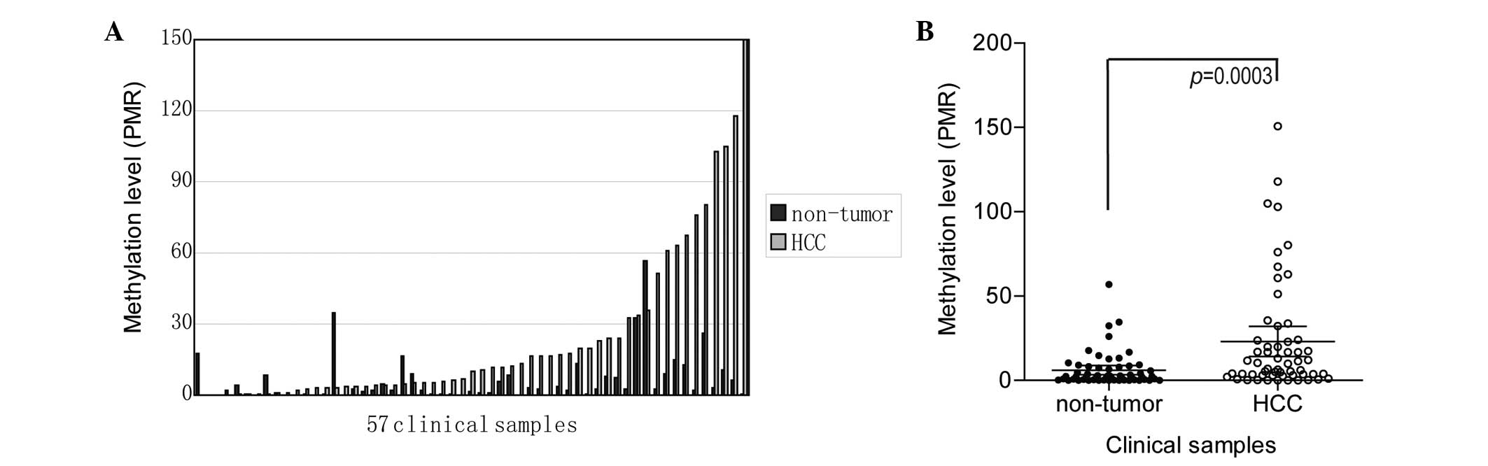
matched non-tumor liver tissues. The data indicated that the levels
of the APC promoter were higher in 45 out of 57 (78.95%) tumor
tissues compared with adjacent non-cancerous liver tissues
(Fig. 1A). The rate of DNA
methylation [(gene sample/Alu-C4 sample)/(gene SssI-treated
sample/Alu-C4 SssI-treated sample) × 100] in the APC gene promoter
was also revealed to be significantly higher in the HCC tissues
compared with the adjacent non-cancerous tissues (paired t-test,
P=0.0003; Fig. 1B). The median rate
of methylation in the HCC tissues was 9.93% (range, 0.02–150.8) and
the median rate in the non-cancerous liver tissues was 2.2% (range,
0.0035–56.84). The median rate of DNA methylation was upregulated
by 4.51-fold in the HCC tissues compared with the non-cancerous
tissues. Occasionally PMR values may be >100%; this occurred in
cases when the M.SssI treatment of the standard DNA was not
complete or in cases of aneuploidy of the gene locus of interest.
Details are shown in Table I.
Overall, these data showed that the rate of DNA methylation in the
APC locus was upregulated in the HCC tissue compared with the
non-cancerous adjacent tissues.
Correlation between clinicopathological
data and methylation level of the APC promoter in HCC
To determine whether hypermethylation of APC can be
a characteristic biomarker of certain types of HCC, the
correlations between the methylation level of the APC promoter in
the HCC samples and the clinicopathological parameters were
analyzed. Notably, the methylation level of the APC promoter in the
HCC samples was higher in older patients when the cut-off was set
at 60 years old. In the 12 patients who were >60 years old, the
mean methylation level was 34.90%, while in the 45 patients who
were <60 years old, the mean methylation level was 20.04%
(P=0.0438; Fig. 2).
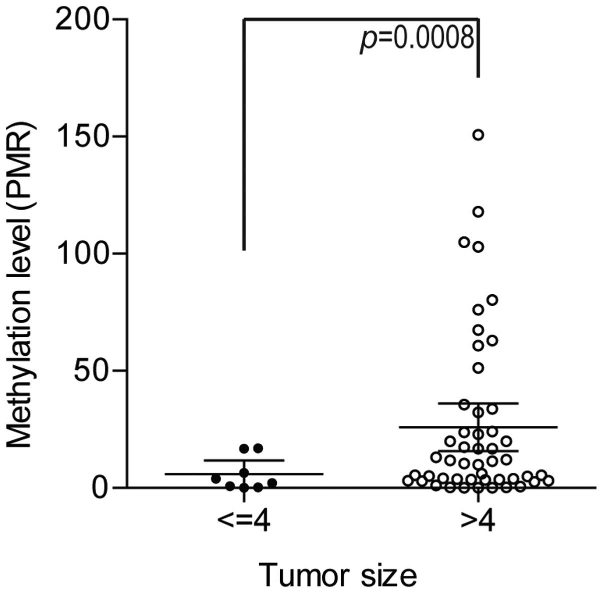
Additionally, the CpG island methylation level of
the APC promoter in the HCC samples was significantly higher in the
patients with larger tumors when the cut-off was set at 4 cm. Of
the 8 tumors with a size of >4 cm, the mean methylation level of
the tumor suppressor gene APC promoter was 13.06%, which is
significantly higher than the mean methylation level of the
remaining 49 patients (2.96%) (P=0.0008; Fig. 3). When correlated with other
clinical parameters, including gender, presence of a tumor embolus
or tumor capsule, tumor-node-metastasis stage, paracirrhosis and
α-fetoprotein level, there was no significant correlation with the
methylation level of the APC promoter in the HCC samples (Table I).
Correlation between survival and
methylation level of the APC promoter in HCC
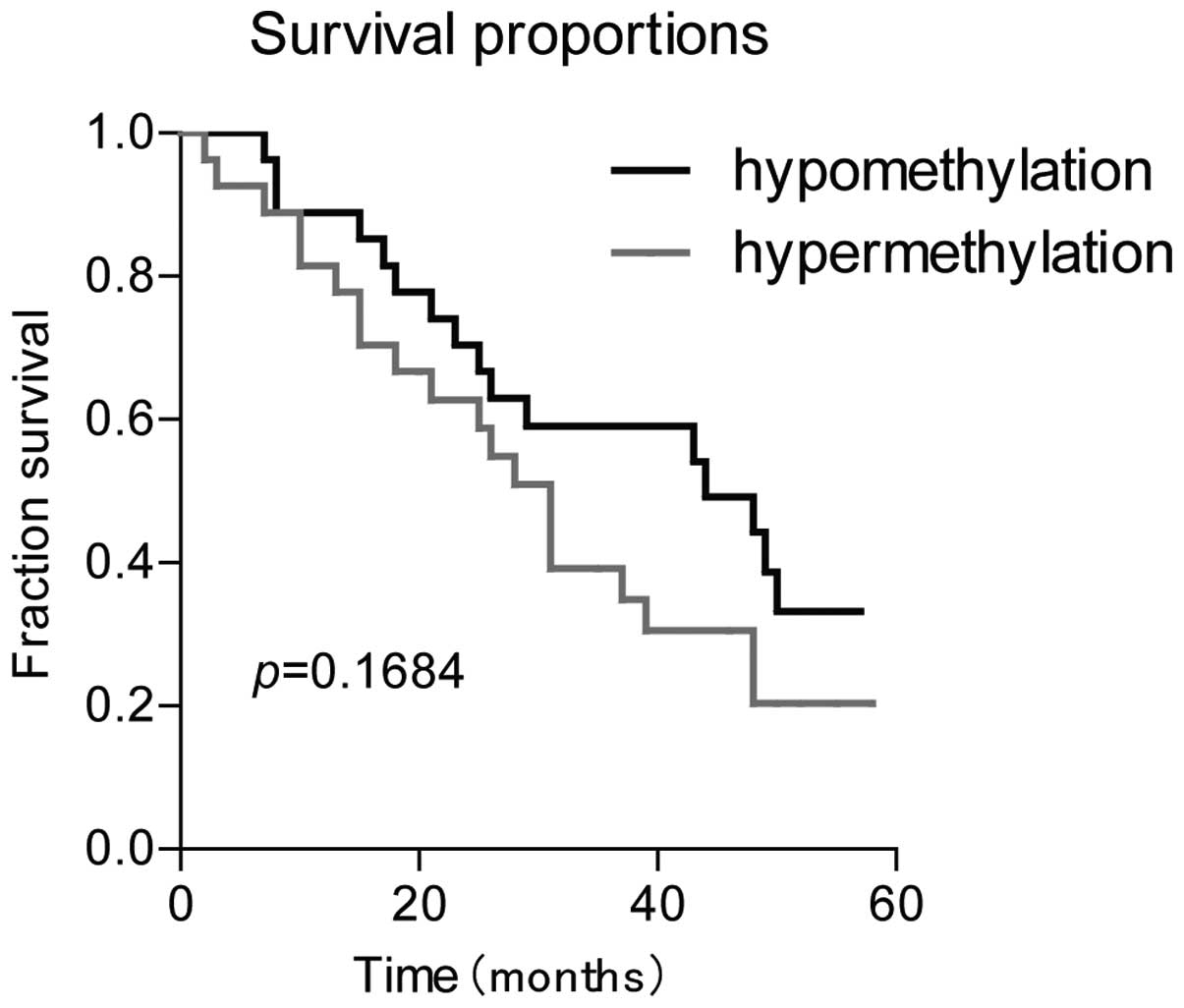
The survival rate of the HCC patients was also
explored and the association with the methylation status of the APC
promoter was evaluated. Complete follow-up data were available in
54/57 patients (94.74%). Of the 54 patients, 36 (66.67%) succumbed
to disease (median follow-up, 50.5 months) and 18 (33.33%) survived
(median, 24 months). Overall, the patients survived between 2 and
58 months, with a median of 30 months. The methylation rate in HCC
tissues above the mean PMR were defined as hypermethylation cases
and HCC tissues with methylation rates below the mean PMR were
defined as hypomethylation cases. The follow-up results did not
reveal any significant difference between the overall survival rate
in the hypomethylation or hypermethylation cases (P=0.1684;
Fig. 4).
Discussion
Promoter hypermethylation of tumor suppressor or
tumor-related genes plays a significant role in tumorigenesis.
Hypermethylation of APC, a well-characterized tumor suppressor, has
been detected frequently in HCC in various studies (2–12).
However, the majority of previous studies used methylation-specific
PCR, which has been considered as an easy-to-use qualitative method
for the detection of DNA methylation. DNA hypermethylation can be
assessed by methylation-specific PCR, which although extremely
sensitive, is dependent on the number of PCR cycles, the mixture
conditions and the amount of DNA (16). Therefore, the reported methylation
frequencies of identical genes in HCC varies between studies
(2,4–12).
Taqman probe-based quantitative methylation-specific PCR
(MethyLight) can overcome issues associated with PCR cycling and
provide reliable quantitative information about the methylation
status of the target CpG island loci (11).
The findings of the present study showed that the
promoter region of the APC gene was methylated in 35.09% of the
non-cancerous liver tissues and 64.91% of HCC samples using a
cut-off set at 4% PMR, which indicates a high methylation frequency
of the APC gene promoter in HCC and a moderate methylation
frequency in the paired non-tumorous liver tissues. The moderate
methylation frequencies observed in the non-tumorous liver tissue
indicate epigenetic evidence for field cancerization involved in
the early stage of liver carcinogenesis (5). The fact that the methylation frequency
of the APC promoter in the HCC samples was much higher than that in
the paired non-tumorous liver tissues indicates that this aberrant
methylation alteration is a quantitative epigenetic change that
accumulated through the process of hepatocarcinogenesis. The
results of the present study were highly consistent with those of
the previously published studies, regardless of the detection
method used (2,4–12).
Aberrant methylated genes can be used as biomarkers
for tumor classification and early detection, and to detect the
response to treatments, including traditional chemotherapy drugs,
target therapy and epigenetic agents (14). In the present study, the CpG island
methylation level of the APC promoter in the HCC samples was
significantly higher in the patients with larger tumors when the
cut-off was set at 4 cm. Of the 8 tumors with a size of >4 cm,
the mean methylation level of the tumor suppressor gene APC
promoter was 13.06%, while for the remaining 49 patients with a
smaller tumor, the mean methylation level was 2.96%. This result
may be due to the inactivity of the APC protein. The functionally
inactive APC protein, created by transcriptional silencing through
promoter methylation, inhibits interactions with β-catenin. This
allows the direct interaction of β-catenin with the lymphoid
enhancer factor-T cell factor family of transcription factors and
the promotion of cells into the cell cycle (3).
In the present study, the methylation level of the
APC promoter in the HCC samples was also identified to be higher in
older patients when the cut-off was set at 60 years old. In the 12
patients who were >60 years old, the mean methylation level was
34.90%, while in the 45 patients who were <60 years old, the
mean methylation level was 20.04%. This is the first APC
hypermethylation study that has shown a correlation between
methylation level and patient age. No similar result has been
reported in previous studies. However, the methylation level of
another frequently detected tumor suppressor gene, Ras association
domain family 1A (RASSF1A), has been reported to be correlated with
patient age. A study by Di Gioia et al demonstrated that the
age-related methylation of the RASSF1A promoter takes place early
in a small subpopulation of cells of the human liver (17). Conversely, Feng et al and
Zhong et al indicated that no association was apparent
between the methylation of the RASSF1A gene promoter and patient
age (18,19). Demonstrations of the association
between the methylation level of the APC promoter and patient age
are further required by other quantitative methylation detection
methods, including pyrosequencing, in prospective studies of
various geographical cohorts.
Last, but not least, the correlation between APC
hypermethylation and survival rate was analyzed in the present
study by univariate analysis using KM and log-rank tests. The
aberrant hypermethylation of the APC promoter had no significant
impact on the overall survival rate. Thus the methylation level of
the APC promoter in HCC may not serve as a promising prognostic
biomarker for the disease. This result was inconsistent with a
previous study in which elevated plasma methylation levels of APC
were associated with a poorer overall survival rate (20).
In summary, using a highly precise and quantitative
tool for detecting epigenetic changes in clinical samples of HCC
and corresponding non-cancerous liver tissues, the present study
identified that the methylation level of the APC promoter in HCC
tissues was significantly higher than in paracancerous liver
tissues. This result indicated that hypermethylation of the APC
promoter is an early event in hepatocarcinogenesis and is
quantitatively accumulated in the development of HCC. In addition,
the methylation level of the APC promoter is correlated with the
age at the time of HCC diagnosis and the tumor size. However, a
higher degree of APC promoter methylation in tumor tissues does not
appear to be responsible for a poorer overall survival rate, as has
been reported in previous studies. Therefore, the methylation level
of the APC promoter in HCC may not serve as a good prognostic
biomarker of HCC. This result requires further elucidation by other
quantitative methylation detection methods, including
pyrosequencing, in prospective studies of various geographical
cohorts.
Acknowledgements
This study was supported by the Natural Science
Foundation of the Shanghai Science and Technology Committee (no.
12ZR1430200), the research fund of the State Key Laboratory of
Oncogenes and Related Genes (91-11-05) and the Young Scientists
Foundation of the Shanghai Cancer Institute (SB11-10).
Reference
|
1
|
Saelee P, Wongkham S, Chariyalertsak S,
Petmitr S and Chuensumran U: RASSF1A promoter hypermethylation as a
prognostic marker for hepatocellular carcinoma. Asian Pac J Cancer
Prev. 11:1677–1681. 2010.PubMed/NCBI
|
|
2
|
Jain S, Chang TT, Hamilton JP, et al:
Methylation of the CpG sites only on the sense strand of the APC
gene is specific for hepatocellular carcinoma. PLoS One.
6:e267992011. View Article : Google Scholar
|
|
3
|
Csepregi A, Röcken C, Hoffmann J, et al:
APC promoter methylation and protein expression in hepatocellular
carcinoma. J Cancer Res Clin Oncol. 134:579–589. 2008. View Article : Google Scholar
|
|
4
|
Um TH, Kim H, Oh BK, et al: Aberrant CpG
island hypermethylation in dysplastic nodules and early HCC of
hepatitis B virus-related human multistep hepatocarcinogenesis. J
Hepatol. 54:939–947. 2011. View Article : Google Scholar
|
|
5
|
Lou C, Du Z, Yang B, Gao Y, Wang Y and
Fang S: Aberrant DNA methylation profile of hepatocellular
carcinoma and surgically resected margin. Cancer Sci. 100:996–1004.
2009. View Article : Google Scholar
|
|
6
|
Nishida N, Nagasaka T, Nishimura T, Ikai
I, Boland CR and Goel A: Aberrant methylation of multiple tumor
suppressor genes in aging liver, chronic hepatitis, and
hepatocellular carcinoma. Hepatology. 47:908–918. 2008. View Article : Google Scholar
|
|
7
|
Nishida N, Kudo M, Nagasaka T, Ikai I and
Goel A: Characteristic patterns of altered DNA methylation predict
emergence of human hepatocellular carcinoma. Hepatology.
56:994–1003. 2012. View Article : Google Scholar : PubMed/NCBI
|
|
8
|
Liu JB, Zhang YX, Zhou SH, et al: CpG
island methylator phenotype in plasma is associated with
hepatocellular carcinoma prognosis. World J Gastroenterol.
17:4718–4724. 2011. View Article : Google Scholar : PubMed/NCBI
|
|
9
|
Hernandez-Vargas H, Lambert MP, Le
Calvez-Kelm F, et al: Hepatocellular carcinoma displays distinct
DNA methylation signatures with potential as clinical predictors.
PLoS One. 5:e97492010. View Article : Google Scholar
|
|
10
|
Moribe T, Iizuka N, Miura T, et al:
Methylation of multiple genes as molecular markers for diagnosis of
a small, well-differentiated hepatocellular carcinoma. Int J
Cancer. 125:388–397. 2009. View Article : Google Scholar
|
|
11
|
Lee HS, Kim BH, Cho NY, et al: Prognostic
implications of and relationship between CpG island
hypermethylation and repetitive DNA hypomethylation in
hepatocellular carcinoma. Clin Cancer Res. 15:812–820. 2009.
View Article : Google Scholar
|
|
12
|
Harder J, Opitz OG, Brabender J, et al:
Quantitative promoter methylation analysis of hepatocellular
carcinoma, cirrhotic and normal liver. Int J Cancer. 122:2800–2804.
2008. View Article : Google Scholar : PubMed/NCBI
|
|
13
|
Friedrich MG, Chandrasoma S, Siegmund KD,
et al: Prognostic relevance of methylation markers in patients with
non-muscle invasive bladder carcinoma. Eur J Cancer. 41:2769–2778.
2005. View Article : Google Scholar
|
|
14
|
Weisenberger DJ, Campan M, Long TI, et al:
Analysis of repetitive element DNA methylation by MethyLight.
Nucleic Acids Res. 33:6823–6836. 2005.PubMed/NCBI
|
|
15
|
Eads CA, Danenberg KD, Kawakami K, et al:
MethyLight: a high-throughput assay to measure DNA methylation.
Nucleic Acids Res. 28:E322000. View Article : Google Scholar : PubMed/NCBI
|
|
16
|
Xu BY, Di JZ, Wang Z, et al: Quantitative
analysis of RASSF1A promoter methylation in hepatocellular
carcinoma and its prognostic implications. Biochem Biophys Res
Commun. 438:324–328. 2013. View Article : Google Scholar
|
|
17
|
Di Gioia S, Bianchi P, Destro A, et al:
Quantitative evaluation of RASSF1A methylation in the non-lesional,
regenerative and neoplastic liver. BMC Cancer. 6:892006.
|
|
18
|
Feng Q, Stern JE, Hawes SE, Lu H, Jiang M
and Kiviat NB: DNA methylation changes in normal liver tissues and
hepatocellular carcinoma with different viral infection. Exp Mol
Pathol. 88:287–292. 2010. View Article : Google Scholar : PubMed/NCBI
|
|
19
|
Zhong S, Yeo W, Tang MW, Wong N, Lai PB
and Johnson PJ: Intensive hypermethylation of the CpG island of Ras
association domain family 1A in hepatitis B virus-associated
hepatocellular carcinomas. Clin Cancer Res. 9:3376–3382. 2003.
|
|
20
|
Huang ZH, Hu Y, Hua D, Wu YY, Song MX and
Cheng ZH: Quantitative analysis of multiple methylated genes in
plasma for the diagnosis and prognosis of hepatocellular carcinoma.
Exp Mol Pathol. 91:702–707. 2011. View Article : Google Scholar
|