Introduction
Hepatocellular carcinoma (HCC) is a common
malignancy with ~600,000 new cases of primary liver cancer each
year and is the third leading cause of cancer-related mortality
worldwide (1,2). The prognosis of HCC depends on the
stage of cancer at diagnosis. Although surgery has resulted in an
improved five-year survival rate of select patients, the majority
of patients with HCC gain no significant benefit from traditional
chemotherapy (3). HCC is mostly
resistant to conventional chemotherapy and radiotherapy, and
commonly metastasizes to lymph nodes, lungs, bones and adrenal
glands, as well as the skull (4).
Therefore, identifying novel therapeutic strategies for the
treatment of HCC is crucial.
Cancer stem cells (CSCs) represent a small subset of
tumor cells with stem cell properties, and are able to initiate and
sustain tumor growth (5,6). Similar to somatic stem cells, CSCs
possess the characteristics of self-renewal, differentiation and
proliferation following a prolonged period of quiescence (7). Furthermore, CSCs are responsible for
the failure of chemotherapy and radiotherapy, as well as the
initiation, progression and recurrence of local and distant
metastasis. Therefore, the clinical corollary currently extends to
proposals of cancer treatment via targeting putative CSCs.
Previous studies have focused on identifying the
characteristics of CSCs in HCC, specifically in liver cancer stem
cells (LCSCs). Markers that characterize putative human LCSCs, such
as cluster of differentiation (CD)133, CD90, CD44, epithelial cell
adhesion molecule, OV6 and CD13 have previously been investigated
(8–13). Cell marker expression is associated
with fetal liver cell marker expression, tumor initiation, in
vitro culture and chemoresistance. Cells lacking such markers
exhibit LCSC properties; therefore, individual markers may not be
sufficient to represent all of the characteristics of CSCs
(14,15).
Novel therapeutic agents are urgently required for
the treatment of HCC. Casticin
(3′,5-dihydroxy-3,4′,6,7-tetramethoxyflavone), also known as
vitexicarpin, is the predominant component of Fructus
Viticis, a traditional Chinese medicine prepared from the fruit
of Vitex trifolia L., which has been widely used in China,
for thousands of years, as an anti-inflammatory agent and for the
treatment of certain cancers (16).
Casticin inhibits prolactin release in vivo and in
vitro (17), induces leukemic
cell death via apoptosis and mitotic catastrophe, and synergizes
with phosphatidylinositol 3-kinase (18). Recent studies have demonstrated the
anticarcinogenic properties of casticin; Yang et al
(19) reported that casticin
significantly induced apoptosis of HCC cells and may affect the
number of glioma stem-like cells that were sorted from U251 cells
(20). However, the function of
casticin in regulating the self-renewal capacity of LCSCs has not
been fully investigated.
The present study aimed to demonstrate that casticin
results in significant inhibition of the self-renewal capacity of
CD133+ sphere-forming cells (SFCs) of the MHCC97 cell
line, namely LCSCs, by downregulating β-catenin expression.
Materials and methods
Cell culture and reagents
The HCC MHCC97 cell line was purchased from Shanghai
Xiangf Biotechnology Co., Ltd. (Shanghai, China). The MHCC97 cells
were maintained in Dulbecco’s modified Eagle’s medium (DMEM)
supplemented with 10% fetal bovine serum (FBS; Hangzhou Sijiqing
Biological Engineering Materials Co., Ltd, Hangzhou, China), 100
U/ml penicillin and 100 μg/ml streptomycin (Invitrogen Life
Technologies, Carlsbad, CA, USA), and incubated in an atmosphere of
5% CO2 at 37°C. Casticin was purchased from Chengdu
Biopurify Phytochemicals Ltd. (Chengdu, China, dissolved in
dimethyl sulfoxide (DMSO) as a 10-mmol/l stock solution, and
diluted in a medium to the indicated concentration. MTT and lithium
chloride were purchased from Sigma-Aldrich (St. Louis, MO, USA).
Trypsin and DMSO were purchased from Amersco Company (Solon, OH,
USA). Mouse anti-human β-catenin, cyclin D1, β-actin antibodies and
horseradish peroxidase-conjugated rabbit anti-mouse secondary
antibody were all purchased from Santa Cruz Biotechnology, Inc.
(Santa Cruz, CA, USA).
Cell sorting and sphere culture
Cell sorting was performed with MHCC97 cells using
the cell surface phenotype, CD133+, through magnetic
activated cell sorting (MACS) separation columns (Miltenyi Biotec,
Bergisch Gladbach, Germany) according to the manufacturer’s
instructions. Cells were trypsinized and washed with
phosphate-buffered saline (PBS) and suspended in PBS containing
0.5% bull serum albumin (BSA). FcR Blocking Reagent (100 μl;
anti-CD133 antibody), 100 μl CD133-conjugated MicroBeads (AC133
Cell Isolation Kit, Miltenyi Biotec), and 108 cells were
subsequently added to the sample and incubated in parallel for 30
min on ice. After washing the cells, CD133-positive and -negative
fractions were isolated through MACS separation columns. The
CD133+ and parental cells were collected and washed to
remove serum, and suspended in serum-free DMEM/F12, which was
supplemented with 20 ng/ml human recombinant epidermal growth
factor (EGFR), 20 ng/ml human recombinant basic fibroblast growth
factor, 2% B27 supplement without vitamin A, 0.4% BSA, 4 ng/ml
insulin, 100 IU/ml penicillin and 100 μg/ml streptomycin. The
single-cell suspensions were suspended at a density of 2,000
cells/ml in stem cell-conditioned medium and seeded into ultra-low
attachment six-well plates (Corning, Inc., Corning, NY, USA). When
the spheroid diameter reached 50 μm, the suspension cultures were
passaged every six days. Colonies were counted in 10 different
views under a microscope (IX71, Olympus, Tokyo, Japan). The volume
of the spheroids (μm3) was estimated using the following
formula: V=(4/3) πR3, where R denotes radius. The
experiments were repeated three times in duplicate.
Flow cytometry (FCM)
The parental cells, and sorted CD133+ and
CD133− cells were resuspended in PBS, sub-packaged in
Eppendorf tubes (density, 1×10⁵ cells/ml) and incubated
directly with the conjugated monoclonal antibodies, mouse
anti-human CD133-R-phycoerythrin (PE) and mouse IgG2b isotype
control-PE for 30 min at 4°C in the dark. The fluorescence value
was measured by FCM with 10,000 cells per tube.
Spheroid passage and sphere formation
assay
The CD133+ SFCs of the MHCC97 cell line
were collected by gentle centrifugation at 80 × g (TL-5-A, Jintan
Shenglan Instrument Manufacturing Co., Ltd., Jintan, China),
dissociated with trypsin-EDTA and mechanically disrupted using a
pipette. The resulting single cells were centrifuged to remove the
enzyme, resuspended in a stem cell-conditioned culture medium and
allowed to reform spheres. The tumorspheres were passaged every six
days until reaching a diameter of 50 μm. The dissociated single
SFCs were diluted to a density of 500 cells/ml, the diluted cell
suspension was plated onto an ultra-low attachment 96-well plate
(Corning Inc.) with 2 μl/well of serum-free medium (150 μl). The
wells containing only one cell were marked, observed and
photographed with an inverted microscope (IX71, Olympus) daily for
approximately nine days.
To examine the effects of casticin on sphere
formation, the resulting single-cell suspension, with a density of
2×10³ cells/ml, was plated onto an ultra-low attachment
six-well plate supplemented with serum-free medium at the same
volume as the primary tumorsphere formation experiment (primary
experiment). As a second experimental process the density was
altered to ~1×10³ cells/ml. In the primary experiment
the medium was supplemented with various concentrations of
casticin, however, this was not the case in the second
experiment.
In order to assess the effect of the Wnt/β-catenin
pathway on the formation of sub-tumorspheres, dissociated MHCC97
CD133+ SFCs were treated with a culture medium
containing casticin-LiCl (0 μmol/l, 20 mmol/l), casticin-LiCl (1
μmol/l, 0 mmol/l), casticin-LiCl (1 μmol/l, 10 mmol/l),
casticin-LiCl (1 μmol/l, 20 mmol/l) or control, 0.1% DMSO
respectively, for 24 h and the formation of sub-tumorspheres was
observed.
In vivo tumorigenicity assay
Twenty pathogen-free male Balb/c-nu mice (age, 5–6
weeks) were purchased from the Animal Institute of the Chinese
Academy of Medical Science. The animal studies were performed in
accordance with standard protocols approved by the Ethics Committee
of Hunan Normal University and the Committee of Experimental Animal
Feeding and Management (Changsha, China). The mice were randomly
divided into five groups (n=4 per group) and maintained under
standard conditions, according to typical protocols. The cells were
suspended in a serum free-DMEM/Matrigel (BD Biosciences, Franklin
Lakes, NJ, USA) mixture (1:1 volume). The mice were inoculated with
different quantities of CD133+ SFCs (5×10²,
1×10³, 5×10³, 1×10⁴ and
5×10⁴ cells) in one flank, and unsorted MHCC97 cells
(5×10⁴, 1×10⁵, 2×10⁵,
5×10⁵ and 1×10⁶ cells) in the other.
Tumorigenicity experiments were terminated two months after cell
inoculation. Tumor size was measured using a caliper and the volume
was calculated as follows: V (mm3) =L × W2 ×
0.5, where L denotes length and W denotes width. The harvested
tumors were photographed and weighed immediately. Specimens from
tumor tissue samples were fixed in 10% neutral-buffered formalin,
processed in paraffin blocks and sectioned. The sections were
stained with hematoxylin and eosin (H&E) and examined under an
inverted microscope (IX71, Olympus).
MTT assay
CD133+ SFCs or parental MHCC97 cells were
seeded in 96-well plates pre-coated with 0.6% agarose at a density
of 5,000 cells/well as described previously (19). One day after plating,
8-bromo-7-methoxychrysin of different concentrations was added to
each well and cultured for 48 h at 37°C. Following removal from the
medium, the cells were incubated with 5 mg/ml MTT for 4 h. The
cells were extracted with acidic isopropanol and the absorbance at
a 570-nm wavelength (A570) was measured using an enzyme-labeling
instrument (ELx800 Absorbance Microplate Reader type, BioTek
Instruments, Inc., Winooski, VT, USA). The relative cell
proliferation inhibition rate was calculated as follows: Average
A570 of the experimental group/average A570 of the control group ×
100%.
Western blot analysis
The preparation of whole cell lysates and western
blot analysis were performed as previously described (19). Mouse anti-human β-catenin, cyclin D1
and β-actin antibodies served as primary antibodies. The signals
were visualized using a chemiluminescent substrate (enhanced
chemiluminescence; Amersham Life Science, Arlington Heights, IL,
USA) and β-actin served as an internal control. Images were scanned
and densitometry analysis was performed with a UN-SCAN-IT graph
digitizer (Silk Scientific, Inc., Orem, UT, USA).
In order to assess the effect of LiCl attenuated
during the casticin-induced downregulation of β-catenin or cyclin
D1 protein expression, dissociated MHCC97 CD133+ SFCs
were treated with a culture medium containing casticin-LiCl (0
μmol/l, 20 mmol/l), casticin-LiCl (1 μmol/l, 0 mmol/l),
casticin-LiCl (1 μmol/l, 10 mmol/l), casticin-LiCl (1 μmol/l, 20
mmol/l) or control, 0.1% DMSO, respectively, for 24 h and the
expression of β-catenin or cyclin D1 was observed.
Statistical analysis
The data are expressed as means ± SD and the data
were analyzed with SPSS software, version 15.0 (SPSS, Inc.,
Chicago, IL, USA). In addition, one-way analysis of variance was
performed. After the equal check of variance, two-two comparisons
of the means between the test and control groups were performed
using the least-significant difference method; or Dunnett’s test
was used. P<0.05 was considered to indicate a statistically
significant difference.
Results
Isolation and identification of LCSCs
from the MHCC97 cell line
CD133 is classified as a CSC marker in HCC (10). Therefore, the CD133+
subpopulation was sorted from the MHCC97 cell line using MACS and
cultured in vitro. The expression of the stem cell marker,
CD133, was examined by FCM. The subpopulation of CD133+
cells showed a high purity of 56.26±2.34% compared with the purity
of CD133− (1.04±0.27%) and parental cells (3.32±0.38%;
Fig. 1A). To establish long-term
cultures enriched in stem cells from sorted CD133+
cells, the tumorsphere formation assay in a stem cell-conditioned
medium was performed. The spheroids from CD133+ and
parental cells were obtained after six days of culture (Fig. 1B). The CD133+
subpopulation exhibited a greater quantity of tumorsphere formation
and increased size compared with the parental cells (Fig. 1B and C). These findings indicate the
existence of LCSCs in sorted CD133+ cells and that LCSCs
are highly enriched in CD133+ tumor-forming cells.
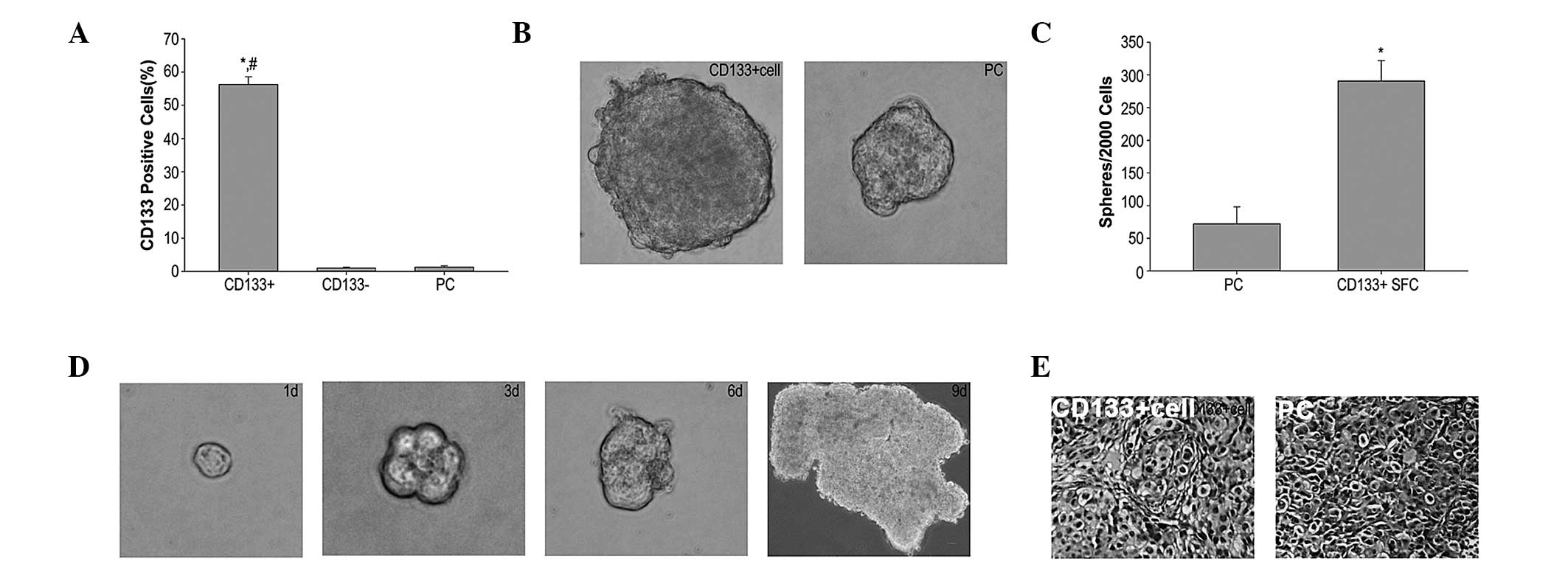 | Figure 1CD133+ SFCs derived from
the MHCC97 cell line possess characteristics of LCSCs. (A)
CD133+ cell subpopulation, sorted from the
hepatocellular carcinoma MHCC97 cell line by magnetic activated
cell sorting, overexpressed the stem cell surface marker, CD133,
detected by flow cytometry using PE-conjugated anti-human CD133
antibody. #P<0.05 compared with the CD133−
cell group. *P<0.05 compared with the PCs(B and C)
CD133+ cells derived from the MHCC97 cells and PCs
formed liver cancer spheroids in stem cell-conditioned medium
(magnification, ×100). Data are expressed as the mean ± standard
deviation (n=3). *P<0.05 compared with the PCs. (D)
The sphere formation of single cells in six-well plates was
detected on the first, third, sixth (magnification, ×400) and ninth
day (magnification, ×40). (E) Hematoxylin and eosin staining
revealed histological characteristics in tumor xenografts derived
from CD133+ SFCs comparable with the PCs (magnification,
×100). CD133, cluster of differentiation 133; SFCs, sphere-forming
cells; LCSCs, liver cancer stem cells; PE, R-phycoerythrin; PC,
parental cell; d, days. |
To further investigate stem-cell properties and the
function of CD133+ SFCs, self-renewal capacity and
tumorigenic potential were analyzed. The capacity of single cells
(obtained from CD133+ dissociated spheres) to form
secondary tumorspheres was measured. Within nine days of culture,
new spheroids of growing undifferentiated CD133+ cells
were observed (Fig. 1D). Thus, the
in vitro CD133+ SFCs from the MHCC97 cell line
demonstrated a self-renewing capacity. Furthermore, the tumorigenic
potential of CD133+ SFCs of the MHCC97 cell line was
investigated in Balb/c-nu mice. Our findings demonstrated that
≤2×10⁵ parental cells were required to initiate stable
tumor formation 37 days after inoculation. By contrast, only
1×10³ CD133+ SFCs were sufficient to generate
visible tumors 27 days after inoculation (Table I). These data indicate that
CD133+ SFCs of the MHCC97 cell line have a greater
tumerogenic capacity compared with parental cells in vivo.
Additionally, H&E staining revealed histological
characteristics in tumor xenografts, which were derived from
CD133+ SFCs, were similar to those of the parental cells
(Fig. 1E). Collectively, these data
demonstrate that CD133+ SFCs possess an ability to
self-renew in vitro and initiate tumor growth in
vivo, indicating that the CD133+ SFCs may provide a
true representation of LCSCs in the HCC MHCC97 cell line.
 | Table ITumorigenicity experiments of
CD133+ SFCs and parental cells in Balb/c-nu mice (n=4
per group). |
Table I
Tumorigenicity experiments of
CD133+ SFCs and parental cells in Balb/c-nu mice (n=4
per group).
| Cell type | Cell number | Tumor
incidence | Latency (days) |
|---|
| Parental cells |
5×10⁴ | 0/4 | - |
|
1×10⁵ | 0/4 | - |
|
2×10⁵ | 3/4 | 37 |
|
5×10⁵ | 4/4 | 29 |
|
1×10⁶ | 4/4 | 7 |
| CD133+
SFCs |
5×10² | 0/4 | - |
|
1×10³ | 3/4 | 27 |
|
5×10³ | 4/4 | 16 |
|
1×10⁴ | 4/4 | 10 |
|
5×10⁴ | 4/4 | 7 |
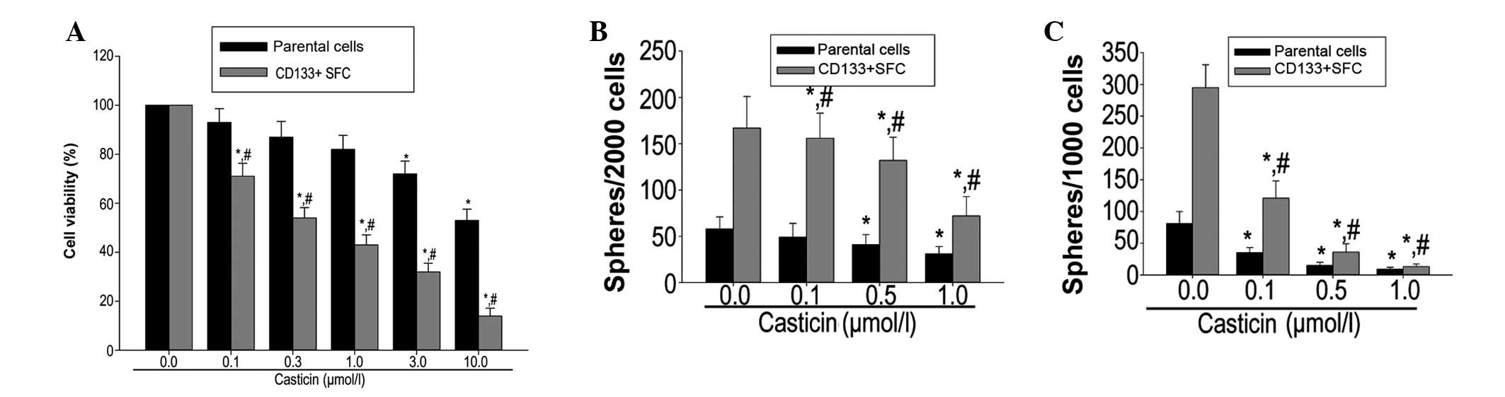
Casticin inhibits proliferation and
self-renewal of LCSCs derived from the MHCC97 cell line
CD133+ SFCs and parental cells were
treated with different concentrations of casticin (0.1, 0.3, 1.0,
3.0 and 10.0 μmol/l) to examine its effect on cell viability of
LCSCs using the MTT assay. Casticin preferentially inhibited cell
viability of CD133+ SFCs derived from MHCC97 cells in a
dose-dependent manner (Fig. 2A).
The half maximal inhibitory concentration of the parental cells and
the CD133+ SFCs was 17.9 and 0.5 μmol/l, respectively
(Fig. 2A).
In order to evaluate whether casticin suppresses the
self-renewal of LCSCs derived from the MHCC97 cell line in
vitro, the primary tumorspheres were treated with various
concentrations of casticin, followed by drug removal and culturing
in another passage to form secondary spheres. Treatment with
casticin resulted in a decreased number of tumorspheres in LCSCs
(Fig. 2B) and a decreased number of
secondary tumorspheres; these findings are consistent with the
reduced self-renewal capacity of LCSCs by casticin treatment
(Fig. 2C).
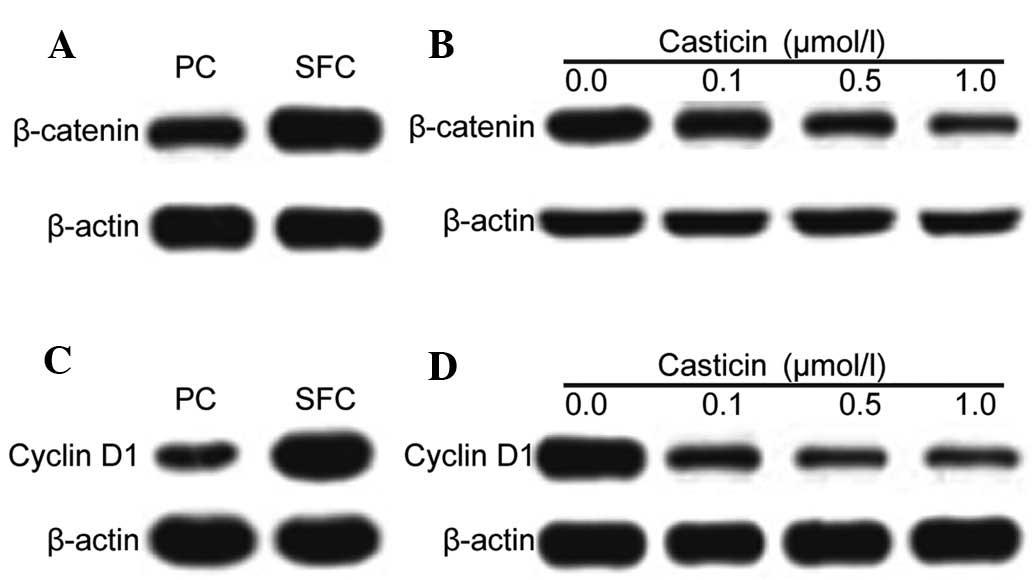
Casticin inhibits self-renewal in LCSCs
through modulating β-catenin expression
The Wnt/β-catenin signaling pathway is a
well-established and significant regulator of stem cell
self-renewal. Wnt/β-catenin signaling has been implicated in the
maintenance of CSCs that are present in liver cancer (21). The expression level of the stem cell
signal molecule, β-catenin, and its downstream target molecule,
cyclin D1, were measured following casticin treatment in LCSCs and
parental cells. Western blot analysis revealed that β-catenin and
cyclin D1 were highly expressed in LCSCs compared with the parental
cells. Additionally, casticin treatment (0.1, 0.5 and 1.0 μM)
resulted in a significant decrease in β-catenin and cyclin D1
expression in LCSCs (Fig. 3).
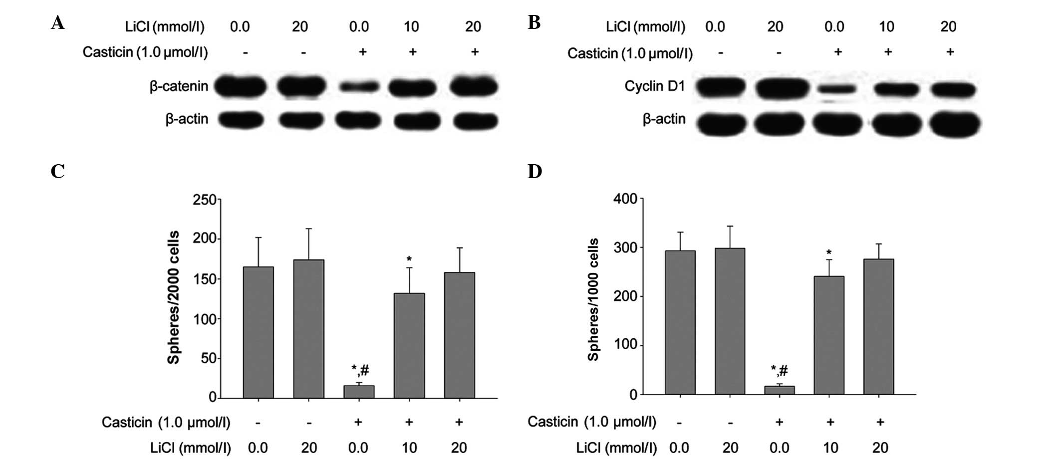
The role of β-catenin in maintaining the
self-renewal characteristics of LCSCs was investigated. LCSCs were
treated with lithium chloride, an agonist known to activate the
Wnt/β-catenin pathway. The addition of lithium chloride resulted in
the upregulation of β-catenin and cyclin D1 in LCSCs. In addition,
lithium chloride antagonized the inhibitory effects of casticin on
the self-renewal of LCSCs and attenuated the casticin-induced
downregulation of β-catenin and cyclin D1 expression in LCSCs
(Fig. 4).
Discussion
Selectively targeting CSCs is a focus of
investigation with emerging evidence demonstrating their role in
the development of cancer (22). A
number of potential CSC therapeutic targets have been identified,
including the ABC superfamily, anti-apoptotic factors, detoxifying
and DNA repair enzymes, and distinct oncogenic cascades (such as
the Wnt/β-catenin, hedgehog, EGFR and Notch pathways) (23,24).
Certain studies have reported a therapeutic strategy that may
successfully kill CSCs; however, certain methods remain under
preclinical and clinical evaluation.
Casticin, a promising candidate agent, has been
reported to effectively eliminate induced apoptosis and exert
antimitotic affects, which results in growth inhibition of cancer
cells in different human malignant tumors in vivo and in
vitro (25,26). Feng et al (20) proposed that casticin inhibits the
proliferation of CSCs. However, the number of studies regarding
casticin-targeting CSCs remains limited. Therefore, the effect of
casticin on CSCs was investigated in the present study.
Abundant evidence has demonstrated the presence of
CSCs in solid tumors. The cell surface marker, CD133, has been used
to isolate and identify populations of LCSCs (27). However, it was proposed that
individual markers should not be used to represent all of the
characteristics of CSCs (15,16).
Thus, in the present study, CD133+ cells were isolated
from the HCC MHCC97 cell line using MACS. The CD133+
cells formed anchorage-independent three-dimensional spheres in the
stem cell-conditioned culture medium. The self-renewal capacity of
CD133+ SFCs of the MHCC97 cell line was assessed using a
sphere formation assay. The standard criterion for estimating
tumorigenicity of tumor cells is with a xenotransplantation assay.
The CD133+ SFCs of the MHCC97 cell line were assessed
for their tumor-initiating ability by subcutaneous inoculation in
nude mice. Our findings demonstrated that only 1×10³
CD133+ SFCs of the MHCC97 cell line were required to
initiate tumor growth compared with 5×10⁵ parental
cells. These findings identified that the ability of
CD133+ SFCs to stimulate tumor growth was higher
compared with parental cells. However, the two cell subpopulations
possessed similar histological characteristics, indicating that
CD133+ SFCs possess the properties of LCSCs. These
findings are consistent with those of Ma et al (28).
In the present study, LCSCs were treated with
various concentrations of casticin and the influence of casticin on
the cell viability and self-renewal capacity were observed.
Casticin preferentially inhibited the viability of lower survival
percentages compared with the parental cells, a finding that is
consistent with that of Feng et al (20). Additionally, when the
CD133+ SFCs were treated with casticin, the
sphere-forming capacity was reduced in the primary and secondary
generations. Therefore, we hypothesized that casticin
preferentially inhibits proliferation and self-renewal of
LCSCs.
The classic Wnt/β-catenin signaling pathway is vital
in the self-renewal and differentiation of LCSCs, and acts as the
predominant factor for chemotherapy resistance. PKF118–310 inhibits
the self-renewal of breast tumor-initiating cells by Wnt/β-catenin
signaling and CDH11 was found to inhibit actin stress fiber
formation, thus, further inhibiting tumor cell migration and
invasion via the regulation of Wnt/β-catenin signaling (29). In the present study, the expression
of β-catenin and cyclin D1 was higher in the parental cells; when
CD133+ SFCs of the MHCC97 cell line were treated with
casticin, the expression of β-catenin and cyclin D1 was
downregulated in a dose-dependent manner. In addition, treatment
with lithium chloride effectively attenuated the inhibition of the
self-renewal capacity by casticin in CD133+ SFCs of the
MHCC97 cell line. Our findings indicate that casticin regulates
self-renewal of LCSCs by downregulating the expression of
β-catenin.
In conclusion, CD133+ SFCs of the MHCC97
cell line possess the characteristics of LCSCs. Moreover, casticin
inhibited the self-renewal capacity of LCSCs, which was a result of
blocking the Wnt/β-catenin signaling pathway. Therefore, casticin,
by targeting LCSCs, may have a therapeutic role in the treatment of
HCC.
Acknowledgements
The authors would like to thank Dr Jian-Guo Cao
(Medical College, Hunan Normal University, Changsha, China) for the
critical reading of this manuscript. This study was supported by
the Project of Scientific Research from the Administration Bureau
of Traditional Chinese Medicine, Hunan Province (grant no.
2010081), the Project of Scientific Research, the Department of
Education, Hunan Province (grant no. 10C0975), the Major Project
Item of Scientific Research, the Department of Education, Hunan
Province (grant no. 09A054), the Project of Scientific Research
from Changsha city Bureau of Science and Technology (grant no.
K1104060-31) and the Hunan province Science and Technology Project
(grant no. 2011FJ4144).
References
|
1
|
Ma S, Chan KW and Guan XY: In search of
liver cancer stem cells. Stem Cell Rev. 4:179–192. 2008. View Article : Google Scholar : PubMed/NCBI
|
|
2
|
Tomuleasa C, Soritau O, Rus-Ciuca D, et
al: Isolation and characterization of hepatic cancer cells with
stem-like properties from hepatocellular carcinoma. J
Gastrointestin Liver Dis. 19:61–67. 2010.PubMed/NCBI
|
|
3
|
Ricci-Vitiani L, Lombardi DG, Pilozzi E,
et al: Identification and expansion of human
colon-cancer-initiating cells. Nature. 445:111–115. 2007.
View Article : Google Scholar : PubMed/NCBI
|
|
4
|
Lee TK, Castilho A, Ma S and Ng IO: Liver
cancer stem cells: implications for a new therapeutic target. Liver
Int. 29:955–965. 2009. View Article : Google Scholar : PubMed/NCBI
|
|
5
|
Mackillop WJ, Ciampi A, Till JE and Buick
RN: A stem cell model of human tumor growth: implications for tumor
cell clonogenic assays. J Natl Cancer Inst. 70:9–16.
1983.PubMed/NCBI
|
|
6
|
Gupta PB, Chaffer CL and Weinberg RA:
Cancer stem cells: mirage or reality? Nat Med. 15:1010–1012. 2009.
View Article : Google Scholar : PubMed/NCBI
|
|
7
|
Chiba T, Kita K, Zheng YW, et al: Side
population purified from hepatocellular carcinoma cells harbors
cancer stem cell-like properties. Hepatology. 44:240–251. 2006.
View Article : Google Scholar : PubMed/NCBI
|
|
8
|
Yang ZF, Ho DW, Ng MN, et al: Significance
of CD90+ cancer stem cells in human liver cancer. Cancer Cell.
13:153–166. 2008.
|
|
9
|
Ma S, Chan KW, Hu L, et al: Identification
and characterization of tumorigenic liver cancer stem/progenitor
cells. Gastroenterology. 132:2542–2556. 2007. View Article : Google Scholar : PubMed/NCBI
|
|
10
|
Zhu Z, Hao X, Yan M, et al: Cancer
stem/progenitor cells are highly enriched in CD133+CD44+ population
in hepatocellular carcinoma. Int J Cancer. 126:2067–2078. 2010.
|
|
11
|
Kimura O, Takahashi T, Ishii N, et al:
Characterization of the epithelial cell adhesion molecule (EpCAM)+
cell population in hepatocellular carcinoma cell lines. Cancer Sci.
101:2145–2155. 2010.
|
|
12
|
Yang W, Yan HX, Chen L, et al:
Wnt/beta-catenin signaling contributes to activation of normal and
tumorigenic liver progenitor cells. Cancer Res. 68:4287–4295. 2008.
View Article : Google Scholar : PubMed/NCBI
|
|
13
|
Haraguchi N, Ishii H, Mimori K, et al:
CD13 is a therapeutic target in human liver cancer stem cells. J
Clin Investig. 120:3326–3339. 2010. View
Article : Google Scholar : PubMed/NCBI
|
|
14
|
Salnikov AV, Kusumawidjaja G, Rausch V, et
al: Cancer stem cell marker expression in hepatocellular carcinoma
and liver metastases is not sufficient as single prognostic
parameter. Cancer Lett. 275:185–193. 2009. View Article : Google Scholar
|
|
15
|
Pellegrino R, Brusasco V, Viegi G, et al:
Definition of COPD: based on evidence or opinion? Eur Respir J.
31:681–682. 2008. View Article : Google Scholar : PubMed/NCBI
|
|
16
|
Pharmacopoeia Commission of People’s
Republic of China. Pharmacopoeia of the Peoples Republic of China.
1. China Chemical Industry Press; Beijing: 2010, (In Chinese).
|
|
17
|
Ye Q, Zhang QY, Zheng CJ, Wang Y and Qin
LP: Casticin, a flavonoid isolated from Vitex rotundifolia,
inhibits prolactin release in vivo and in vitro. Acta Pharmacol
Sin. 31:1564–1568. 2010. View Article : Google Scholar : PubMed/NCBI
|
|
18
|
Shen JK, Du HP, Yang M, Wang YG and Jin J:
Casticin induces leukemic cell death through apoptosis and mitotic
catastrophe. Ann Hematol. 88:743–752. 2009. View Article : Google Scholar : PubMed/NCBI
|
|
19
|
Yang J, Yang Y, Tian L, Sheng XF, Liu F
and Cao JG: Casticin-induced apoptosis involves death receptor 5
upregulation in hepatocellular carcinoma cells. World J
Gastroenterol. 17:4298–4307. 2011. View Article : Google Scholar : PubMed/NCBI
|
|
20
|
Feng X, Zhou Q, Liu C and Tao ML: Drug
screening study using glioma stem-like cells. Mol Med Rep.
6:1117–1120. 2012.PubMed/NCBI
|
|
21
|
Wang F, He L, Dai W-Q, et al: Salinomycin
inhibits proliferation and induces apoptosis of human
hepatocellular carcinoma cells in vitro and in vivo. PloS One.
7:e506382012. View Article : Google Scholar : PubMed/NCBI
|
|
22
|
Naujokat C and Steinhart R: Salinomycin as
a drug for targeting human cancer stem cells. J Biomed Biotechnol.
2012:9506582012. View Article : Google Scholar : PubMed/NCBI
|
|
23
|
Wang Z, Li Y, Ahmad A, et al: Targeting
miRNAs involved in cancer stem cell and EMT regulation: an emerging
concept in overcoming drug resistance. Drug Resist Updat.
13:109–118. 2010. View Article : Google Scholar : PubMed/NCBI
|
|
24
|
Liu J, Kopecková P, Bühler P, et al:
Biorecognition and subcellular trafficking of HPMA
copolymer-anti-PSMA antibody conjugates by prostate cancer cells.
Mol Pharm. 6:959–970. 2009. View Article : Google Scholar : PubMed/NCBI
|
|
25
|
Shen JK, Du HP, Yang M, Wang YG and Jin J:
Casticin induces leukemic cell death through apoptosis and mitotic
catastrophe. Ann Hematol. 88:743–752. 2009. View Article : Google Scholar : PubMed/NCBI
|
|
26
|
Kobayakawa J, Sato-Nishimori F, Moriyasu M
and Matsukawa Y: G2-M arrest and antimitotic activity mediated by
casticin, a flavonoid isolated from Viticis Fructus (Vitex
rotundifolia Linne fil). Cancer Lett. 208:59–64. 2004.
View Article : Google Scholar : PubMed/NCBI
|
|
27
|
Mizrak D, Brittan M and Alison M: CD133:
molecule of the moment. J Pathol. 214:3–9. 2008. View Article : Google Scholar : PubMed/NCBI
|
|
28
|
Ma S, Tang KH, Chan YP, et al: miR-130b
promotes CD133(+) liver tumor-initiating cell growth and
self-renewal via tumor protein 53-induced nuclear protein 1. Cell
Stem Cell. 7:694–707. 2010.PubMed/NCBI
|
|
29
|
Li L, Ying J, Li H, et al: The human
cadherin 11 is a pro-apoptotic tumor suppressor modulating cell
stemness through Wnt/beta-catenin signaling and silenced in common
carcinomas. Oncogene. 31:3901–3912. 2012. View Article : Google Scholar : PubMed/NCBI
|