Introduction
Salivary adenoid cystic carcinoma (SACC) is a
frequent subtype of salivary gland malignancy accounting for 25% of
malignant tumors in the major salivary glands (1) and 50% in the minor glands (2). The neoplasm is characterized by
heterogeneous phenotypic features and persistently progressive
biological behavior. Although various treatment options have been
extensively investigated, the poor long-term prognosis for patients
with SACC is primarily due to local recurrence associated with
perineural invasion (PNI) and delayed onset of distant metastasis,
particularly to the lungs (3,4).
Therefore, it is necessary to identify and
understand the diverse mechanisms behind metastasis so as to
improve the treatment strategies for SACC patients. Genomic and
proteomic studies have yielded various novel insights into
molecular targets and the mechanisms of SACC metastasis (5,6). A
number of mechanisms underlying the distant metastasis of SACC have
been described, including the Notch gene family, matrix
metalloproteinases, nerve growth factor, and vascular endothelial
growth factor (7–12). However, the mechanisms of distant
metastasis are complicated and remain obscure; therefore, further
elucidation is required.
The changes in gene expression that accompany
metastasis include DNA, mRNA and protein levels, however, the
changes in the levels of mRNA and the encoded proteins are often
disproportionate. This may be due to a number of reasons, among
which post-transcriptional regulation by microRNAs (miRNAs) is
particularly important. miRNAs are small non-coding RNAs of 21–25
nt that negatively modulate protein expression (13). miRNAs are involved in various
biological processes, including development, proliferation,
apoptosis and differentiation (14,15).
In addition, studies have demonstrated that aberrant microRNA
expression is correlated with malignant transformation and tumor
development (16,17).
Thus, it was hypothesized that miRNAs may be
significant in mediating metastatic progression in SACC. In order
to investigate this hypothesis, a microRNA array was employed to
detect a distinctive miRNA expression pattern between an SACC cell
line, ACC-2, and a highly metastatic SACC cell line, ACC-M, which
was screened from ACC-2 by a combination of in vivo
selection and cloning in vitro (18). Since ACC-2 and ACC-M cells have an
identical genetic background (with the exception of different
metastatic behavior), it was presumed that differentially expressed
miRNAs were metastasis-related miRNAs, which are involved directly
or indirectly in the progression of metastasis. The results of the
miRNA microarray analysis were further verified by quantitative
polymerase chain reaction (qPCR). In addition, bioinformatic
methods were employed in the analysis of the miRNA expression
arrays to identify the target genes, which are regulated by miRNAs,
and analyze the gene functions that are associated with the
metastasis of tumors. The miRNA signature of metastatic progression
may aid with the development of novel therapeutic strategies for
SACC patients.
Materials and methods
Cell lines and cell culture
condition
The cell lines, ACC-2 and ACC-M, were provided by
the Department of Oral Biology, School of Stomatology, Fourth
Military Medical University (Xi’an, China). The two types of cells
were cultured in Dulbecco’s modified Eagle’s medium (Life
Technologies, Carlsbad, CA, USA) supplemented with 10% fetal bovine
serum (Life Technologies), 2.05 mM L-glutamine, 100 μg/ml
penicillin and 100 μg/ml streptomycin at 37°C with 5%
CO2.
miRNA microarray
Total RNA from the SACC cell line, ACC-2 and the
highly metastatic SACC cell line, ACC-M was isolated using TRIzol
reagent (Life Technologies). The quality and quantity of the RNA
samples was assessed by standard electrophoresis and ultraviolet
(UV) spectrophotometry methods (19). The miRNA microarray analysis was
performed using the Human miRNA OneArray v4 (Phalanx Biotech Group,
Hsinchu, Taiwan), which contains 1,884 unique microRNA probes that
are complementary to human microRNA sequences (Sanger miRBase
Release 18.0). The data from the microarray were collected and
analyzed in accordance with the Minimum Information About a
Microarray Experiment guidelines (20). The differential miRNA expression was
determined using a two-tailed t-test on a single miRNA basis. The
signals were considered to be different when P<0.01 and these
miRNAs were subsequently selected for cluster analysis. The miRNA
microarray analyses were performed in duplicate and the miRNAs that
exhibited common differential expression levels were selected.
qPCR analysis of miRNA expression
qPCR assays were performed on two samples. Using the
TaqMan MicroRNA assay kit (Life Technologies), which utilizes
miRNA-specific primers, each RNA sample (10 μg) was reverse
transcribed according to the manufacturer’s instructions. The
resulting cDNA was semi-quantitatively amplified in 45 cycles on an
ABI 7500 Real-Time PCR system, using TaqMan® Universal
PCR Master Mix, No AmpErase® UNG and TaqMan MicroRNA
assays for hsa-miR-4487, hsa-miR-4430, hsa-miR-486-3p,
hsa-miR-5191, hsa-miR-3131, hsa-miR-211-3p and U6 snRNA (all
Applied Biosystems, Carlsbad, CA, USA). These miRNAs were selected
as they exhibited the greatest fold change (hsa-miR-4487 and
hsa-miR-4430 were upregulated, and hsa-miR-5191 and hsa-miR-3131
were downregulated), or were of potential importance as indicated
by previous studies (hsa-miR-486-3p was upregulated and
hsa-miR-211-3p was downregulated). Each qPCR assay was performed at
least three times (21,22).
miRNA target prediction
Candidate miRNAs that were selected for the qPCR
analysis were utilized for target prediction. The potential miRNA
targets among the genes, which were negatively correlated with
miRNA expression, were determined using the publicly available
TargetScan (http://www.targetscan.org), miRBase
(http://www.ebi.ac.uk/enright-srv/microcosm/htdocs/targets/v5)
and PicTar (http://pictar.mdc-berlin.de) algorithms. The genes
that were predicted as candidate miRNA targets and those that were
selected on the basis of Gene Ontology (http://www.geneontology.org) were compared, and the
genes that appeared in the two lists were included in the present
study.
Statistical analysis
Statistical analysis was performed using SPSS 17.0
statistical software (SPSS, Inc., Chicago, IL, USA). All numerical
data from the qPCR assays were analyzed following the derivation of
standard curves for each miRNA of interest. Statistical analysis
was performed with Student’s t-test and P<0.05 was considered to
indicate a statistically significant difference. Each experiment
was performed in triplicate and each individual sample was run in
triplicate.
Results
Analysis of the quality of RNA isolated
from ACC-2 and ACC-M cells
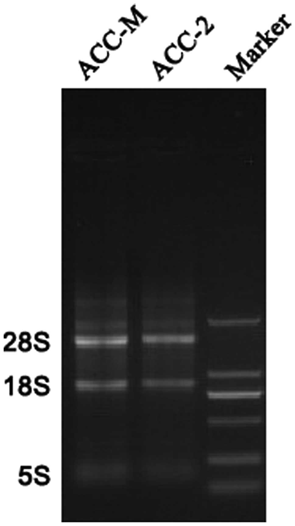
The quality of total RNA was analyzed via denaturing
agarose gel electrophoresis and UV spectrophotometry (UV-2600;
Shimadzu Corporation, Kyoto, Japan). RNA that was isolated from the
ACC-2 and ACC-M cells exhibited clear bands of 28S rRNA and 18S
rRNA (Fig. 1). The
OD260/OD280 values of each sample were 2.05
and 2.06, respectively, and the OD260/OD230
values of each sample were 2.05 and 2.17, respectively, as
determined by the UV spectrophotometer. The RNA quality was
confirmed by the Agilent 2100 Bioanalyzer (Agilent Technologies,
Santa Clara, CA, USA) prior to further hybridization. All these
results indicated that the obtained high quality RNA was suitable
for microRNA microarray analysis and the following qPCR analysis to
verify the results of the microarray.
miRNA expression profiles in the SACC
cell line, ACC-2 and the highly metastatic SACC cell line,
ACC-M
To identify the changes in the miRNA expression
profiles between the SACC cell line, ACC-2 and the highly
metastatic SACC cell line, ACC-M, miRNA microarray analysis was
conducted. The miRNA expression profile of the two SACC cell lines
revealed that 38 out of 1,884 human miRNAs were differentially
expressed between the ACC-2 and ACC-M cell lines. Of the 38 miRNAs
identified as differentially expressed, 20 were upregulated
(Table I) and 18 were downregulated
(Table II) in the ACC-M cell line
compared with the control ACC-2 cell line. In the average
fold-change analysis, 17 of the 38 flagged miRNAs exhibited
>2-fold change in expression level, while there were 21 miRNAs
that exhibited <2-fold change in expression level (Table I and II).
 | Table ImiRNAs upregulated in the ACC-M cell
line compared with the control ACC-2 cell line. |
Table I
miRNAs upregulated in the ACC-M cell
line compared with the control ACC-2 cell line.
| Phalanx ID | Name | Average fold
change |
|---|
| PH_mr_0004751 | hsa-miR-4487a | 32.24 |
| PH_mr_0004716 | hsa-miR-4430a | 4.05 |
| PH_mr_0004876 | hsa-miR-5096a | 3.61 |
| PH_mr_0000840 |
hsa-miR-1285-3pa | 2.85 |
| PH_mr_0008030 |
hsa-miR-1273g-3pa | 2.60 |
| PH_mr_0004900 |
hsa-miR-3150b-3pa | 2.59 |
| PH_mr_0004874 | hsa-miR-1273fa | 2.46 |
| PH_mr_0001931 | hsa-miR-1273aa | 2.43 |
| PH_mr_0004658 | hsa-miR-1273ea | 2.25 |
| PH_mr_0000379 | hsa-miR-574-5p | 1.98 |
| PH_mr_0004873 | hsa-miR-5095 | 1.88 |
| PH_mr_0004549 | hsa-miR-4638-5p | 1.88 |
| PH_mr_0004565 | hsa-miR-4688 | 1.87 |
| PH_mr_0001461 | hsa-miR-877-5p | 1.86 |
| PH_mr_0004257 | hsa-miR-3154 | 1.82 |
| PH_mr_0003162 | hsa-miR-766-3p | 1.80 |
| PH_mr_0004018 | hsa-miR-1972 | 1.78 |
| PH_mr_0000953 | hsa-miR-486-3p | 1.78 |
| PH_mr_0004577 | hsa-miR-4728-5p | 1.74 |
| PH_mr_0004187 | hsa-miR-1976 | 1.74 |
| Table IImiRNAs downregulated in the ACC-M cell
line compared with the control ACC-2 cell line. |
Table II
miRNAs downregulated in the ACC-M cell
line compared with the control ACC-2 cell line.
| Phalanx ID | Name | Average fold
change |
|---|
| PH_mr_0008120 |
hsa-miR-5191a | 0.028 |
| PH_mr_0004035 |
hsa-miR-3131a | 0.22 |
| PH_mr_0004331 |
hsa-miR-4278a | 0.26 |
| PH_mr_0004534 |
hsa-miR-4498a | 0.39 |
| PH_mr_0008014 |
hsa-miR-211-3pa | 0.40 |
| PH_mr_0004731 |
hsa-miR-4450a | 0.41 |
| PH_mr_0000102 |
hsa-miR-373-5pa | 0.44 |
| PH_mr_0000010 |
hsa-miR-7-5pa | 0.48 |
| PH_mr_0008044 |
hsa-miR-5010-5p | 0.52 |
| PH_mr_0004714 | hsa-miR-4428 | 0.53 |
| PH_mr_0001673 | hsa-miR-18b-5p | 0.53 |
| PH_mr_0001728 | hsa-miR-18a-5p | 0.53 |
| PH_mr_0004543 | hsa-miR-1587 | 0.54 |
| PH_mr_0001696 | hsa-miR-20a-5p | 0.55 |
| PH_mr_0000136 | hsa-miR-1265 | 0.55 |
| PH_mr_0000436 | hsa-miR-92a-3p | 0.56 |
| PH_mr_0000747 | hsa-miR-186-5p | 0.57 |
| PH_mr_0000812 | hsa-miR-92b-3p | 0.57 |
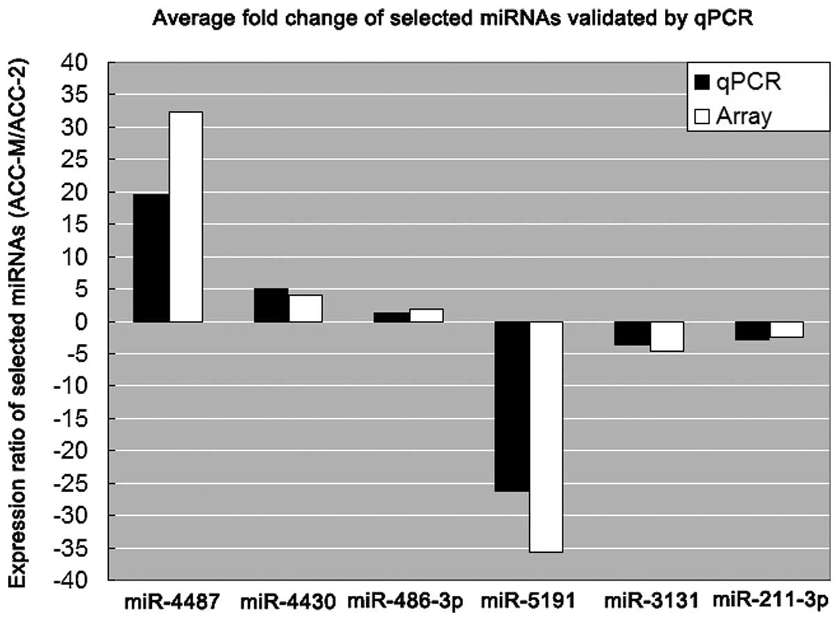
Confirmation of miRNA microarray data by
qPCR
To confirm the miRNA microarray data, a TaqMan
MicroRNA assay kit was used to perform qPCR analyses of the
expression levels of miR-4487, -4430, -486-3p, -5191, -3131 and
-211-3p in the ACC-2 and ACC-M cell lines. It is generally accepted
that gene expression levels should be normalized by a carefully
selected, stable internal control gene. To validate the assumption
of stable expression of a given control gene, prior knowledge of a
reliable measure to normalize this gene, in order to eliminate any
non-specific variation, is required. For each sample, the
expression values were normalized to the U6 snRNA gene (a stable
housekeeping gene) and the expression levels relative to the ACC-2
cell line were calculated. The expression pattern that was
identified in the arrays was confirmed by this additional analysis.
Compared with the ACC-2 cells, three miRNAs (miR-4487, -4430 and
-486-3p) were significantly upregulated, while three miRNAs
(miR-5191, -3131 and -211-3p) were significantly downregulated in
the highly metastatic SACC cell line, ACC-M. For these candidate
miRNAs, the qPCR analysis revealed similar patterns of up- or
downregulation to those revealed by the results of the microarray
analysis (Table III; Fig. 2), although the magnitude of the
reported changes in expression differed. Thus, miRNA expression
profiles clearly exhibit significant differences between ACC-2 and
ACC-M, indicating that miRNAs are significant during the metastatic
progression of SACC.
| Table IIIValidation of microarray analysis
data by qPCR for candidate microRNAs. |
Table III
Validation of microarray analysis
data by qPCR for candidate microRNAs.
| | Average expression
ratio |
|---|
| |
|
|---|
| Phalanx ID | Name | qPCR | Array |
|---|
| PH_mr_0004751 | hsa-miR-4487 | 19.560 | 32.240 |
| PH_mr_0004716 | hsa-miR-4430 | 4.960 | 4.050 |
| PH_mr_0000953 | hsa-miR-486-3p | 1.210 | 1.780 |
| PH_mr_0008120 | hsa-miR-5191 | 0.038 | 0.028 |
| PH_mr_0004035 | hsa-miR-3131 | 0.280 | 0.220 |
| PH_mr_0008014 | hsa-miR-211-3p | 0.340 | 0.400 |
Prediction of miRNA targets
The identification of potential target genes of
miRNAs that are associated with the metastatic progression of SACC
is essential to investigate their biological functions. Previous
studies have shown that miRNAs are able to regulate expression of
their target genes by decreasing mRNA stability in addition to
translational inhibition (23). The
candidate miRNAs, which were used for qPCR, were selected for
target prediction and analyzed with the following three target
prediction tools: TargetScan 6.2 (Whitehead Institute for
Biomedical Research website; http://www.targetscan.org); miRBase Targets Release v5
(the Enright Group; http://www.ebi.ac.uk/enright-srv/microcosm/htdocs/targets/v5);
and PicTar (http://pictar.mdc-berlin.de). By analysis of these
databases, human genes, which were known to be involved in cell
proliferation, apoptosis, cell cycle, DNA damage, DNA repair and
signaling pathways were selected from the Gene Ontology website
(http://www.ebi.ac.uk/GOA). Those genes that were
predicted to be targets of the candidate miRNAs were selected and
are listed in Table IV. These data
may provide the foundation for further analysis of the mechanisms
of metastatic progression in SACC.
| Table IVPredicted target genes of miRNAs. |
Table IV
Predicted target genes of miRNAs.
| Putative targets of
miRNAs according to function |
|---|
|
|
|---|
| miRNA name | Apoptosis and
proliferation | Cell cycle | DNA damage and
repair | Signaling
pathways |
|---|
| hsa-miR-4487 | GIPC3, URGCP,
ITGA5, VASH1, FGD1, HECA, FLT1 | GALNTL2, NAP1L5,
ZNF740, TBC1D4, CDKN1A | PSMA5, HIPK2,
USH1G, BOLL | EFNB3, SORT1, RORC,
FMOD, FAM123C, CUL3, CXCR3, WNT4 |
| hsa-miR-4430 | MAP3K9, MAP2K7,
SHROOM4, KRT85, MNT, GLI2, IREB2 | SEZ6, GRIN3A,
MOCS1, PARP16 | POMT2, RPP14,
ZDHHC22 | POP4, EIF2C1,
POFUT1, EFNB3, CD72, LPHN2, EIF2C3 |
| hsa-miR-486-3p | NAT15, GDI1, DMBX1,
CNP, PAK6, CD276, MAP4, VASH1, PLA2G6, SPTBN4, SAP30BP, COL4A2,
HS6ST1, DCAF7, TANC2 | SF3A1, ATXN7L3,
SDC3, TTYH3, STX1B, GATAD2B | FYCO1, STIM1,
CACNA2D2 | FLOT2, CTNNBIP1,
MARK2, MAPKBP1, LOC388630 |
| hsa-miR-5191 | FOXL1, AHI1,
PERP | MAP1B, CDH12,
ABCA9, WWP2, HOXC6, EIF4E, CD1D | PPEF2, ZNF547,
RANBP10, FLG2, NIPA2, PARP9, ZNF32, ZFP28 | RBPJL, GNB1, GNB4,
SOX9, YWHAZ, NOTCH2 |
| hsa-miR-3131 | SEMA6C, MEF2D,
TMEM119, TRAF3, EIF4G1 | RIMBP2, SYN1,
FBXO22 | NFIX | PPARGC1B |
| hsa-miR-211-3p | NFASC, SEMA5A,
DMC1, MAFG, PITPNA, ERLIN2, PDXK, SMARCD1, SOCS5, TNFAIP1 | MAU2, PDS5A, NOS1,
ATP11A, XPO4 | SMCR7L, ZNF217,
OBFC1, RBFOX2, ATG10 | PIK3CG, LONP2,
PPP2R5E |
Discussion
SACC is a common malignant tumor that arises from
the secretory epithelial cells in the salivary glands of the head
and neck. SACC accounts for 1% of all head and neck malignancies
and 10% of all salivary neoplasms (24). Although SACC tend to grow slowly,
this neoplasm has a poor prognosis owing to its insidious invasion
into adjacent tissues and hematogenous spread to distant organs
(lungs, bone and liver) (24,25).
The five-year disease-free survival rate is ≤90%, however, this
reduces to 40% after 15 years (27). Advanced tumor stage, distant
metastasis to the lungs, PNI, and solid subtype are among the
clinicopathological parameters that have been proposed as useful
predictors of the clinical course of SACC (28). However, accurate prediction is often
challenging, and in-depth studies on invasion and metastatic
mechanisms are of great significance for prognosis and evaluation,
and the selection of appropriate treatment protocols.
miRNAs are short non-coding RNAs of 21–25 nt that
regulate gene expression in multicellular organisms. Numerous
studies have indicated that miRNAs are significant in
carcinogenesis and tumor progression in certain types of cancer,
including SACC (29–31). However, to the best of our
knowledge, the altered expression of miRNAs, which is associated
with metastatic progression in SACC, has not been investigated.
Therefore, in the present study, high-throughput miRNA microarray
technology, with 1,884 miRNAs selected from the miRBase, was used
to detect miRNA expression in parental ACC-2 cells and highly
metastatic ACC-M cells. The microarray data revealed that 38 miRNAs
exhibited significant differences in expression between ACC-2 and
ACC-M cell lines; 20 were upregulated and 18 were downregulated in
the ACC-M cells. In the present analysis, 17 miRNAs in the ACC-M
cells exhibited >2-fold changes compared with the control cells
(ACC-2). Although not all miRNAs in ACC-M cells exhibit similar
regulation patterns, numerous miRNAs with ≥2-fold expression
changes were identified in the present study for further
investigation. Therefore, to confirm the expression of the six
miRNAs that were identified in the microarray analysis, a qPCR
assay was performed. The results of the qPCR analysis of the
upregulated (miR-4487, -4430 and -486-3p) and downregulated
(miR-5191, -3131 and -211-3p) miRNAs are demonstrated in Fig. 2. For all six miRNAs, the qPCR
experiment confirmed their down- or upregulation, however, the
magnitude of the changes in the expression levels was found to
differ between the microarray and qPCR analyses.
Thus far, to the best of our knowledge, there have
been no reports of a correlation between the six miRNAs discussed
in the present study and the metastatic progression of SACC. To
elucidate the regulatory mechanisms of those miRNAs, which are
associated with the metastatic progression of SACC, various
bioinformatic methods were employed to identify potential target
genes of those miRNAs. The target genes, which have been reported
to be associated with the metastatic progression of human tumors,
were divided into four groups according to function (as described
by the Gene Ontology website, http://www.geneontology.org): Apoptosis and
proliferation (46 genes); cell cycle (30 genes); DNA damage and
repair (24 genes); and signaling pathway (30 genes; Table IV).
Computational predictions indicate that one miRNA is
able to target numerous mRNAs and each mRNA can also be targeted by
numerous miRNAs. Thus, a one-to-one association between miRNAs and
target genes should not be expected, rather a one-to-many or
many-to-one association. Previous studies have demonstrated that
functional miRNA binding sites can lie in the coding or
5′-untranslated regions of endogenous mRNAs (31). Therefore, the number of target genes
that are regulated by miRNAs may be larger than what was identified
by the bioinformatic methods used in the present study. Although
the expression levels of miRNAs may demonstrate inverse
correlations with the protein expression levels of their potential
target genes, further studies are required to confirm a regulatory
association between miRNAs and the predicted target genes.
In conclusion, SACC is a common subtype of salivary
gland malignancy, which exhibits biological behavior that
facilitates hematogenous spread to lungs. In the present study, the
experimental data demonstrated that alteration of the miRNA
expression profile in the ACC-M cell line may be associated with
metastatic progression. These findings may aid with elucidating the
potential mechanisms that underlie the metastatic progression of
SACC and provide a novel molecular therapeutic target for the
treatment of SACC.
Acknowledgements
The present study was supported by grants from the
National Natural Science Foundation of China (grant no. 81102051)
and the Natural Science Foundation of Jiangsu Province (grant no.
BK2011659).
Abbreviations:
|
SACC
|
salivary adenoid cystic carcinoma
|
|
miRNA
|
microRNA
|
|
PNI
|
perineural invasion
|
|
qPCR
|
quantitative PCR
|
References
|
1
|
Renehan A, Gleave EN, Hancock BD, Smith P
and McGurk M: Long-term follow-up of over 1000 patients with
salivary gland tumours treated in a single centre. Br J Surg.
83:1750–1754. 1996. View Article : Google Scholar : PubMed/NCBI
|
|
2
|
Anderson JN Jr, Beenken SW, Crowe R, Soong
SJ, Peters G, Maddox WA and Urist MM: Prognostic factors in minor
salivary gland cancer. Head Neck. 17:480–486. 1995. View Article : Google Scholar : PubMed/NCBI
|
|
3
|
van der Wal JE, Becking AG, Snow GB and
van der Waal I: Distant metastases of adenoid cystic carcinoma of
the salivary glands and the value of diagnostic examinations during
follow-up. Head Neck. 24:779–783. 2002.PubMed/NCBI
|
|
4
|
Ramer N, Wu H, Sabo E, Ramer Y, Emanuel P,
Orta L and Burstein DE: Prognostic value of quantitative p63
immunostaining in adenoid cystic carcinoma of salivary gland
assessed by computerized image analysis. Cancer. 116:77–83.
2010.PubMed/NCBI
|
|
5
|
Chen W, Zhang HL, Shao XJ, Jiang YG, Zhao
XG, Gao X, Li JH, Yang J, Zhang YF, Liu BL and Sun MY: Gene
expression profile of salivary adenoid cystic carcinoma associated
with perineural invasion. Tohoku J Exp Med. 212:319–334. 2007.
View Article : Google Scholar : PubMed/NCBI
|
|
6
|
Nakashima D, Uzawa K, Kasamatsu A, Koike
H, Endo Y, Saito K, Hashitani S, Numata T, Urade M and Tanzawa H:
Protein expression profiling identifies maspin and stathmin as
potential biomarkers of adenoid cystic carcinoma of the salivary
glands. Int J Cancer. 118:704–713. 2006. View Article : Google Scholar
|
|
7
|
Chen W, Zhang H, Wang J, Cao G, Dong Z, Su
H, Zhou X and Zhang S: Lentiviral-mediated gene silencing of
Notch-4 inhibits in vitro proliferation and perineural
invasion of ACC-M cells. Oncol Rep. 29:1794–1804. 2013.
|
|
8
|
Ding LC, She L, Zheng DL, Huang QL, Wang
JF, Zheng FF and Lu YG: Notch-4 contributes to the metastasis of
salivary adenoid cystic carcinoma. Oncol Rep. 24:363–368.
2010.PubMed/NCBI
|
|
9
|
Yang X, Zhang P, Ma Q, Kong L, Li Y, Liu B
and Lei D: EMMPRIN silencing inhibits proliferation and perineural
invasion of human salivary adenoid cystic carcinoma cells in
vitro and in vivo. Cancer Biol Ther. 13:85–91. 2012.
View Article : Google Scholar : PubMed/NCBI
|
|
10
|
Luukkaa H, Klemi P, Hirsimäki P, Vahlberg
T, Kivisaari A, Kähäri VM and Grénman R: Matrix metalloproteinase
(MMP)-1, -9 and -13 as prognostic factors in salivary gland cancer.
Acta Otolaryngol. 128:482–490. 2008. View Article : Google Scholar : PubMed/NCBI
|
|
11
|
Hao L, Xiao-lin N, Qi C, Yi-ping Y,
Jia-quan L and Yan-ning L: Nerve growth factor and vascular
endothelial growth factor: retrospective analysis of 63 patients
with salivary adenoid cystic carcinoma. Int J Oral Sci. 2:35–44.
2010. View Article : Google Scholar : PubMed/NCBI
|
|
12
|
Costa AF, Tasso MG, Mariano FV, Soares AB,
Chone CT, Crespo AN, Fresno MF, Llorente JL, Suárez C, de Araújo
VC, Hermsen M and Altemani A: Levels and patterns of expression of
hypoxia-inducible factor-1α, vascular endothelial growth factor,
glucose transporter-1 and CD105 in adenoid cystic carcinomas with
high-grade transformation. Histopathology. 60:816–825. 2012.
|
|
13
|
Chekanova JA and Belostotsky DA: MicroRNAs
and messenger RNA turnover. Methods Mol Biol. 342:73–85.
2006.PubMed/NCBI
|
|
14
|
Lages E, Ipas H, Guttin A, Nesr H, Berger
F and Issartel JP: MicroRNAs: molecular features and role in
cancer. Front Biosci (Landmark Ed). 17:2508–2540. 2012. View Article : Google Scholar
|
|
15
|
Li C, Hashimi SM, Good DA, Cao S, Duan W,
Plummer PN, Mellick AS and Wei MQ: Apoptosis and microRNA
aberrations in cancer. Clin Exp Pharmacol Physiol. 39:739–46. 2012.
View Article : Google Scholar : PubMed/NCBI
|
|
16
|
Nishizawa T and Suzuki H: The role of
microRNA in gastric malignancy. Int J Mol Sci. 14:9487–9496. 2013.
View Article : Google Scholar : PubMed/NCBI
|
|
17
|
Wu WK, Lee CW, Cho CH, Fan D, Wu K, Yu J
and Sung JJ: MicroRNA dysregulation in gastric cancer: a new player
enters the game. Oncogene. 29:5761–5771. 2010. View Article : Google Scholar : PubMed/NCBI
|
|
18
|
Guan XF, Qiu WL, He RG and Zhou XJ:
Selection of adenoid cystic carcinoma cell clone highly metastatic
to the lung: an experimental study. Int J Oral Maxillofac Surg.
26:116–119. 1997. View Article : Google Scholar : PubMed/NCBI
|
|
19
|
Vela J, Vitorica J and Ruano D: Rapid
PCR-mediated synthesis of competitor molecules for accurate
quantification of beta(2) GABA(A) receptor subunit mRNA. Brain Res
Brain Res Protoc. 8:184–190. 2001. View Article : Google Scholar : PubMed/NCBI
|
|
20
|
Brazma A, Hingamp P, Quackenbush J,
Sherlock G, Spellman P, Stoeckert C, Aach J, Ansorge W, Ball CA,
Causton HC, et al: Minimum information about a microarray
experiment (MIAME)-toward standards for microarray data. Nat Genet.
29:365–371. 2001. View Article : Google Scholar : PubMed/NCBI
|
|
21
|
Liu SG, Qin XG, Zhao BS, Qi B, Yao WJ,
Wang TY, Li HC and Wu XN: Differential expression of miRNAs in
esophageal cancer tissue. Oncol Lett. 5:1639–1642. 2013.PubMed/NCBI
|
|
22
|
Margue C, Philippidou D, Reinsbach SE,
Schmitt M, Behrmann I and Kreis S: New target genes of MITF-induced
microRNA-211 contribute to melanoma cell invasion. PLoS One.
8:e734732013. View Article : Google Scholar : PubMed/NCBI
|
|
23
|
Nikitina EG, Urazova LN and Stegny VN:
MicroRNAs and human cancer. Exp Oncol. 34:2–8. 2012.
|
|
24
|
Matsuba HM, Spector GJ, Thawley SE,
Simpson JR, Mauney M and Pikul FJ: Adenoid cystic salivary gland
carcinoma. A histopathologic review of treatment failure patterns.
Cancer. 57:519–524. 1986. View Article : Google Scholar : PubMed/NCBI
|
|
25
|
Kim KH, Sung MW, Chung PS, Rhee CS, Park
CI and Kim WH: Adenoid cystic carcinoma of the head and neck. Arch
Otolaryngol Head Neck Surg. 120:721–726. 1994. View Article : Google Scholar : PubMed/NCBI
|
|
26
|
Wiseman SM, Popat SR, Rigual NR, Hicks WL
Jr, Orner JB, Wein RO, McGary CT and Loree TR: Adenoid cystic
carcinoma of the paranasal sinuses or nasal cavity: a 40-year
review of 35 cases. Ear Nose Throat J. 81:510–517. 2002.PubMed/NCBI
|
|
27
|
Fordice J, Kershaw C, El-Naggar A and
Goepfert H: Adenoid cystic carcinoma of the head and neck:
predictors of morbidity and mortality. Arch Otolaryngol Head Neck
Surg. 125:149–152. 1999. View Article : Google Scholar : PubMed/NCBI
|
|
28
|
Sequeiros-Santiago G, García-Carracedo D,
Fresno MF, Suarez C, Rodrigo JP and Gonzalez MV: Oncogene
amplification pattern in adenoid cystic carcinoma of the salivary
glands. Oncol Rep. 21:1215–1222. 2009.PubMed/NCBI
|
|
29
|
He Q, Zhou X, Li S, Jin Y, Chen Z, Chen D,
Cai Y, Liu Z, Zhao T and Wang A: MicroRNA-181a suppresses salivary
adenoid cystic carcinoma metastasis by targeting MAPK-Snai2
pathway. Biochim Biophys Acta. 1830:5258–5266. 2013. View Article : Google Scholar : PubMed/NCBI
|
|
30
|
Mitani Y, Roberts DB, Fatani H, Weber RS,
Kies MS, Lippman SM and El-Naggar AK: MicroRNA profiling of
salivary adenoid cystic carcinoma: association of miR-17–92
upregulation with poor outcome. PLoS One. 8:e667782013.PubMed/NCBI
|
|
31
|
Liu L, Hu Y, Fu J, Yang X and Zhang Z:
MicroRNA155 in the growth and invasion of salivary adenoid cystic
carcinoma. J Oral Pathol Med. 42:140–147. 2013. View Article : Google Scholar : PubMed/NCBI
|
|
32
|
Pinós T, Barbosa-Desongles A, Hurtado A,
Santamaria-Martínez A, de Torres I, Reventós J and Munell F: Human
SHBG mRNA translation is modulated by alternative 5′-non-coding
exons 1A and 1B. PLoS One. 5:e138442010.PubMed/NCBI
|