Introduction
Urethral leiomyomas are rare mesenchymal benign
tumors of smooth muscle origin that occur almost exclusively in
females (1). To the best of our
knowledge, only ~100 cases have been reported in the literature
(1–8). The etiology and pathogenesis of these
tumors are unclear, although, some are hypothesized to have a
hormonal influence (5,6). Surgical excision is the first choice
of treatment for these tumors and the prognosis has been good with
the exception of one reported case of local recurrence (9). In the present study, a rare case of
huge urethral leiomyoma recurrence in a Chinese female is reported.
Patient provided written informed consent.
Case report
A 47-year-old Chinese female was admitted to the
Department of Urology (The First Affiliated Hospital, College of
Medicine, Zhejiang University, Hangzhou, Zheijiang, China), due to
a solid mass detected during a gynecological examination. The
patient was asymptomatic and had undergone two surgeries for
urethral leiomyoma, which occurred six and nine years ago,
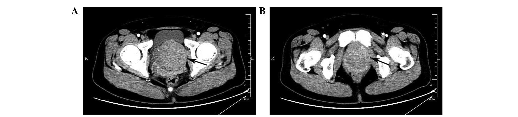
respectively. Computed tomography demonstrated that the bladder and
uterine were compressed by a 7.5×7.0-cm mass with well-defined
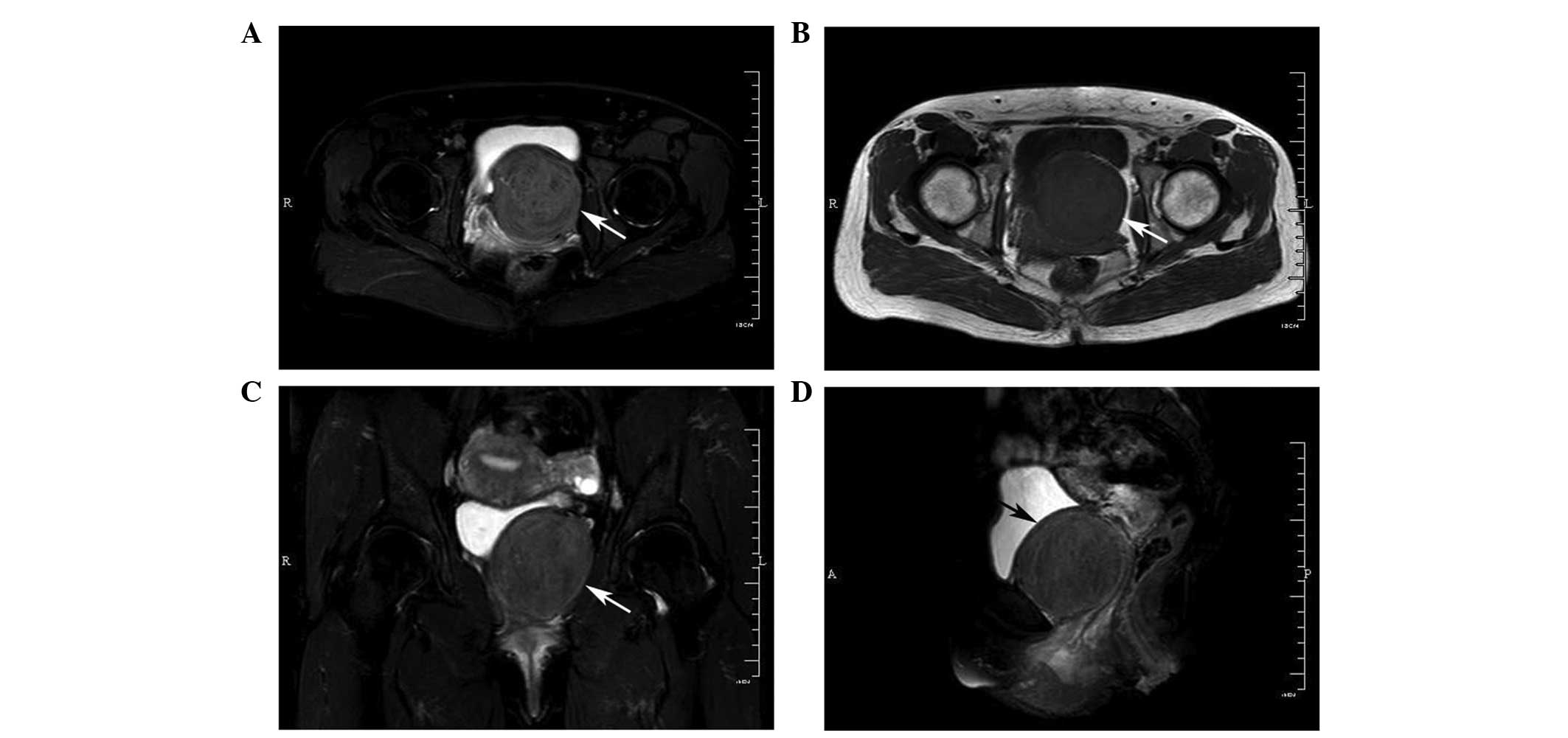
outlines (Fig. 1). Magnetic
resonance imaging was later performed to improve the definition of
the structure and the association of the lesion with the urethra
and vagina. The mass was isointense to muscle on T1-weighted images
and slightly hyperintense on T2-weighted images, indicating a solid
mass (Fig. 2).
The urethral tumor was completely excised and
removed as close to the bladder neck as possible by transabdominal
surgery. The detachment in the paraurethral region was meticulous
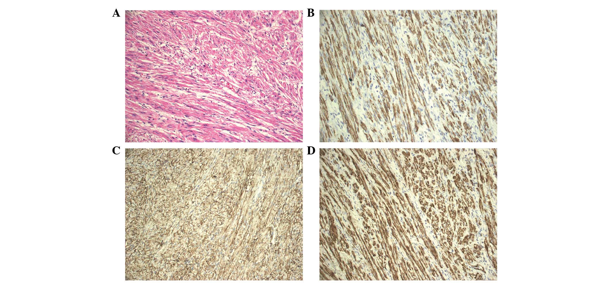
to reduce injury to the urethra. The subsequent pathological
diagnosis was of a leiomyoma. Immunohistological analysis
demonstrated that the tumor cells were positive for desmin, cluster
of differentiation 10 (CD10), smooth muscle actin and caldesmon,
and negative for CD117 (Fig. 3).
The patient was carefully followed up without any other treatment,
and no sign of recurrence was observed in the first year
post-surgery.
Discussion
Leiomyomas are benign tumors of smooth muscle origin
that occur throughout the genitourinary system, most commonly in
the renal capsule (7). Urethral
leiomyomas are rare benign tumors affecting females significantly
more than males (1–8). In 1894, Buttner (10) described the first urethral
leiomyoma. Thus far, only ~100 cases have been reported in the
literature (1–8). The posterior wall of the urethra is
the site of predilection, although any wall may be affected
(11). Additionally, the distal
urethra can be affected, but the proximal segment is the most
common site (12).
Urethral leiomyomas are usually asymptomatic when
they are small. As they grow in size, patients may complain of
urinary tract infection, dyspareunia, urinary retention or
irritative lower urinary tract symptoms (13). Physical examination may reveal a
mass in the anterior vaginal wall or one that protrudes from the
urethral meatus (12).
Ultrasonography and magnetic resonance imaging have been shown to
provide useful pre-operative information regarding the morphology
and structure of the mass (14,15).
However, a pathological examination is indispensable to exclude the
possibility of a malignancy.
In the present study, the huge pelvic tumor should
be differentiated from the urethral carcinoma or masses that have
originated from other tissues, although the patient had a past
history of urethral leiomyoma present six and nine years ago,
respectively. In all previously published cases, the urethral
leiomyomas have been treated surgically, with surgical excision as
the first choice of treatment. The present patient was treated by
transabdominal excision and the tumor was found to originate from
the proximal segment of the urethra, which is close to the bladder
neck.
All previously reported vesical and urethral
leiomyomas have followed a benign biological course (7), with only a single reported recurrence
treated by a repeat excision (9).
The present study is the second reported case of urethral leiomyoma
recurrence, and the patient has undergone three surgical procedures
to date.
Acknowledgements
This study was supported by grants from the National
Natural Science Foundation of China (grant no. 81101718) and the
National Key Clinical Specialty Construction Project of China.
References
|
1
|
Shield DE and Weiss RM: Leiomyoma of the
female urethra. J Urol. 109:430–431. 1973.PubMed/NCBI
|
|
2
|
Cheng C, Mac-Moune Lai F and Chan PS:
Leiomyoma of the female urethra: a case report and review. J Urol.
148:1526–1527. 1992.PubMed/NCBI
|
|
3
|
Leidinger RJ and Das S: Leiomyoma of the
female urethra. A report of two cases. J Reprod Med. 40:229–231.
1995.PubMed/NCBI
|
|
4
|
Leung YL, Lee F and Tam PC: Leiomyoma of
female urethra causing acute urinary retention and acute renal
failure. J Urol. 158:1911–1912. 1997. View Article : Google Scholar : PubMed/NCBI
|
|
5
|
Alvarado-Cabrero I, Candanedo-González F
and Sosa-Romero A: Leiomyoma of the urethra in a Mexican woman: a
rare neoplasm associated with the expression of estrogen receptors
by immunohistochemistry. Arch Med Res. 32:88–90. 2001. View Article : Google Scholar
|
|
6
|
Kato T, Kobayashi T, Ikeda R, Nakamura T,
Akakura K, Hikage T and Inoue T: Urethral leiomyoma expressing
estrogen receptors. Int J Urol. 11:573–575. 2004. View Article : Google Scholar : PubMed/NCBI
|
|
7
|
Goldman HB, McAchran SE and MacLennan GT:
Leiomyoma of the urethra and bladder. J Urol. 177:18902007.
View Article : Google Scholar : PubMed/NCBI
|
|
8
|
Garrido Abad P, Fernández Arjona M,
Herranz Fernández LM, Muñoz-Delgado Salmerón J and Capote LF:
Leiomyoma of the male urethra: case report and review of the
literature. Arch Esp Urol. 63:71–74. 2010.PubMed/NCBI
|
|
9
|
Merrell RW and Brown HE: Recurrent
urethral leiomyoma presenting as stress incontinence. Urology.
17:588–589. 1981. View Article : Google Scholar : PubMed/NCBI
|
|
10
|
Buttner: A case of myoma of the female
urethra. Z Geburshe Gynak. 28:1351894.(In German).
|
|
11
|
Wani NA, Bhan BL, Guru AA and Garyali RK:
Leiomyoma of the female urethra: a case report. J Urol.
116:120–121. 1976.PubMed/NCBI
|
|
12
|
Lee MC, Lee SD, Kuo HT and Huang TW:
Obstructive leiomyoma of the female urethra: report of a case. J
Urol. 153:420–421. 1995. View Article : Google Scholar : PubMed/NCBI
|
|
13
|
Pahwa M, Saifee Y, Pahwa AR and Gupta M:
Leiomyoma of the female urethra-a rare tumor: case report and
review of the literature. Case Rep Urol. 2012:2808162012.PubMed/NCBI
|
|
14
|
Pavlica P, Bartolone A, Gaudiano C and
Barozzi L: Female paraurethral leiomyoma: ultrasonographic and
magnetic resonance imaging findings. Acta Radiol. 45:796–798. 2004.
View Article : Google Scholar : PubMed/NCBI
|
|
15
|
Ikeda R, Suga K and Suzuki K: MRI
appearance of a leiomyoma of the female urethra. Clin Radiol.
56:76–79. 2001. View Article : Google Scholar : PubMed/NCBI
|