Introduction
In recent years, growing numbers of patients with
breast cancer have been treated with neoadjuvant chemotherapy (NAC)
to shrink the tumor size for an improved chance of breast
conservation. In fact, it has been reported by specific studies
that more patients treated with NAC undergo breast-conserving
surgery than those not treated (1,2).
Another advantage of NAC is that the patient prognosis can be
estimated by means of a pathological evaluation of the surgical
specimens, i.e., if a pathological complete response (pCR) is
achieved, the patient prognosis can be expected to be excellent,
while the prognosis of patients who do not achieve a pCR is
reportedly worse, with a 5-year relapse-free survival rate of
50–70% (3,4). Since prognostic evaluation is
extremely important in deciding whether or not further adjuvant
therapy should be used, more effective prognostic factors are
required for those patients who cannot achieve a pCR.
It has been hypothesized and is currently accepted
that the detection of tumor-specific DNA methylation in serum is
useful for prognosis prediction and for monitoring responses to
systemic therapy in patients with breast cancer (5–13).
Certain studies have assessed the prognostic value of the presence
of methylated DNA (met-DNA) in the serum of patients with breast
cancer (5–9). It has also been reported that breast
cancer patients with met-DNA in the serum detected by one-step
methylation-specific polymerase chain reaction (PCR) show a poorer
prognosis compared with those without it (10,11).
However, other studies have reported that met-DNA in the serum of
patients with breast cancer treated with adjuvant therapy
correlates with the pathological response (12,13). A
study by Sharma et al (12)
detected methylation of the glutathione S-transferase pi 1
(GSTP1) and breast cancer 1, early onset (BRCA1)
genes in serum more frequently in non-responders to NAC than in
responders, while Avraham et al (13) reported that none of the responders
to NAC showed methylated Ras association (RalGDS/AF-6) domain
family member 1 (RASSF1A) in the serum. However, no studies
have been published on the correlation between met-DNA in serum and
the prognosis for patients with breast cancer treated with NAC.
Therefore, the present study investigated whether the presence of
met-DNA in the serum, as detected by one-step methylation-specific
PCR (OS-MSP), may be associated with a poor prognosis for patients
with breast cancer treated with NAC. In addition, the prognostic
significance of high total DNA levels in the serum was also
investigated.
Materials and methods
Patients
Patients with invasive breast cancer (n=120) who
underwent breast conserving surgery or mastectomy following NAC at
the Osaka University Hospital (Suita-shi, Osaka, Japan) between
March 2000 and May 2007 were retrospectively included in the
present study. Informed consent was obtained from each patient. A
total of 44 patients had been treated with paclitaxel (80
mg/m2) weekly for 12 cycles followed by 5-fluorouracil
(500 mg/m2), epirubicin (75 mg/m2) and
cyclophosphamide (500 mg/m2) every three weeks for 4
cycles (P-FEC). Another 37 patients had been treated with docetaxel
(75 mg/m2) every three weeks for 4 cycles, while 29 had
been treated with cyclophosphamide (600 mg/m2) and
epirubicin (60 mg/m2) every three weeks for 4 cycles,
followed by docetaxel (60 mg/m2) every three weeks for 4
cycles. Finally, 10 patients had been treated with other types of
chemotherapy consisting of docetaxel (60 mg/m2) or
cyclophosphamide (600 mg/m2) and epirubicin (60
mg/m2) every three weeks or paclitaxel (80
mg/m2) weekly.
Serum samples were obtained following NAC and prior
to surgery, and stored at −80°C until use. The median follow-up
period was 73 months (range, 3–134 months) and the median age of
the patients at the time of surgery was 51 years (range, 26–75
years). The clinicopathological characteristics of the patients are
summarized in Table I. Adjuvant
hormonal therapy was administered to 79 patients: Tamoxifen for 27,
goserelin plus tamoxifen for 13 and anastrozole for 39 patients,
all essentially in accordance with the St. Gallen recommendations
(14–16). Subsequent to the surgery, the
patients were followed up every 3 months for 1–2 years, every 6
months for 3–5 years and once every year thereafter. This study was
approved by the Ethics Committee of Osaka University Graudate
School of Medicine (Suita, Osaka, Japan).
 | Table ICorrelations between the presence of
methylated DNA or levels of total DNA in serum and the
clinicopathological parameters prior to NACa. |
Table I
Correlations between the presence of
methylated DNA or levels of total DNA in serum and the
clinicopathological parameters prior to NACa.
| | Met-DNAb in serum | Total DNA levels in
serum |
|---|
| |
|
|
|---|
| n | Met (−)c, n (%) | Met (+)d, n (%) | P-valuee | Low, n (%) | High, n (%) | P-valuef |
|---|
| Total patients | 120 | 113 (94) | 7 (6) | 80 (67) | 40 (33) | | |
| Menopausal
status |
| Premenopausal | 62 | 60 (97) | 2 (3) | 0.261 | 44 (71) | 18 (29) | 0.301 |
| Postmenopausal | 58 | 53 (91) | 5 (9) | | 36 (62) | 22 (38) | |
| Tumor size |
| T1 | 3 | 3 (100) | 0 (0) | 0.424g | 2 (67) | 1 (33) | 0.763g |
| T2 | 71 | 68 (96) | 3 (4) | | 47 (66) | 24 (34) | |
| T3 | 22 | 21 (95) | 1 (5) | | 15 (68) | 7 (32) | |
| T4 | 23 | 20 (87) | 3 (13) | | 16 (70) | 7 (30) | |
| Unknown | 1 | 1 (100) | 0 (0) | | 0 (0) | 1 (100) | |
| Lymph node
metastasis |
| N0 | 35 | 35 (100) | 0 (0) | 0.105 | 24 (69) | 11 (31) | 0.776 |
| N1 | 85 | 78 (92) | 7 (8) | | 56 (66) | 29 (34) | |
| Histological
grade |
| G1 | 26 | 26 (100) | 0 (0) | 0.116h | 20 (77) | 6 (23) | 0.031h |
| G2 | 64 | 61 (95) | 3 (5) | | 44 (69) | 20 (31) | |
| G3 | 25 | 22 (88) | 3 (12) | | 12 (48) | 13 (52) | |
| Unknown | 5 | 4 (80) | 1 (20) | | 4 (80) | 1 (20) | |
| Histological
type |
| Invasive ductal
carcinoma | 111 | 106 (95) | 5 (5) | 0.125 | 73 (66) | 38 (34) | 0.811e |
| Invasive lobular
carcinoma | 8 | 6 (75) | 2 (25) | | 6 (75) | 2 (25) | |
| Others | 1 | 1 (100) | 0 (0) | | 1 (100) | 0 (0) | |
| ER |
| Negative | 53 | 48 (91) | 5 (9) | 0.239 | 35 (66) | 18 (34) | 0.897 |
| Positive | 67 | 65 (97) | 2 (3) | | 45 (67) | 22 (33) | |
| PR |
| Negative | 72 | 66 (92) | 6 (8) | 0.240 | 46 (64) | 26 (36) | 0.429 |
| Positive | 48 | 47 (98) | 1 (2) | | 34 (71) | 14 (29) | |
| HER2 |
| Negative | 88 | 83 (94) | 5 (6) | 1.000 | 61 (69) | 27 (31) | 0.398 |
| Positive | 28 | 27 (96) | 1 (4) | | 17 (61) | 11 (39) | |
| Unknown | 4 | 3 (75) | 1 (25) | | 2 (50) | 2 (50) | |
| CEA |
| Negative | 103 | 97 (94) | 6 (6) | 1.000 | 70 (68) | 33 (32) | 0.263d |
| Positive | 15 | 14 (93) | 1 (7) | | 8 (53) | 7 (47) | |
| Unknown | 2 | 2 (100) | 0 (0) | | 2 (100) | 0 (0) | |
| CA15-3 |
| Negative | 107 | 101 (94) | 6 (6) | 0.505 | 71 (66) | 36 (34) | 1.000d |
| Positive | 11 | 10 (91) | 1 (9) | | 7 (64) | 4 (36) | |
| Unknown | 2 | 2 (100) | 0 (0) | | 2 (100) | 0 (0) | |
| Pathological
response |
| Non-pCR | 101 | 94 (93) | 7 (7) | 0.595 | 68 (67) | 33 (33) | 0.724 |
| pCR | 19 | 19 (100) | 0 (0) | | 12 (63) | 7 (37) | |
OS-MSP assay for GSTP1, RASSF1A and
retinoic acid receptor β2 (RARβ2) promoter hypermethylation in
serum
The OS-MSP assay was conducted as previously
described (10). In brief, a total
of 1,000 μl of serum from each patient was solubilized by
incubation with lysis buffer (5 M guanidine-HCl and 0.65 mg/ml
proteinase K) at 50°C for 1 h and then incubated with a 5 M
bisulfite solution at 80°C for 40 min. Bisulfite-modified DNA was
purified with a DNA purification kit (Qiagen, Valencia, CA, USA)
and eluted in 50 μl dH2O. Modification by bisulfite was
completed by treatment with 0.3 M NaOH for 5 min at room
temperature, subsequent to which bisulfite-modified DNA was
extracted by gel filtration (GE Healthcare, Princeton, NJ, USA) for
use as the PCR template. Promoter hypermethylation of GSTP1,
RASSF1A and RARβ2 was then evaluated by OS-MSP. The
primer and probe sequences were as follows: GSTP1 forward
primer, 5′-CGTCGTGATTTAGTATTGGGGC-3′ and reverse primer,
5′-CTAATAACGAAAACTACGACGACGAAA-3′; GSTP1 probe,
5′-FAM-ATAAGGTTCGGAGGTCGCGAGGTTTTC GT-DDQ1-3′; RASSF1A
forward primer, 5′-ATAGTTTTTGTA TTTAGGTTTTTATTGCGC-3′ and reverse
primer, 5′-GCT AACAAACGCGAACCG-3′; RASSF1A probe, 5′-FAM-TTG
AAGTCGGGGTTCGTTTTGTGGTTTCGT-DDQ1-3′; RARβ2 forward primer,
5′-GAATATCGTTTTTTAAGTTAAGTC GTC-3′ and reverse primer,
5′-GAAACGCTACTCCTAACT CACG-3′; and RARβ2 probe,
5′-FAM-AGGCGTAAAGGG AGAGAAGTTGGTGTTTA-DDQ1-3′. For the PCR
amplifications, the Light Cycler 480 Real-Time PCR System (Roche
Applied Science, Madison, WI, USA) was used under the following
conditions: 1 cycle at 95°C for 10 min, followed by 50 cycles of
95°C for 30 sec, 60°C for 30 sec and 72°C for 30 sec. Every sample
was analyzed in a single assay for each gene. Finally, the PCR
products were also analyzed by means of 3% agarose gel
electrophoresis, staining with ethidium bromide and visualization
under UV illumination.
Assay for total DNA levels in serum
Total DNA levels were quantified as previously
described (10). In brief, in order
to quantify total DNA levels in serum following bisulfite
treatment, a genomic locus lacking a cytosine base was selected,
since such a locus is not affected by bisulfite treatment. This
locus was amplified by PCR using the primers, probe and standard
oligoDNA. The primer and probe sequences were as follows: Forward
primer, 5′-AGGGAGTAGAGAAAAAGT AGGAAGATGAGT-3′ and reverse primer,
5′-TCCAACATC ACATCCAATCCA-3′; probe, 5′-FAM-AGGGTGATAATG
AGTGTGTTGGGAAATAGA-DDQ1-3′; and standard oligo DNA,
5′-AGGGAGTAGAGAAAAAGTAGGAAGATGAGT
CCAGGGTGATAATGAGTGTGTTGGGAAATAGACCTG GATTGGATGTGATGTTGGA-3′. The
PCR conditions were as aforementioned. The patients were divided
into three tertiles (low, middle and high) according to the level
of total DNA in serum. Those in the low and middle tertiles were
then combined and treated as the low total DNA group and those in
the high tertile were treated as the high total DNA group.
DNA extraction from tumor tissues
DNA was also extracted as previously described
(17) from breast tumor tissues
obtained prior to (core-needle biopsy specimens) or following
(surgical specimens) NAC from the patients who showed positive
results for the OS-MSP assay in at least one of the three genes.
The specimens were then examined as to whether the corresponding
genes were methylated in the tumor tissues. For this examination, 1
μg of DNA was subjected to sodium bisulfite treatment using the
EpiTect Bisulfite kit (48; Qiagen) according to the manufacturer’s
instructions, and analyzed by the OS-MSP assay for promoter
hypermethylation of GSTP1, RASSF1A and RARβ2, as
previously described (10).
Histological grade and estrogen receptor
(ER), progesterone receptor (PR) and human epidermal growth factor
receptor 2 (HER2) expression
Histological grade was determined using the
Scarff-Bloom-Richardson grading system (18). ER and PR were classified as positive
when ≥10% of the tumor cells showed immunohistochemically-positive
staining (ER, clone 6F11; and PR, clone 16; Ventana Japan K.K.,
Yokohama and SRL, Inc., Tokyo, Japan, respectively). HER-2
expression was determined immunohistochemically using anti-human
c-erbB-2 polyclonal antibody (Nichirei Biosciences, Inc., Tokyo,
Japan) or by means of fluorescence in situ hybridization
(FISH) using PathVysion Her2 DNA Probe kits (SRL, Inc.). When a
tumor showed +3 immunostaining (positive tumor cells >30%) or a
FISH ratio (HER2 gene signals to chromosome 17 centromere signals)
of ≥2.0, it was considered HER2-positive.
Evaluation of the response to
chemotherapy
The pathological response to chemotherapy was
evaluated by examining the surgical specimens obtained during the
surgery. The specimens were cut at 5-mm intervals for the
preparation of hematoxylin and eosin sections. A complete loss of
invasive tumor cells in the primary tumor site without any lymph
node metastasis was classified as a pCR.
Statistical analysis
Associations of met-DNA or total DNA levels in serum
with various clinicopathological parameters were assessed by means
of Pearson’s χ2 test. Disease-free survival (DFS) was
calculated as the time from surgery until the date of any
recurrence of breast cancer (local or distant) or mortality from
any cause. Overall survival (OS) was calculated as the time from
surgery to the date of mortality from any cause. DFS and OS were
assessed with the Kaplan-Meier method and log-rank tests, while
univariate and multivariate analyses (Cox regression models) were
used to evaluate the prognostic significance of various parameters.
Multivariate analyses included the parameters P<0.1 or known
prognostic factor (pCR). All the statistical analyses were
two-sided and P<0.05 was considered to indicate a statistically
significant difference. SPSS software (SPSS, Inc., Chicago, IL,
USA) was used for all statistical analyses.
Results
Associations between clinicopathological
parameters and met-DNA or total DNA levels in serum
Promoter hypermethylation of GSTP1, RASSF1A
and RARβ2 in serum was detected with the OS-MSP assay in 4,
1 and 2% of the 120 patients, respectively. When promoter
hypermethylation was found in at least one of these three genes,
the result of the OS-MSP assay for met-DNA in serum was considered
to be positive for the subsequent analysis. A total of seven
patients (6%) were found to be positive for met-DNA in serum. No
significant association was observed between met-DNA in serum and
any clinicopathological characteristics (Table I). The promoter methylation status
of the genes methylated in the serum of the seven patients who were
positive for met-DNA was also examined in the tumor specimens
obtained from the same patients. The same genes that were
methylated in the serum were also found to be methylated in the
tumor tissue of each of the seven patients.
The median total DNA level in the serum was 1.9
ng/ml (range, 0–63.3 ng/ml). The patients were divided into
tertiles (high, middle and low) according to the level of total DNA
in the serum. The middle and low tertiles were combined and treated
as the low total DNA group (n=80), and the high tertile was treated
as the high total DNA group (n=40). The cut-off value for the high
and low total DNA groups was 3.3 ng/ml. Correlations between the
total DNA levels in the serum and the clinicopathological
characteristics are shown in Table
I. The patients in the high total DNA group were significantly
more likely to have tumors of high histological grade (52 vs. 29%,
P=0.031) and to be positive for met-DNA in the serum (71 vs. 31%,
P=0.028). By contrast, pCR was not significantly associated with
either met-DNA or total DNA levels in the serum.
Association of prognosis with met-DNA or
total DNA levels in serum
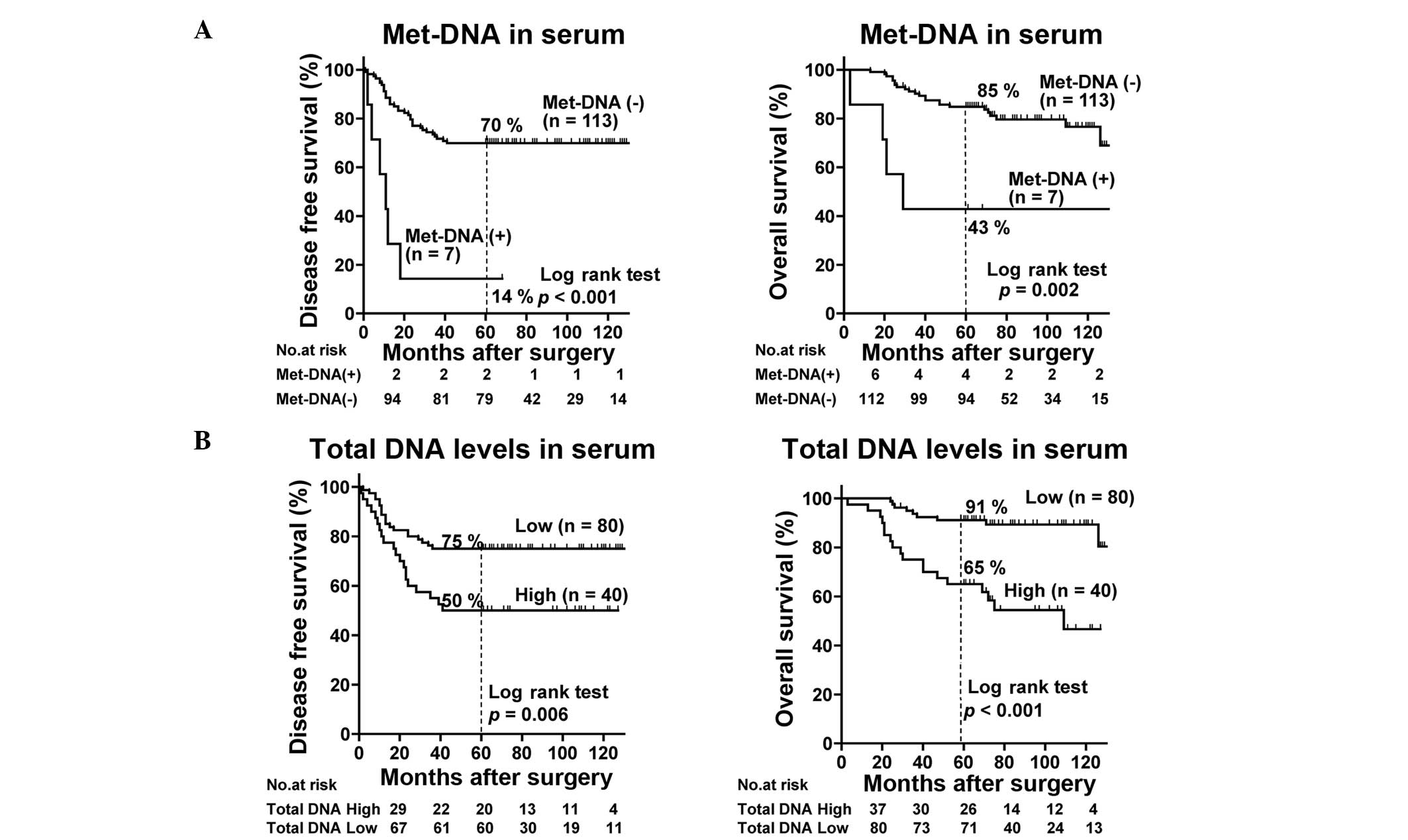
The seven patients with serum positive for met-DNA
exhibited significantly worse DFS and OS rates compared with those
negative for met-DNA (n=113) (P<0.001 and P=0.002, respectively;
Fig. 1A), while the patients with
high total DNA levels in the serum (n=40) also showed significantly
worse DFS and OS rates compared with those with low levels of total
DNA (n=80) (P=0.006 and P<0.001, respectively; Fig. 1B). Subsequently, univariate and
multivariate analyses were conducted to determine whether met-DNA
and total DNA levels in the serum are independent prognostic
factors for the other parameters (Tables II and III). The multivariate analysis showed
that met-DNA in the serum was significantly associated with DFS
(P=0.003) and OS (P=0.009), as was total DNA levels in the serum
(P=0.045 and P=0.001, respectively).
| Table IIUnivariate and multivariate analysis
of various prognostic factors for DFS of 120 patients. |
Table II
Univariate and multivariate analysis
of various prognostic factors for DFS of 120 patients.
| Univariate
analysis | Multivariate
analysis |
|---|
|
|
|
|---|
| Parameters | HR | 95% CI | P-value | HR | 95% CI | P-value |
|---|
| Met-DNA in serum
(positive vs. negative) | 6.650 | 2.74–16.17 | <0.001 | 4.226 | 1.63–10.95 | 0.003 |
| Total DNA levels in
serum (high vs. low) | 2.336 | 1.26–4.34 | 0.007 | 1.926 | 1.02–3.654 | 0.045 |
| Tumor size (3,4 vs.
1,2) prior to NAC | 1.422 | 0.76–2.68 | 0.276 | | | |
| Histological grade
(3 vs. 1,2) prior to NAC | 1.560 | 0.76–3.21 | 0.227 | | | |
| LN (positive vs.
negative) prior to NAC | 2.856 | 1.20–6.81 | 0.018 | 2.864 | 1.17–7.01 | 0.021 |
| CEA (positive vs.
negative) prior to NAC | 2.050 | 0.94–4.45 | 0.070 | 2.074 | 0.94–4.59 | 0.072 |
| CEA (positive vs.
negative) following NAC | 1.815 | 0.56–5.89 | 0.321 | | | |
| CA15-3 (positive
vs. negative) prior to NAC | 1.099 | 0.39–3.09 | 0.858 | | | |
| CA15-3 (positive
vs. negative) following NAC | 0.433 | 0.06–3.15 | 0.408 | | | |
| NAC (P-FEC vs.
others) | 0.528 | 0.26–1.08 | 0.080 | 0.589 | 0.28–1.24 | 0.163 |
| Pathological
response (pCR vs. non-pCR) | 0.234 | 0.06–0.97 | 0.045 | 0.260 | 0.06–1.12 | 0.071 |
| Table IIIUnivariate and multivariate analysis
of various prognostic factors for OS of 120 patients. |
Table III
Univariate and multivariate analysis
of various prognostic factors for OS of 120 patients.
| Univariate
analysis | Multivariate
analysis |
|---|
|
|
|
|---|
| Parameters | HR | 95% CI | P-value | HR | 95% CI | P-value |
|---|
| Met-DNA in serum
(positive vs. negative) | 4.834 | 1.65–14.15 | 0.004 | 4.805 | 1.49–15.52 | 0.009 |
| Total DNA levels in
serum (high vs. low) | 5.119 | 2.28–11.49 | <0.001 | 4.112 | 1.77–9.57 | 0.001 |
| Tumor size (3,4 vs.
1,2) | 1.366 | 0.63–2.97 | 0.430 | | | |
| Histological grade
(3 vs. 1,2) prior to NAC | 2.530 | 1.16–5.53 | 0.020 | 2.302 | 1.02–5.21 | 0.046 |
| LN (positive vs.
negative) prior to NAC | 2.179 | 0.82–5.77 | 0.117 | | | |
| CEA (positive vs.
negative) prior to NAC | 2.298 | 0.93–5.70 | 0.073 | 3.497 | 1.34–9.11 | 0.010 |
| CEA (positive vs.
negative) following NAC | 0.643 | 0.09–4.75 | 0.666 | | | |
| CA15-3 (positive
vs. negative) prior to NAC | 2.087 | 0.72–6.04 | 0.175 | | | |
| CA15-3 (positive
vs. negative) following NAC | 0.046 | 0.00–65.53 | 0.405 | | | |
| NAC (P-FEC vs.
others) | 0.744 | 0.31–1.80 | 0.510 | | | |
| Pathological
response (pCR vs. non-pCR) | 0.418 | 0.10–1.77 | 0.235 | 0.258 | 0.06–1.18 | 0.081 |
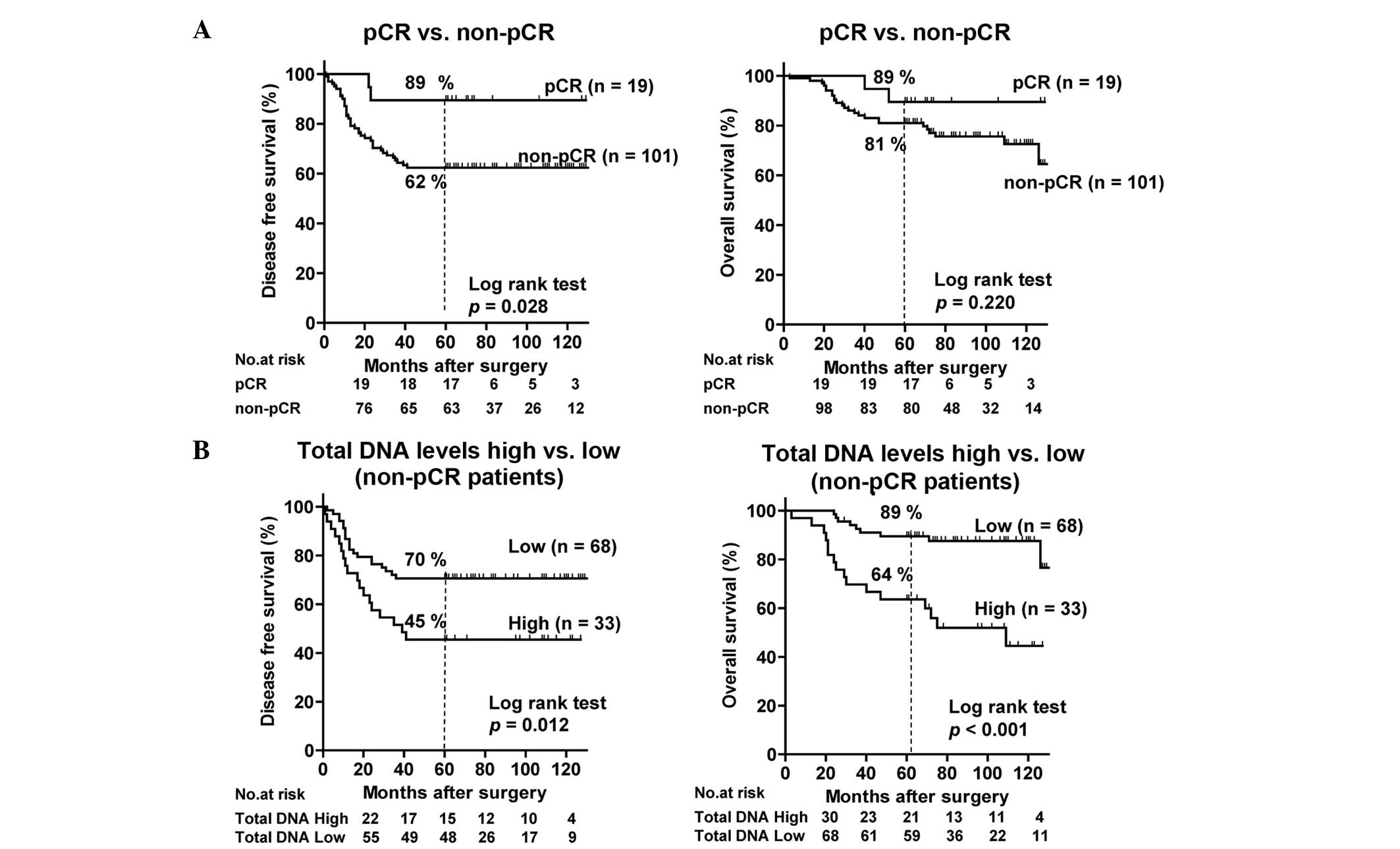
The patients who achieved pCR (n=19) showed
significantly improved DFS rates compared with those who did not
(n=101) (P=0.028; Fig. 2A), and the
prognostic significance for the latter group of total DNA levels in
the serum was evaluated. The patients with high total DNA levels in
the serum (n=33) again showed significantly worse DFS and OS rates
compared with those with low total DNA levels (n=68) (P=0.012 and
P<0.001, respectively; Fig.
2B).
Discussion
The present study investigated whether detection of
met-DNA and high total DNA levels in the serum by means of the
OS-MSP assay could serve as novel prognostic factors for patients
with breast cancer treated with NAC. By using multivariate analysis
it was shown that met-DNA and total DNA levels in the serum
following NAC are independent prognostic factors for DFS and OS,
independent of pCR, which is a well-established prognostic factor
for NAC-treated patients. The results indicate that met-DNA and
total DNA levels in the serum may be clinically useful prognostic
factors.
Met-DNA is believed to represent circulating tumor
genomes, but not necessarily total DNA levels, as it can not only
originate from tumor cells, but also from inflammatory cells,
endothelial cells and fibroblasts in tumor tissues (19–21).
Although the reason why total DNA levels in the serum is associated
with prognosis remains unclear, it is possible that high total DNA
levels in the serum may reflect specific tumor biology that is
associated with tumor metastasis and tumor-induced inflammation. It
is also possible that circulating tumor cells (22,23)
and micrometastatic deposits of distant organs, including the bone
marrow and liver, may contribute to total DNA levels (19,24).
The OS-MSP assay showed that the serum was positive
for met-DNA in 6% of patients in the present study. This positivity
appears to be slightly lower compared with our previous study, in
which it was found that met-DNA was positively detected in 10% of
stage I and II patients without NAC treatment (11). These results indicate that
positivity of met-DNA in serum decreases following NAC. In fact,
studies by Sharma et al (12) and Avraham et al (13) reported a significant decline in
met-DNA levels following NAC. However, in the present study, the
serum samples were not obtained prior to NAC and thus the change in
the positivity of met-DNA prior to and following NAC could not be
investigated.
While a significant correlation between met-DNA and
pCR was not detected, none of the patients that were positive for
met-DNA achieved pCR, which is consistent with the findings of
other studies (12,13). Sharma et al (12) reported that all non-responders were
positive for met-DNA (GSTP1 and BRCA1) following NAC,
and Avraham et al (13)
reported that all patients that were positive for met-DNA
(RASSF1A) following NAC could not achieve pCR. These results
appear to indicate that positivity for met-DNA following NAC is
associated with non-pCR.
A limitation of the present study is that the serum
samples could be obtained following, but not prior to, NAC, so that
the prognostic significance of met-DNA and total DNA levels in
serum prior to NAC could not be addressed. However, the available
data appears to indicate that met-DNA following NAC is a highly
significant prognostic indicator for poor prognosis, as none of the
patients that were positive for met-DNA attained pCR, and as many
as 86% developed recurrence. High total DNA levels in the serum
following NAC were also significantly associated with a poor
prognosis. It has been reported that the quantity of disseminated
or circulating tumor cells detected following, but not prior to,
chemotherapy can predict prognosis, as the numbers detected
following chemotherapy may reflect the residual presence of
micro-metastatic tumors (25,26).
In analogy to this finding, the presence of met-DNA and high total
DNA levels may represent the residual presence of micro-metastatic
tumors. Patients with met-DNA and/or high total DNA levels in the
serum can thus be considered to be essentially resistant to NAC and
to have a poor prognosis, necessitating the use of non-cross
resistant, post-operative adjuvant chemotherapy.
In conclusion, the present study was able to
demonstrate that the presence of met-DNA and high levels of total
DNA detected with the OS-MSP assay is a significant and independent
prognostic factor for patients with breast cancer treated with NAC.
The OS-MSP assay may thus be clinically useful for the selection of
NAC-treated patients who are at a high risk of relapse and thus
require treatment with additional post-operative adjuvant
chemotherapy. To the best of our knowledge, this is the first study
to assess the association between met-DNA or total DNA levels in
the serum and prognosis following NAC. However, future studies
including larger numbers of patients with sufficient follow-up
times are required to further validate the clinical utility of the
OS-MSP assay for patients treated with NAC.
Acknowledgements
This study was supported in part by a Grants-in-Aid
from the Comprehensive 10-Year Strategy for Cancer Control Program
of the Ministry of Health, Labor and Welfare, Japan and a research
fund from Sysmex Corporation. The funders had no role in the study
design, data analysis, decision to publish or preparation of the
manuscript. Shinzaburo Noguchi received research funding from
Pfizer, Bristol-Myers Squibb and Sysmex Corporation, and Honoraria
from Pfizer. Seung Jin Kim received honoraria from Sysmex
Corporation, Pfizer and Bristol-Myers Squibb.
References
|
1
|
van der Hage JA, van de Velde CJ, Julien
JP, Tubiana-Hulin M, Vandervelden C and Duchateau L: Preoperative
chemotherapy in primary operable breast cancer: results from the
European Organization for Research and Treatment of Cancer trial
10902. J Clin Oncol. 19:4224–4237. 2001.
|
|
2
|
Singletary SE: Neoadjuvant chemotherapy in
the treatment of stage II and III breast cancer. Am J Surg.
182:341–346. 2001.
|
|
3
|
Wolmark N, Wang J, Mamounas E, Bryant J
and Fisher B: Preoperative chemotherapy in patients with operable
breast cancer: nine-year results from National Surgical Adjuvant
Breast and Bowel Project B-18. J Natl Cancer Inst Monogr. 96–102.
2001.
|
|
4
|
Bear HD, Anderson S, Smith RE, et al:
Sequential preoperative or postoperative docetaxel added to
preoperative doxorubicin plus cyclophosphamide for operable breast
cancer: National Surgical Adjuvant Breast and Bowel Project
Protocol B-27. J Clin Oncol. 24:2019–2027. 2006.
|
|
5
|
Müller HM, Widschwendter A, Fiegl H, et
al: DNA methylation in serum of breast cancer patients: an
independent prognostic marker. Cancer Res. 63:7641–7645. 2003.
|
|
6
|
Göbel G, Auer D, Gaugg I, et al:
Prognostic significance of methylated RASSF1A and PITX2 genes in
blood- and bone marrow plasma of breast cancer patients. Breast
Cancer Res Treat. 130:109–117. 2011.
|
|
7
|
Sharma G, Mirza S, Parshad R, et al:
Clinical significance of promoter hypermethylation of DNA repair
genes in tumor and serum DNA in invasive ductal breast carcinoma
patients. Life Sci. 87:83–91. 2010.
|
|
8
|
Lo PK and Sukumar S: Epigenomics and
breast cancer. Pharmacogenomics. 9:1879–1902. 2008.
|
|
9
|
Martens JW, Margossian AL, Schmitt M,
Foekens J and Harbeck N: DNA methylation as a biomarker in breast
cancer. Future Oncol. 5:1245–1256. 2009.
|
|
10
|
Yamamoto N, Nakayama T, Kajita M, et al:
Detection of aberrant promoter methylation of GSTP1, RASSF1A, and
RARβ2 in serum DNA of patients with breast cancer by a newly
established one-step methylation-specific PCR assay. Breast Cancer
Res Treat. 132:165–173. 2012.
|
|
11
|
Fujita N, Nakayama T, Yamamoto N, et al:
Methylated DNA and total DNA in serum detected by one-step
methylation-specific PCR is predictive of poor prognosis for breast
cancer patients. Oncology. 83:273–282. 2012.
|
|
12
|
Sharma G, Mirza S, Parshad R, Gupta SD and
Ralhan R: DNA methylation of circulating DNA: a marker for
monitoring efficacy of neoadjuvant chemotherapy in breast cancer
patients. Tumour Biol. 33:1837–1843
|
|
13
|
Avraham A, Uhlmann R, Shperber A, et al:
Serum DNA methylation for monitoring response to neoadjuvant
chemotherapy in breast cancer patients. Int J Cancer.
131:E1166–E1172
|
|
14
|
Goldhirsch A, Glick JH, Gelber RD, Coates
AS and Senn HJ: Meeting highlights: International Consensus Panel
on the treatment of primary breast cancer. Seventh International
Conference on Adjuvant Therapy of Primary Breast Cancer. J Clin
Oncol. 19:3817–3827. 2001.
|
|
15
|
Goldhirsch A, Wood WC, Gelber RD, Coates
AS, Thürlimann B and Senn HJ: Meeting highlights: updated
international expert consensus on the primary therapy of early
breast cancer. J Clin Oncol. 21:3357–3365. 2003.
|
|
16
|
Goldhirsch A, Glick JH, Gelber RD, Coates
AS, Thürlimann B, et al; Panel members. Meeting highlights:
international expert consensus on the primary therapy of early
breast cancer 2005. Ann Oncol. 16:1569–1583. 2005.
|
|
17
|
Arai T, Miyoshi Y, Kim SJ, et al:
Association of GSTP1 expression with resistance to docetaxel and
paclitaxel in human breast cancers. Eur J Surg Oncol. 34:734–738.
2008.
|
|
18
|
Robbins P, Pinder S, de Klerk N, et al:
Histological grading of breast carcinomas: a study of interobserver
agreement. Hum Pathol. 26:873–879. 1995.
|
|
19
|
Schwarzenbach H, Hoon DS and Pantel K:
Cell-free nucleic acids as biomarkers in cancer patients. Nat Rev
Cancer. 11:426–437. 2011.
|
|
20
|
Fleischhacker M and Schmidt B: Circulating
nucleic acids (CNAs) and cancer - a survey. Biochim Biophys Acta.
1775:181–232. 2007.
|
|
21
|
Kowalewska M, Nowak R and Chechlinska M:
Implications of cancer-associated systemic inflammation for
biomarker studies. Biochim Biophys Acta. 1806:163–171. 2010.
|
|
22
|
Matuschek C, Bölke E, Lammering G, et al:
Methylated APC and GSTP1 genes in serum DNA correlate with the
presence of circulating blood tumor cells and are associated with a
more aggressive and advanced breast cancer disease. Eur J Med Res.
15:277–286. 2010.
|
|
23
|
Van der Auwera I, Elst HJ, Van Laere SJ,
et al: The presence of circulating total DNA and methylated genes
is associated with circulating tumour cells in blood from breast
cancer patients. Br J Cancer. 100:1277–1286. 2009.
|
|
24
|
Schwarzenbach H, Alix-Panabières C, Müller
I, et al: Cell-free tumor DNA in blood plasma as a marker for
circulating tumor cells in prostate cancer. Clin Cancer Res.
15:1032–1038. 2009.
|
|
25
|
Mathiesen RR, Borgen E, Renolen A, et al:
Persistence of disseminated tumor cells after neoadjuvant treatment
for locally advanced breast cancer predicts poor survival. Breast
Cancer Res. 14:R117
|
|
26
|
Xenidis N, Ignatiadis M, Apostolaki S, et
al: Cytokeratin-19 mRNA-positive circulating tumor cells after
adjuvant chemotherapy in patients with early breast cancer. J Clin
Oncol. 27:2177–2184. 2009.
|