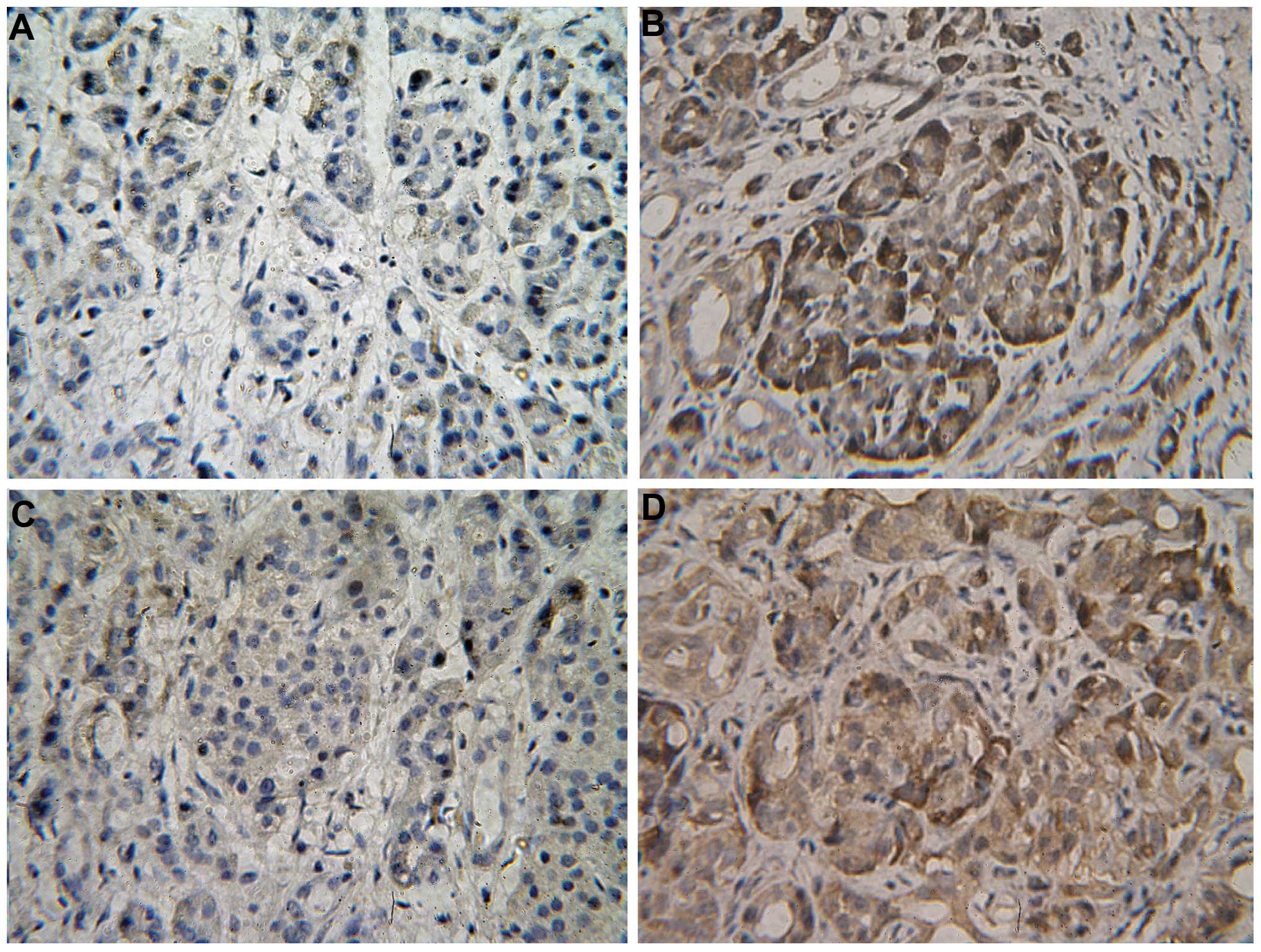
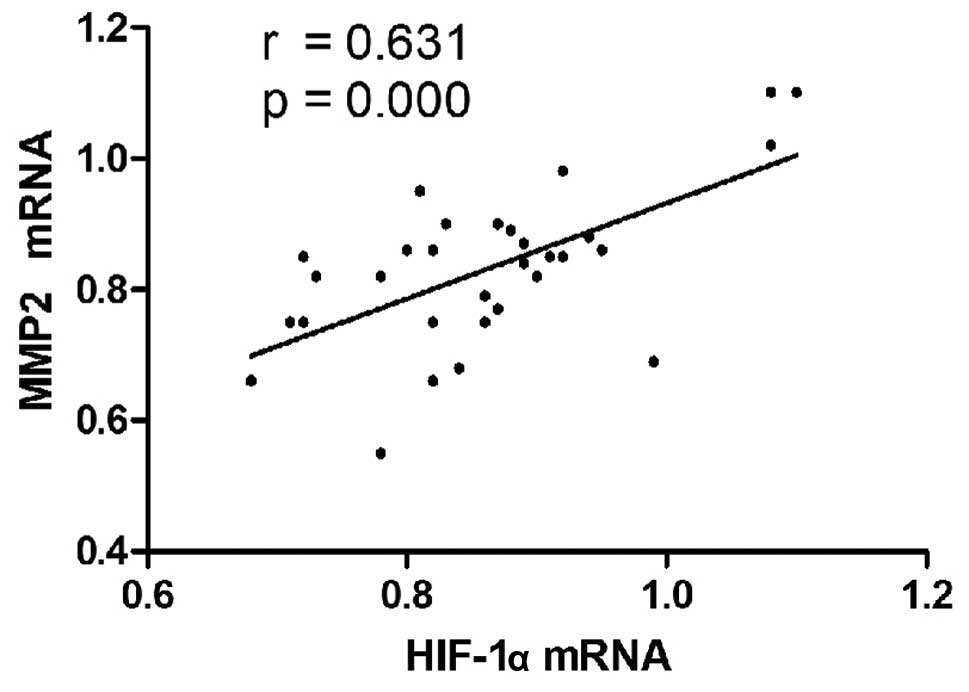
|
1
|
Marín-Hernández A, Gallardo-Pérez JC,
Ralph SJ, Rodríguez-Enríquez S and Moreno-Sánchez R: HIF-1alpha
modulates energy metabolism in cancer cells by inducing
over-expression of specific glycolytic isoforms. Mini Rev Med Chem.
9:1084–1101. 2009.
|
|
2
|
Littlepage LE, Sternlicht MD, Rougier N,
Phillips J, Gallo E, Yu Y, Williams K, Brenot A, Gordon JI and Werb
Z: Matrix metalloproteinases contribute distinct roles in
neuroendocrine prostate carcinogenesis, metastasis, and
angiogenesis progression. Cancer Res. 6:2224–2234. 2010.
|
|
3
|
Hajitou A, Sounni NE, Devy L,
Grignet-Debrus C, Lewalle JM, Li H, Deroanne CF, Lu H, Colige A,
Nusgens BV, Frankenne F, Maron A, Yeh P, Perricaudet M, Chang Y,
Soria C, Calberg-Bacq CM, Foidart JM and Noël A: Down-regulation of
vascular endothelial growth factor by tissue inhibitor of
metalloproteinase-2: effect on in vivo mammary tumor growth and
angiogenesis. Cancer Res. 8:3450–3457. 2001.
|
|
4
|
Yamasaki M, Nagatomo T, Matsuyama T, Ikeho
Y, Kato E and Nishiyama K, Sakakibara Y, Suiko M and Nishiyama K:
Conjugated linoleic acids inhibit hypoxia inducible factor-1α
stabilization under hypoxic condition in human hepatocellular
carcinoma cells. J Oleo Sci. 9:491–496. 2012.
|
|
5
|
Birner P, Schindl M, Obermair A, Plank C,
Breitenecker G and Oberhuber G: Overexpression of hypoxia-inducible
factor 1alpha is a marker for an unfavorable prognosis in
early-stage invasive cervical cancer. Cancer Res. 17:4693–4696.
2000.
|
|
6
|
Karahan N, Güney M, Baspinar S, Oral B,
Kapucuoglu N and Mungan T: Expression of gelatinase (MMP-2 and
MMP-9) and cyclooxygenase-2 (COX-2) in endometrial carcinoma. Eur J
Gynaecol Oncol. 28:184–188. 2007.
|
|
7
|
Sakata H, Fujimura M, Watanabe M and
Tominaga T: Association of cavernous malformation within vestibular
schwannoma: immunohistochemical analysis of matrix
metalloproteinase-2 and -9. Neurol Med Chir. 47:509–512. 2007.
|
|
8
|
Du R, Petritsch C, Lu K, Liu P, Haller A,
Ganss R, Song H, Vandenberg S and Bergers G: Matrix
metalloproteinase-2 regulates vascular patterning and growth
affecting tumor cell survival and invasion in GBM. Neuro Oncol.
3:254–264. 2008.
|
|
9
|
Xiang ZL, Zeng ZC, Fan J, Tang ZY, Zeng HY
and Gao DM: Gene expression profiling of fixed tissues identified
hypoxia-inducible factor-1α, VEGF, and matrix metalloproteinase-2
as biomarkers of lymph node metastasis in hepatocellular carcinoma.
Clin Cancer Res. 16:5463–5472. 2011.
|
|
10
|
Varotti G, Ramacciato G, Ercolani G, Grazi
GL, Vetrone G, Cescon M, Del Gaudio M, Ravaioli M, Ziparo V, Lauro
A and Pinna A: Comparison between the fifth and sixth editions of
the AJCC/UICC TNM staging systems for hepatocellular carcinoma:
multicentric study on 393 cirrhotic resected patients. Eur J Surg
Oncol. 31:760–767. 2005.
|
|
11
|
Nishida Y, Miyamori H, Thompson EW, Takino
T, Endo Y and Sato H: Activation of matrix metalloproteinase-2
(MMP-2) by membrane type 1 matrix metalloproteinase through an
artificial receptor for proMMP-2 generates active MMP-2. Cancer
Res. 21:9096–9104. 2008.
|
|
12
|
Schoedel KE, Tyner VZ, Kim TH,
Michalopoulos GK and Mars WM: HGF, MET, and matrix-related
proteases in hepatocellular carcinoma, fibrolamellar variant,
cirrhotic and normal liver. Mod Pathol. 1:14–21. 2003.
|
|
13
|
Kato T, Kure T, Chang JH, Gabison EE, Itoh
T, Itohara S and Azar DT: Diminished corneal angiogenesis in
gelatinase A-deficient mice. FEBS Lett. 2:187–190. 2001.
|
|
14
|
Semenza GL: Targeting HIF-1 for cancer
therapy. Nat Rev Cancer. 10:721–732. 2003.
|
|
15
|
Hao J, Song X, Song B, Liu Y, Wei L, Wang
X and Yu J: Effects of lentivirus-mediated HIF-1alpha knockdown on
hypoxia-related cisplatin resistance and their dependence on p53
status in fibrosarcoma cells. Cancer Gene Ther. 7:449–455.
2008.
|
|
16
|
Burrows N, Resch J, Cowen RL, von
Wasielewski R, Hoang-Vu C, West CM, Williams KJ and Brabant G:
Expression of hypoxia-inducible factor 1 alpha in thyroid
carcinomas. Endocr Relat Cancer. 1:61–72. 2010.
|
|
17
|
Seeber LM, Horrée N, van der Groep P, van
der Wall E, Verheijen RH and van Diest PJ: Necrosis related
HIF-1alpha expression predicts prognosis in patients with
endometrioid endometrial carcinoma. BMC Cancer. 10:3072010.
|
|
18
|
Samulitis BK, Landowski TH and Dorr RT:
Inhibition of protein synthesis by imexon reduces HIF-1alpha
expression in normoxic and hypoxic pancreatic cancer cells. Invest
New Drugs. 1:89–98. 2009.
|
|
19
|
Milosevic M, Fyles A, Hedley D and Hill R:
The human tumor microenvironment: invasive (needle) measurement of
oxygen and interstitial fluid pressure. Semin Radiat Oncol.
3:249–258. 2004.
|
|
20
|
Vumbaca F, Phoenix KN, Rodriguez-Pinto D,
Han DK and Claffey KP: Double-stranded RNA-binding protein
regulates vascular endothelial growth factor mRNA stability,
translation, and breast cancer angiogenesis. Mol Cell Biol.
2:772–783. 2008.
|
|
21
|
Jing SW, Wang YD, Kuroda M, Su JW, Sun GG,
Liu Q, Cheng YJ and Yang CR: HIF-1α contributes to hypoxia-induced
invasion and metastasis of esophageal carcinoma via inhibiting
E-cadherin and promoting MMP-2 expression. Acta Med Okayama.
5:399–407. 2012.
|
|
22
|
Mimeault M and Batra SK: Hypoxia-inducing
factors as master regulators of stemness properties and altered
metabolism of cancer- and metastasis-initiating cells. J Cell Mol
Med. 1:30–54. 2013.
|
|
23
|
Nurwidya F, Takahashi F, Minakata K,
Murakami A and Takahashi K: From tumor hypoxia to cancer
progression: the implications of hypoxia-inducible factor-1
expression in cancers. Anat Cell Biol. 2:73–78. 2012.
|
|
24
|
Krishnamachary B, Berg-Dixon S, Kelly B,
Agani F, Feldser D, Ferreira G, Iyer N, LaRusch J, Pak B, Taghavi P
and Semenza GL: Regulation of colon carcinoma cell invasion by
hypoxia-inducible factor 1. Cancer Res. 63:1138–1143. 2003.
|
|
25
|
Xie T, Yuan XL, Yu SY, Yang B and Dong LL:
Interference of HIF-1alpha by RNA reduces the expression of matrix
metalloproteinase-2 in human cervical carcinoma HeLa cells. Ai
Zheng. 6:600–605. 2008.(In Chinese).
|
|
26
|
Giannelli G, Bergamini C, Fransvea E,
Marinosci F, Quaranta V and Antonaci S: Human hepatocellular
carcinoma (HCC) cells require both alpha3beta1 integrin and matrix
metalloproteinases activity for migration and invasion. Lab Invest.
4:613–627. 2001.
|
|
27
|
Choi SH, Shin HW, Park JY, Yoo JY, Kim do
Y, Ro WS, Yun CO and Han KH: Effects of the knockdown of hypoxia
inducible factor-1alpha expression by adenovirus-mediated shRNA on
angiogenesis and tumor growth in hepatocellular carcinoma cell
lines. Korean J Hepatol. 3:280–287. 2010.
|