Cytological features of pure micropapillary carcinoma of various organs: A report of eight cases

  • Authors:
    • Gülben Erdem Huq
    • Şule Canberk
    • Meltem Öznur
    • Pelin Yıldız
    • Burak Bahadır
    • Kemal Behzatoğlu
  • View Affiliations

  • Published online on: May 29, 2014     https://doi.org/10.3892/ol.2014.2198
  • Pages: 705-710
Metrics: Total Views: 0 (Spandidos Publications: | PMC Statistics: )
Total PDF Downloads: 0 (Spandidos Publications: | PMC Statistics: )


Abstract

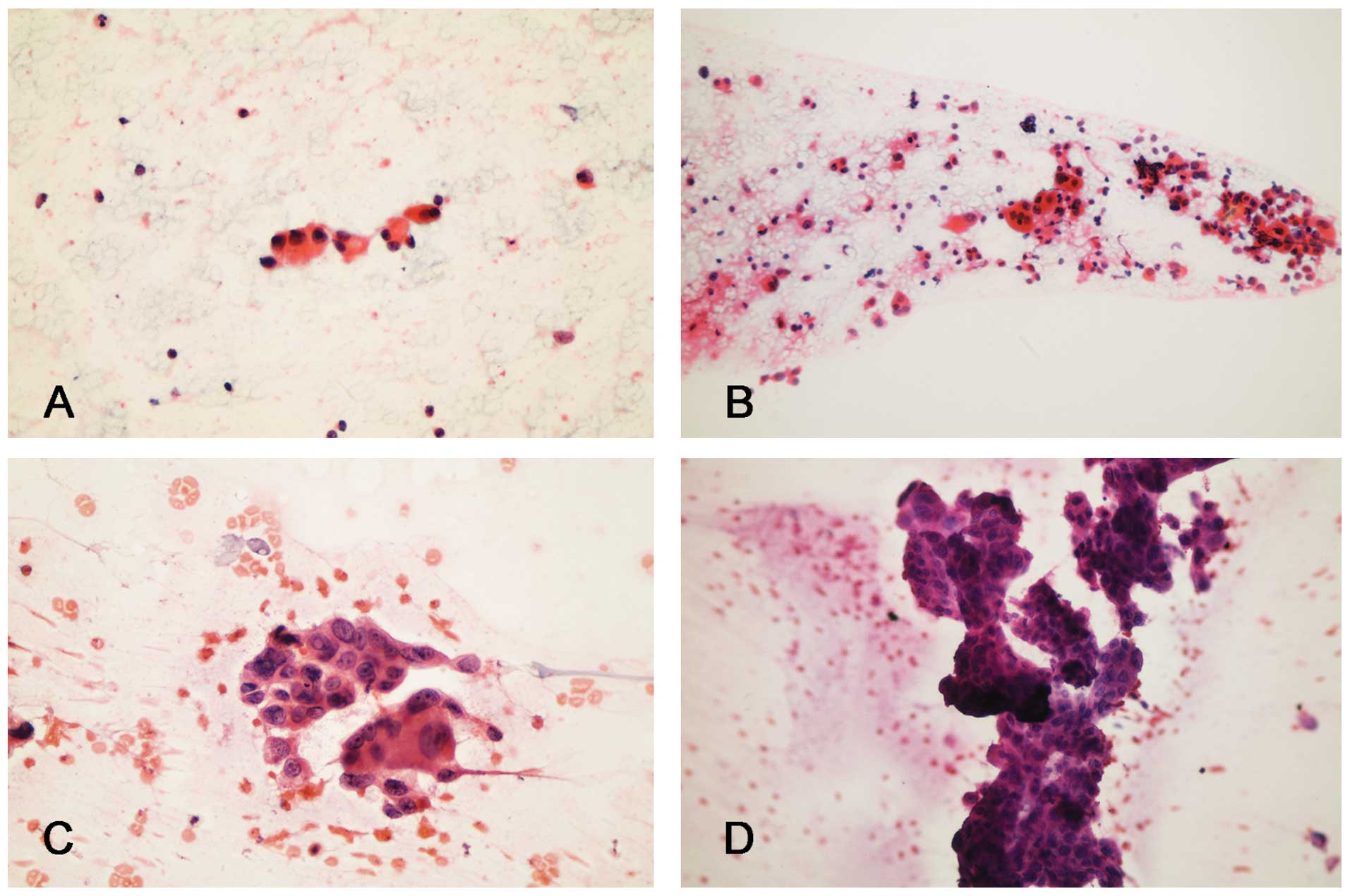
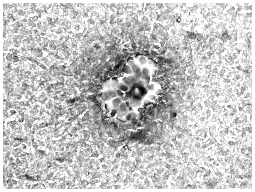
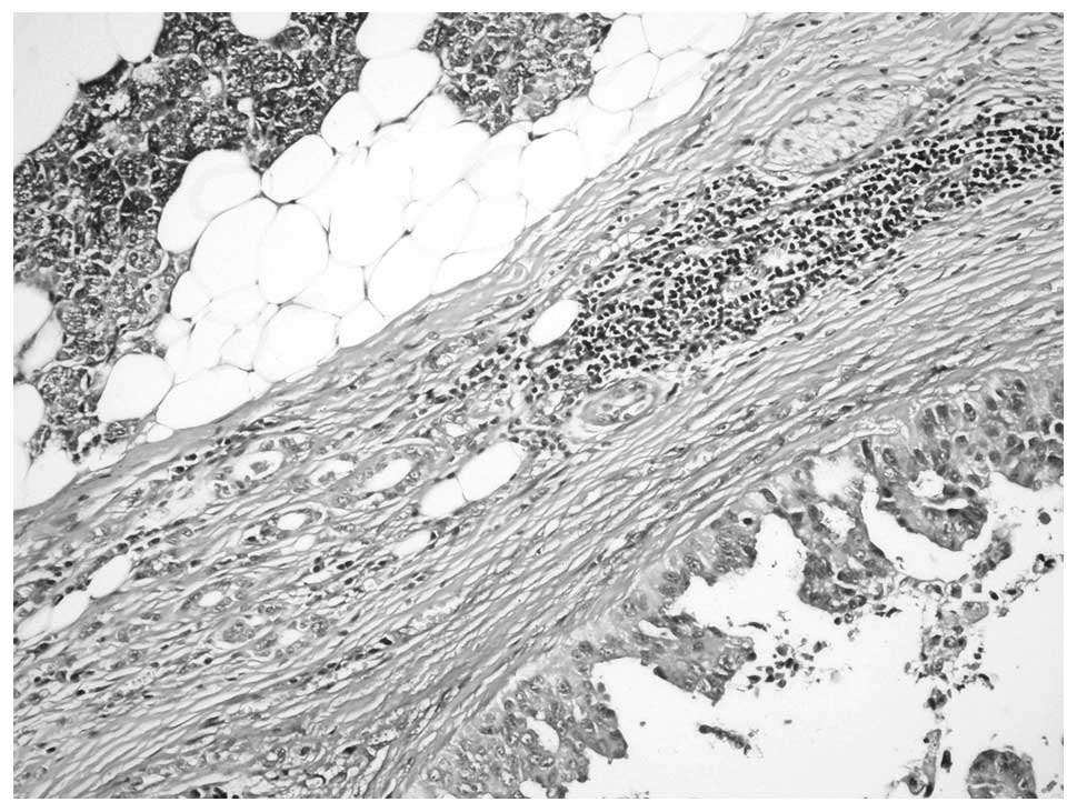
Micropapillary carcinoma (MPC) is a rare aggressive tumor, which generally accompanies the primary carcinoma of the organ of its origin, while the pure form is extremely uncommon. Angiolymphatic involvement is widespread and a considerable proportion of the cases present with metastases. The current study presents eight pure MPC cases arising from the breast (n=3), urinary bladder (n=3), parotid gland (n=1) and lung (n=1, presenting with pericardial effusion), with the cytological findings. The eight patients included three female and five male cases aged between 48 and 74 years. The most common cytological findings were three‑dimensional aggregates, cell clusters with angulated or scalloped borders, single cells with a columnar configuration and eccentric nuclei, and high‑grade nuclear features. Histopathological sections showed accompanying in situ ductal carcinoma in the cases of MPC arising in the parotid gland and breast (n=3), and one case in the bladder exhibited only in situ MPC. The average follow‑up period was 20 months (range, 6‑54 months) and, during this period, three patients succumbed to the disease. At present, four patients are alive with disease and one patient is alive and disease‑free. In conclusion, cytology is an important tool for the diagnosis and management of MPC.
View Figures
View References

Related Articles

Journal Cover

August-2014
Volume 8 Issue 2

Print ISSN: 1792-1074
Online ISSN:1792-1082

Sign up for eToc alerts

Recommend to Library

Copy and paste a formatted citation
x
Spandidos Publications style
Huq GE, Canberk Ş, Öznur M, Yıldız P, Bahadır B and Behzatoğlu K: Cytological features of pure micropapillary carcinoma of various organs: A report of eight cases. Oncol Lett 8: 705-710, 2014
APA
Huq, G.E., Canberk, Ş., Öznur, M., Yıldız, P., Bahadır, B., & Behzatoğlu, K. (2014). Cytological features of pure micropapillary carcinoma of various organs: A report of eight cases. Oncology Letters, 8, 705-710. https://doi.org/10.3892/ol.2014.2198
MLA
Huq, G. E., Canberk, Ş., Öznur, M., Yıldız, P., Bahadır, B., Behzatoğlu, K."Cytological features of pure micropapillary carcinoma of various organs: A report of eight cases". Oncology Letters 8.2 (2014): 705-710.
Chicago
Huq, G. E., Canberk, Ş., Öznur, M., Yıldız, P., Bahadır, B., Behzatoğlu, K."Cytological features of pure micropapillary carcinoma of various organs: A report of eight cases". Oncology Letters 8, no. 2 (2014): 705-710. https://doi.org/10.3892/ol.2014.2198