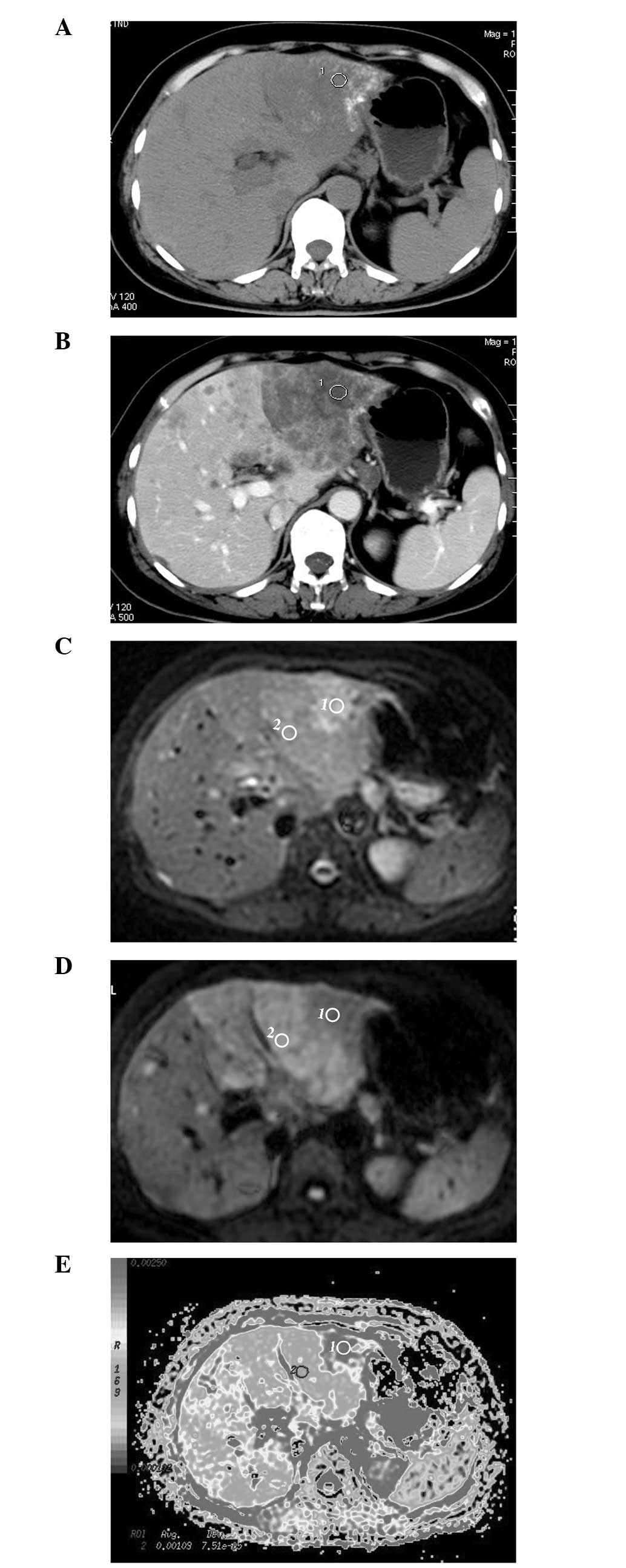
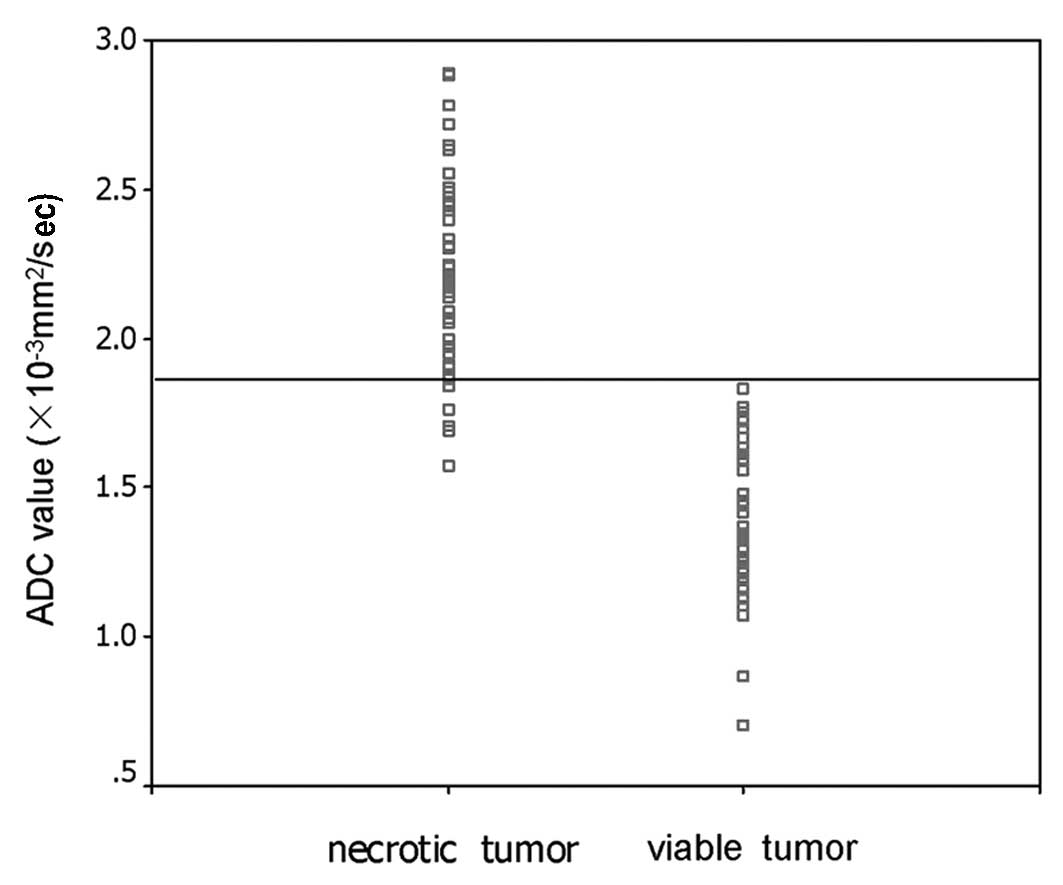
|
1
|
Jemal A, Bray F, Center MM, et al: Global
cancer statistics. CA Cancer J Clin. 61:69–90. 2011.
|
|
2
|
Llovet JM, Real MI, Montaña X, et al:
Arterial embolization or chemoembolisation versus symptomatic
treatment in patients with unresectable hepatocellular carcinoma: a
randomized controlled trial. Lancet. 359:1734–1739. 2002.
|
|
3
|
Takayama T, Makuuchi M, Hirohashi S, et
al: Malignant transformation of adenomatous hyperplasia to
hepatocellular carcinoma. Lancet. 336:1150–1153. 1990.
|
|
4
|
Kloeckner R, Otto G, Biesterfeld S, et al:
MDCT versus MRI assessment of tumor response after transarterial
chemoembolization for the treatment of hepatocellular carcinoma.
Cardiovasc Intervent Radiol. 33:532–540. 2010.
|
|
5
|
Yuan Z, Ye XD, Dong S, et al: Role of
magnetic resonance diffusion-weighted imaging in evaluating
response after chemoembolization of hepatocellular carcinoma. Eur J
Radiol. 75:e9–e14. 2010.
|
|
6
|
Kamel IR, Bluemke DA, Ramsey D, et al:
Role of diffusion-weighted imaging in estimating tumor necrosis
after chemoembolization of hepatocellular carcinoma. AJR Am J
Roentgenol. 181:708–710. 2003.
|
|
7
|
Dong S, Ye XD, Yuan Z, Xu LC and Xiao XS:
Relationship of apparent diffusion coefficient to survival for
patients with unresectable primary hepatocellular carcinoma after
chemoembolization. Eur J Radiol. 81:472–477. 2012.
|
|
8
|
Bruix J, Sherman M, Llovet JM, et al:
Clinical management of hepatocellular carcinoma: conclusions of the
Barcelona 2000 EASL Conference. European Association for the Study
of the Liver. J Hepatol. 35:421–430. 2001.
|
|
9
|
Lim HS, Jeong YY, Kang HK, Kim JK and Park
JG: Imaging features of hepatocellular carcinoma after
transcatheter arterial chemoembolization and radiofrequency
ablation. AJR Am J Roentgenol. 187:W341–W349. 2006.
|
|
10
|
Mannelli L, Kim S, Hajdu CH, et al:
Assessment of tumor necrosis of hepatocellular carcinoma after
chemoembolization: diffusion-weighted and contrast-enhanced MRI
with histopathologic correlation of the explanted liver. AJR Am J
Roentgenol. 193:1044–1052. 2009.
|
|
11
|
Koh DM and Collins DJ: Diffusion-weighted
MRI in the body: applications and challenges in oncology. AJR Am J
Roentgenol. 188:1622–1635. 2007.
|
|
12
|
Sadowski EA, Bennett LK, Chan MR, et al:
Nephrogenic systemic fibrosis: risk factors and incidence
estimation. Radiology. 243:148–157. 2007.
|
|
13
|
Koh DM, Scurr E, Collins D, et al:
Predicting response of colorectal hepatic metastasis: value of
pretreatment apparent diffusion coefficients. AJR Am J Roentgenol.
188:1001–1008. 2007.
|
|
14
|
Theilmann RJ, Borders R, Trouard TP, et
al: Changes in water mobility measured by diffusion MRI predict
response of metastatic breast cancer to chemotherapy. Neoplasia.
6:831–837. 2004.
|
|
15
|
Kamel IR, Liapi E, Reyes DK, et al:
Unresectable hepatocellular carcinoma: serial early vascular and
cellular changes after transarterial chemoembolization as detected
with MR imaging. Radiology. 250:466–473. 2009.
|
|
16
|
Galea N, Cantisani V and Taouli B: Liver
lesion detection and characterization: role of diffusion-weighted
imaging. J Magn Reson Imaging. 37:1260–1276. 2013.
|
|
17
|
Goshima S, Kanematsu M, Kondo H, et al:
Evaluating local hepatocellular carcinoma recurrence
post-transcatheter arterial chemoembolization: is
diffusion-weighted MRI reliable as an indicator? J Magn Reson
Imaging. 27:834–839. 2008.
|
|
18
|
Szafer A, Zhong J, Anderson AW and Gore
JC: Diffusion-weighted imaging in tissues: theoretical models. NMR
Biomed. 8:289–296. 1995.
|
|
19
|
Parikh T, Drew SJ, Lee VS, et al: Focal
liver lesion detection and characterization with diffusion-weighted
MR imaging: comparison with standard breath-hold T2-weighted
imaging. Radiology. 246:812–822. 2008.
|
|
20
|
Taouli B, Sandberg A, Stemmer A, et al:
Diffusion-weighted imaging of the liver: comparison of navigator
triggered and breath-hold acquisitions. J Magn Reson Imaging.
30:561–568. 2009.
|
|
21
|
Nasu K, Kuroki Y, Nawano S, et al: Hepatic
metastases: diffusion-weighted sensitivity-encoding versus
SPIO-enhanced MR Imaging. Radiology. 239:122–130. 2006.
|
|
22
|
Murtz P, Flacke S, Traber F, et al:
Abdomen: diffusion-weighted MR imaging with pulse-triggered
single-shot sequences. Radiology. 224:258–264. 2002.
|
|
23
|
Koh DM, Takahara T, Imai Y and Collins DJ:
Practical aspects of assessing tumors using clinical
diffusion-weighted imaging in the body. Magn Reson Med Sci.
6:211–224. 2007.
|
|
24
|
Coenegrachts K, Delanote J, Ter Beek L, et
al: Improved focal liver lesion detection: comparison of
single-shot diffusion-weighted echoplanar and single-shot T2
weighted turbo spin echo techniques. Br J Radiol. 80:524–531.
2007.
|
|
25
|
Zech CJ, Herrmann KA, Dietrich O, et al:
Black-blood diffusion-weighted EPI acquisition of the liver with
parallel imaging: comparison with a standard T2-weighted sequence
for detection of focal liver lesions. Invest Radiol. 43:261–266.
2008.
|