Colorectal laterally spreading tumors show characteristic expression of cell polarity factors, including atypical protein kinase C λ/ι, E‑cadherin, β‑catenin and basement membrane component

  • Authors:
    • Yasushi Ichikawa
    • Yoji Nagashima
    • Kaori Morioka
    • Kazunori Akimoto
    • Yasuyuki Kojima
    • Takashi Ishikawa
    • Ayumu Goto
    • Noritoshi Kobayashi
    • Kazuteru Watanabe
    • Mitsuyoshi Ota
    • Shoichi Fujii
    • Mayumi Kawamata
    • Ryo Takagawa
    • Chikara Kunizaki
    • Hirokazu Takahashi
    • Atsushi Nakajima
    • Shin Maeda
    • Hiroshi Shimada
    • Yoshiaki Inayama
    • Shigeo Ohno
    • Itaru Endo
  • View Affiliations

  • Published online on: June 20, 2014     https://doi.org/10.3892/ol.2014.2271
  • Pages: 977-984
Metrics: Total Views: 0 (Spandidos Publications: | PMC Statistics: )
Total PDF Downloads: 0 (Spandidos Publications: | PMC Statistics: )


Abstract

Colorectal flat‑type tumors include laterally spreading tumors (LSTs) and flat depressed‑type tumors. The former of which shows a predominant lateral spreading growth rather than an invasive growth. The present study examined the morphological characteristics of LSTs, in comparison with polypoid‑ or flat depressed‑type tumors, along with the expression of atypical protein kinase C (aPKC) λ/ι, a pivotal cell polarity regulator, and the hallmarks of cell polarity, as well as with type IV collagen, β‑catenin and E‑cadherin. In total, 37 flat‑type (24 LSTs and 13 flat depressed‑type tumors) and 20 polypoid‑type colorectal tumors were examined. The LSTs were classified as 15 LST adenoma (LST‑A) and nine LST cancer in adenoma (LST‑CA). An immunohistochemical examination was performed on aPKC λ/ι, type IV collagen, β‑catenin and E‑cadherin. The LST‑A and ‑CA showed a superficial replacing growth pattern, with expression of β‑catenin and E‑cadherin in the basolateral membrane and type IV collagen along the basement membrane. In addition, 86.6% of LST‑A and 55.6% of LST‑CA showed aPKC λ/ι expression of 1+ (weak to normal intensity staining in the cytoplasm compared with the normal epithelium). Furthermore, ~45% of the polypoid‑type adenomas showed 2+ (moderate intensity staining in the cytoplasm and/or nucleus) and 66.7% of the polypoid‑type cancer in adenoma were 3+ (strong intensity staining in the cytoplasm and nucleus). A statistically significant positive correlation was observed between the expression of aPKC λ/ι and β‑catenin (r=0.842; P<0.001), or type IV collagen (r=0.823; P<0.001). The LSTs showed a unique growth pattern, different from the expanding growth pattern presented by a polypoid tumor and invasive cancer. The growth characteristics of LST appear to be caused by adequate coexpression of β‑catenin, type IV collagen and aPKC λ/ι.
View Figures
View References

Related Articles

Journal Cover

September-2014
Volume 8 Issue 3

Print ISSN: 1792-1074
Online ISSN:1792-1082

Sign up for eToc alerts

Recommend to Library

Copy and paste a formatted citation
x
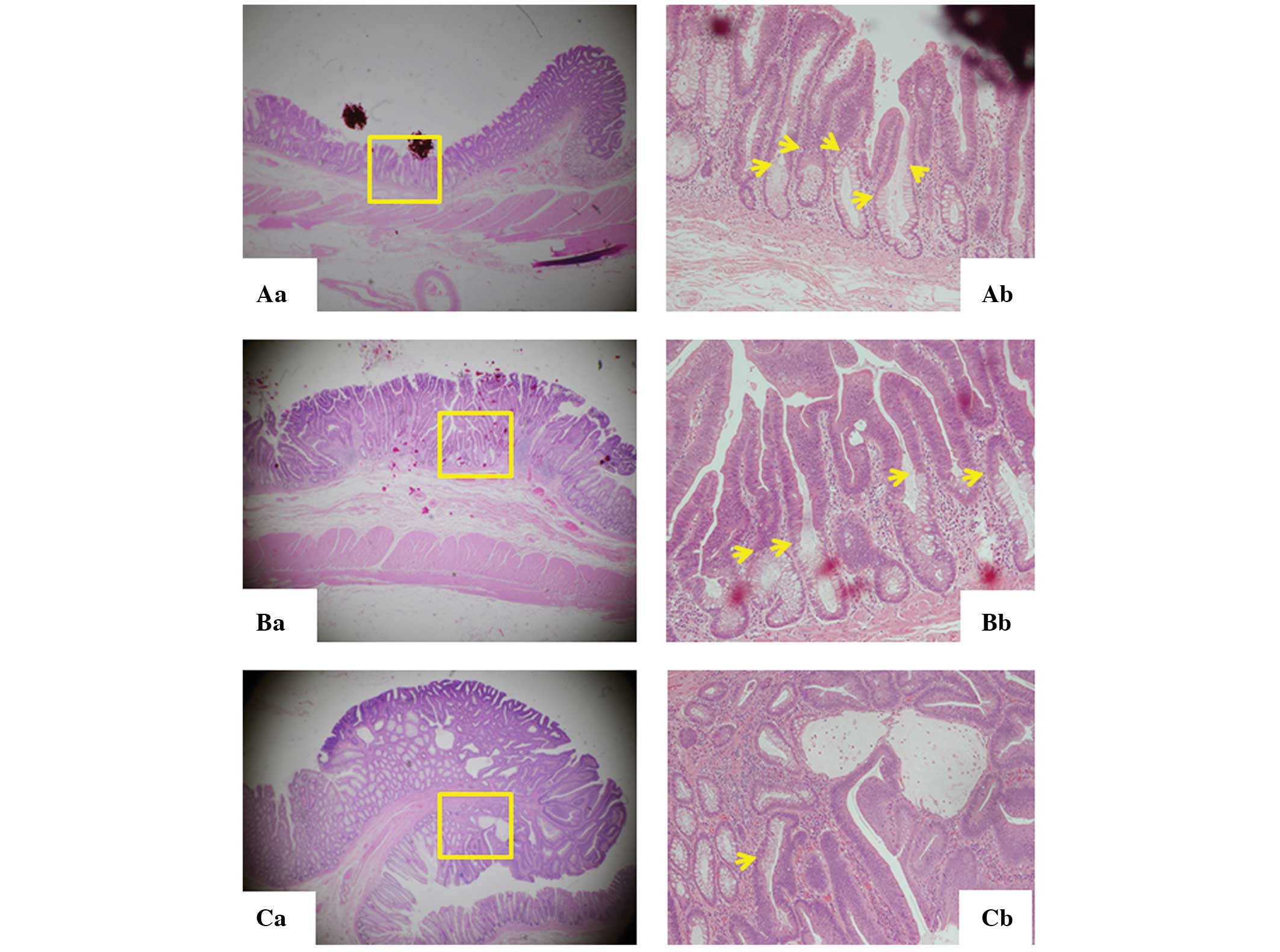
Spandidos Publications style
Ichikawa Y, Nagashima Y, Morioka K, Akimoto K, Kojima Y, Ishikawa T, Goto A, Kobayashi N, Watanabe K, Ota M, Ota M, et al: Colorectal laterally spreading tumors show characteristic expression of cell polarity factors, including atypical protein kinase C λ/ι, E‑cadherin, β‑catenin and basement membrane component. Oncol Lett 8: 977-984, 2014
APA
Ichikawa, Y., Nagashima, Y., Morioka, K., Akimoto, K., Kojima, Y., Ishikawa, T. ... Endo, I. (2014). Colorectal laterally spreading tumors show characteristic expression of cell polarity factors, including atypical protein kinase C λ/ι, E‑cadherin, β‑catenin and basement membrane component. Oncology Letters, 8, 977-984. https://doi.org/10.3892/ol.2014.2271
MLA
Ichikawa, Y., Nagashima, Y., Morioka, K., Akimoto, K., Kojima, Y., Ishikawa, T., Goto, A., Kobayashi, N., Watanabe, K., Ota, M., Fujii, S., Kawamata, M., Takagawa, R., Kunizaki, C., Takahashi, H., Nakajima, A., Maeda, S., Shimada, H., Inayama, Y., Ohno, S., Endo, I."Colorectal laterally spreading tumors show characteristic expression of cell polarity factors, including atypical protein kinase C λ/ι, E‑cadherin, β‑catenin and basement membrane component". Oncology Letters 8.3 (2014): 977-984.
Chicago
Ichikawa, Y., Nagashima, Y., Morioka, K., Akimoto, K., Kojima, Y., Ishikawa, T., Goto, A., Kobayashi, N., Watanabe, K., Ota, M., Fujii, S., Kawamata, M., Takagawa, R., Kunizaki, C., Takahashi, H., Nakajima, A., Maeda, S., Shimada, H., Inayama, Y., Ohno, S., Endo, I."Colorectal laterally spreading tumors show characteristic expression of cell polarity factors, including atypical protein kinase C λ/ι, E‑cadherin, β‑catenin and basement membrane component". Oncology Letters 8, no. 3 (2014): 977-984. https://doi.org/10.3892/ol.2014.2271