Spandidos Publications Logo
  • About
    • About Spandidos
    • Aims and Scopes
    • Abstracting and Indexing
    • Editorial Policies
    • Reprints and Permissions
    • Job Opportunities
    • Terms and Conditions
    • Contact
  • Journals
    • All Journals
    • Oncology Letters
      • Oncology Letters
      • Information for Authors
      • Editorial Policies
      • Editorial Board
      • Aims and Scope
      • Abstracting and Indexing
      • Bibliographic Information
      • Archive
    • International Journal of Oncology
      • International Journal of Oncology
      • Information for Authors
      • Editorial Policies
      • Editorial Board
      • Aims and Scope
      • Abstracting and Indexing
      • Bibliographic Information
      • Archive
    • Molecular and Clinical Oncology
      • Molecular and Clinical Oncology
      • Information for Authors
      • Editorial Policies
      • Editorial Board
      • Aims and Scope
      • Abstracting and Indexing
      • Bibliographic Information
      • Archive
    • Experimental and Therapeutic Medicine
      • Experimental and Therapeutic Medicine
      • Information for Authors
      • Editorial Policies
      • Editorial Board
      • Aims and Scope
      • Abstracting and Indexing
      • Bibliographic Information
      • Archive
    • International Journal of Molecular Medicine
      • International Journal of Molecular Medicine
      • Information for Authors
      • Editorial Policies
      • Editorial Board
      • Aims and Scope
      • Abstracting and Indexing
      • Bibliographic Information
      • Archive
    • Biomedical Reports
      • Biomedical Reports
      • Information for Authors
      • Editorial Policies
      • Editorial Board
      • Aims and Scope
      • Abstracting and Indexing
      • Bibliographic Information
      • Archive
    • Oncology Reports
      • Oncology Reports
      • Information for Authors
      • Editorial Policies
      • Editorial Board
      • Aims and Scope
      • Abstracting and Indexing
      • Bibliographic Information
      • Archive
    • Molecular Medicine Reports
      • Molecular Medicine Reports
      • Information for Authors
      • Editorial Policies
      • Editorial Board
      • Aims and Scope
      • Abstracting and Indexing
      • Bibliographic Information
      • Archive
    • World Academy of Sciences Journal
      • World Academy of Sciences Journal
      • Information for Authors
      • Editorial Policies
      • Editorial Board
      • Aims and Scope
      • Abstracting and Indexing
      • Bibliographic Information
      • Archive
    • International Journal of Functional Nutrition
      • International Journal of Functional Nutrition
      • Information for Authors
      • Editorial Policies
      • Editorial Board
      • Aims and Scope
      • Abstracting and Indexing
      • Bibliographic Information
      • Archive
    • International Journal of Epigenetics
      • International Journal of Epigenetics
      • Information for Authors
      • Editorial Policies
      • Editorial Board
      • Aims and Scope
      • Abstracting and Indexing
      • Bibliographic Information
      • Archive
    • Medicine International
      • Medicine International
      • Information for Authors
      • Editorial Policies
      • Editorial Board
      • Aims and Scope
      • Abstracting and Indexing
      • Bibliographic Information
      • Archive
  • Articles
  • Information
    • Information for Authors
    • Information for Reviewers
    • Information for Librarians
    • Information for Advertisers
    • Conferences
  • Language Editing
Spandidos Publications Logo
  • About
    • About Spandidos
    • Aims and Scopes
    • Abstracting and Indexing
    • Editorial Policies
    • Reprints and Permissions
    • Job Opportunities
    • Terms and Conditions
    • Contact
  • Journals
    • All Journals
    • Biomedical Reports
      • Information for Authors
      • Editorial Policies
      • Editorial Board
      • Aims and Scope
      • Abstracting and Indexing
      • Bibliographic Information
      • Archive
    • Experimental and Therapeutic Medicine
      • Information for Authors
      • Editorial Policies
      • Editorial Board
      • Aims and Scope
      • Abstracting and Indexing
      • Bibliographic Information
      • Archive
    • International Journal of Epigenetics
      • Information for Authors
      • Editorial Policies
      • Editorial Board
      • Aims and Scope
      • Abstracting and Indexing
      • Bibliographic Information
      • Archive
    • International Journal of Functional Nutrition
      • Information for Authors
      • Editorial Policies
      • Editorial Board
      • Aims and Scope
      • Abstracting and Indexing
      • Bibliographic Information
      • Archive
    • International Journal of Molecular Medicine
      • Information for Authors
      • Editorial Policies
      • Editorial Board
      • Aims and Scope
      • Abstracting and Indexing
      • Bibliographic Information
      • Archive
    • International Journal of Oncology
      • Information for Authors
      • Editorial Policies
      • Editorial Board
      • Aims and Scope
      • Abstracting and Indexing
      • Bibliographic Information
      • Archive
    • Medicine International
      • Information for Authors
      • Editorial Policies
      • Editorial Board
      • Aims and Scope
      • Abstracting and Indexing
      • Bibliographic Information
      • Archive
    • Molecular and Clinical Oncology
      • Information for Authors
      • Editorial Policies
      • Editorial Board
      • Aims and Scope
      • Abstracting and Indexing
      • Bibliographic Information
      • Archive
    • Molecular Medicine Reports
      • Information for Authors
      • Editorial Policies
      • Editorial Board
      • Aims and Scope
      • Abstracting and Indexing
      • Bibliographic Information
      • Archive
    • Oncology Letters
      • Information for Authors
      • Editorial Policies
      • Editorial Board
      • Aims and Scope
      • Abstracting and Indexing
      • Bibliographic Information
      • Archive
    • Oncology Reports
      • Information for Authors
      • Editorial Policies
      • Editorial Board
      • Aims and Scope
      • Abstracting and Indexing
      • Bibliographic Information
      • Archive
    • World Academy of Sciences Journal
      • Information for Authors
      • Editorial Policies
      • Editorial Board
      • Aims and Scope
      • Abstracting and Indexing
      • Bibliographic Information
      • Archive
  • Articles
  • Information
    • For Authors
    • For Reviewers
    • For Librarians
    • For Advertisers
    • Conferences
  • Language Editing
Login Register Submit
  • This site uses cookies
  • You can change your cookie settings at any time by following the instructions in our Cookie Policy. To find out more, you may read our Privacy Policy.

    I agree
Search articles by DOI, keyword, author or affiliation
Search
Advanced Search
presentation
Oncology Letters
Join Editorial Board Propose a Special Issue
Print ISSN: 1792-1074 Online ISSN: 1792-1082
Journal Cover
September-2014 Volume 8 Issue 3

Full Size Image

Sign up for eToc alerts
Recommend to Library

Journals

International Journal of Molecular Medicine

International Journal of Molecular Medicine

International Journal of Molecular Medicine is an international journal devoted to molecular mechanisms of human disease.

International Journal of Oncology

International Journal of Oncology

International Journal of Oncology is an international journal devoted to oncology research and cancer treatment.

Molecular Medicine Reports

Molecular Medicine Reports

Covers molecular medicine topics such as pharmacology, pathology, genetics, neuroscience, infectious diseases, molecular cardiology, and molecular surgery.

Oncology Reports

Oncology Reports

Oncology Reports is an international journal devoted to fundamental and applied research in Oncology.

Experimental and Therapeutic Medicine

Experimental and Therapeutic Medicine

Experimental and Therapeutic Medicine is an international journal devoted to laboratory and clinical medicine.

Oncology Letters

Oncology Letters

Oncology Letters is an international journal devoted to Experimental and Clinical Oncology.

Biomedical Reports

Biomedical Reports

Explores a wide range of biological and medical fields, including pharmacology, genetics, microbiology, neuroscience, and molecular cardiology.

Molecular and Clinical Oncology

Molecular and Clinical Oncology

International journal addressing all aspects of oncology research, from tumorigenesis and oncogenes to chemotherapy and metastasis.

World Academy of Sciences Journal

World Academy of Sciences Journal

Multidisciplinary open-access journal spanning biochemistry, genetics, neuroscience, environmental health, and synthetic biology.

International Journal of Functional Nutrition

International Journal of Functional Nutrition

Open-access journal combining biochemistry, pharmacology, immunology, and genetics to advance health through functional nutrition.

International Journal of Epigenetics

International Journal of Epigenetics

Publishes open-access research on using epigenetics to advance understanding and treatment of human disease.

Medicine International

Medicine International

An International Open Access Journal Devoted to General Medicine.

Journal Cover
September-2014 Volume 8 Issue 3

Full Size Image

Sign up for eToc alerts
Recommend to Library

  • Article
  • Citations
    • Cite This Article
    • Download Citation
    • Create Citation Alert
    • Remove Citation Alert
    • Cited By
  • Similar Articles
    • Related Articles (in Spandidos Publications)
    • Similar Articles (Google Scholar)
    • Similar Articles (PubMed)
  • Download PDF
  • Download XML
  • View XML
Case Report

Imaging observations of a schwannoma of low malignant potential in the anterior abdominal wall: A case report

  • Authors:
    • Yongkang Liu
    • Xiao Chen
    • Tianyao Wang
    • Zhongqiu Wang
  • View Affiliations / Copyright

    Affiliations: Department of Radiology, Affiliated Hospital of Nanjing University of Traditional Chinese Medicine, Nanjing, Jiansu 210029, P.R. China, Department of Radiology, Shanghai East Hospital, Tongji University School of Medicine, Shanghai 200120, P.R. China
  • Pages: 1159-1162
    |
    Published online on: July 2, 2014
       https://doi.org/10.3892/ol.2014.2305
  • Expand metrics +
Metrics: Total Views: 0 (Spandidos Publications: | PMC Statistics: )
Metrics: Total PDF Downloads: 0 (Spandidos Publications: | PMC Statistics: )
Cited By (CrossRef): 0 citations Loading Articles...

This article is mentioned in:


Abstract

Neurilemmoma, also known as schwannoma, is an uncommon benign neoplasm that is most commonly found in the trunk and head and neck regions. The present study reports the case of a 67‑year‑old female with schwannoma localized in the anterior abdominal wall and analyzes the ultrasound and computed tomography (CT) imaging observations of the schwannoma. A dynamic time‑intensity curve was also recorded in the study. A well‑defined, elliptic low echo level, heterogeneous mass was observed during ultrasound examination. The CT scan revealed a solid, heterogeneous, low‑density mass in the abdominal wall. Contrast‑enhanced scans showed a heterogeneously enhanced mass during the arterial and venous phase. Centripetal fill‑in was demonstrated and the mass was markedly, homogenously enhanced relative to the muscles during the delayed phase. Peak enhancement was observed during the venous phase and then slowly declined. However, the mass was hyperattenuated during the delayed phase. The lesion was completely excised and no evidence of recurrence has been identified during the 3 months of follow‑up. The present study suggested that a diagnosis of schwannoma should be considered for certain patients with masses in the abdominal wall. Peripheral enhancement during the arterial and venous phases and homogeneous enhancement in the delayed phase are the significant imaging findings of a schwannoma.

Introduction

Neurilemmoma, also known as schwannoma, is an uncommon benign neoplasm originating from the Schwann cells in the peripheral nerve sheaths (1,2). The majority of the tumors commonly localize in the trunk, head and extremities (3). However, occurrence in the anterior abdominal wall is extremely rare. The present study reports the case of a 67-year-old female with a schwannoma of low malignant potential in the right anterior abdominal wall and analyzes the main ultrasound and computed tomography (CT) imaging observations. This study highlights the clinical features and imaging findings of schwannoma in the abdominal wall, and may aid in the future diagnosis and differential diagnosis of this condition. Patient provided written informed consent.

Case report

Patient and clinical data

A 67-year-old female was admitted to the Affiliated Hospital of Nanjing University of Traditional Chinese Medicine (Nanjing, China) complaining of a disconcerting mass in the right anterior abdominal wall. A small mass had incidentally been found 10 years ago, with no restriction of movement present in the right abdominal wall. As the patient did not exhibit any evident uncomfortable symptoms, not enough attention was drawn to the lesion. Over the year prior to the present hospital admittance, the lump gradually increased in size and the patient felt soreness and suffered untimely gas pains. To acquire a diagnosis and treatment, the patient was referred to the outpatient clinic of the the Affiliated Hospital of Nanjing University of Traditional Chinese Medicine. There was no history of anergy, fever, anorexia, weight loss, trauma or surgery, and there was no family history of a similar complaint.

A physical examination showed a 6×4-cm mass protruding through the right anterior abdominal wall. The mass was firm, non-tender and not fixed to the skin of the abdominal wall. All routine laboratory tests were within the normal ranges.

Ultrasound and CT imaging findings

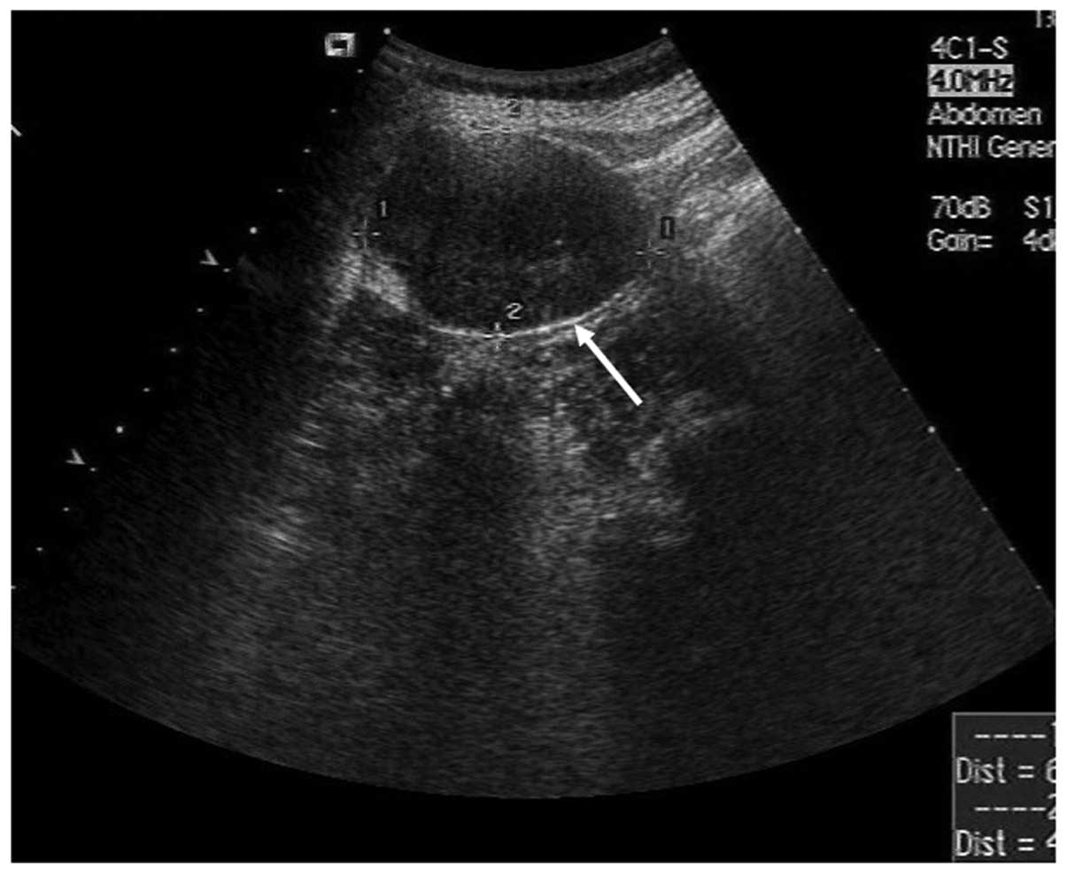
An abdominal ultrasound examination showed a well-defined, elliptic, low echo level, heterogeneous mass just beneath the abdominal skin (Fig. 
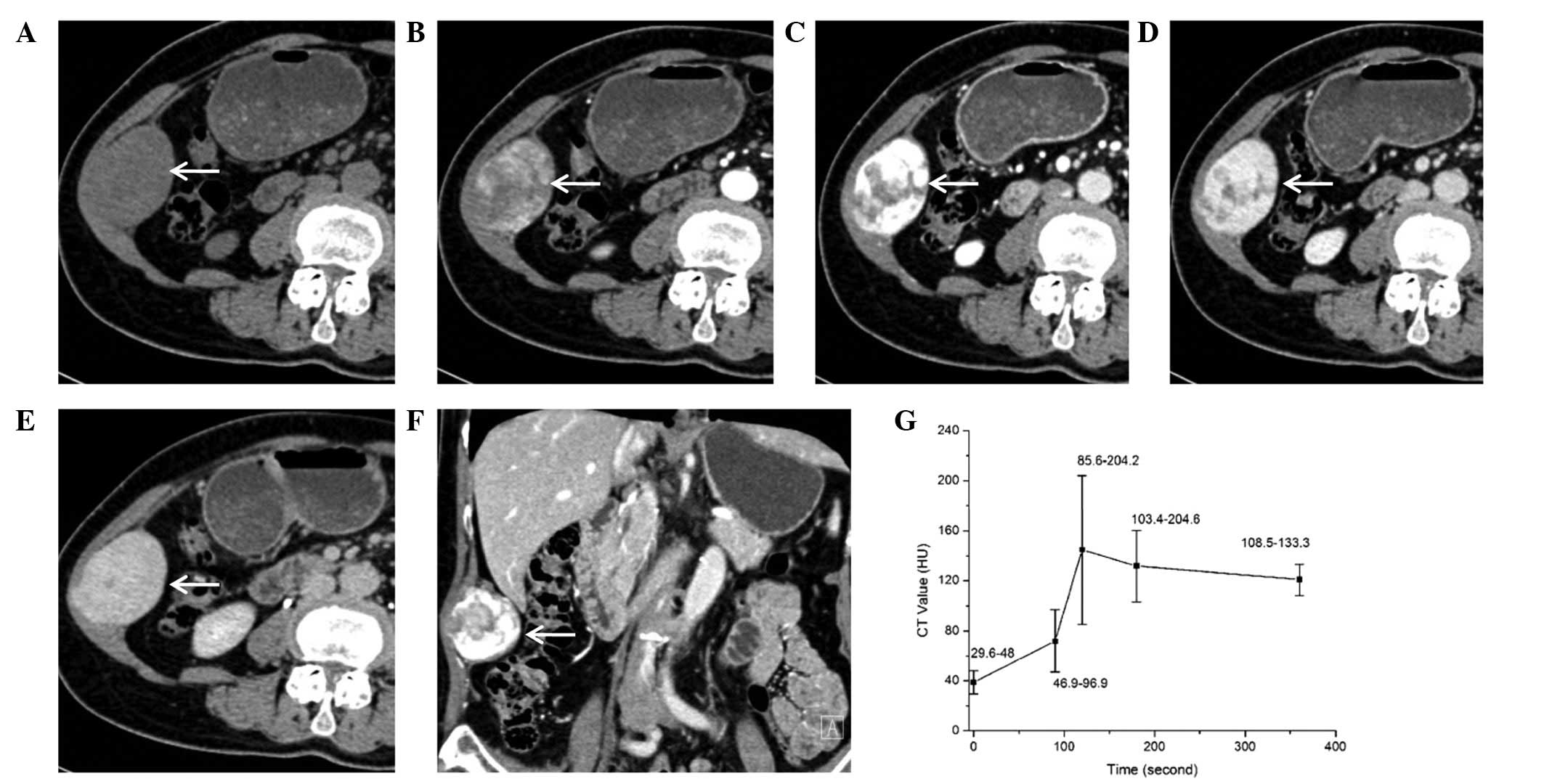
1). The diameter of the tumor was 5.6 cm. A CT scan without contrast enhancement revealed a solid, homogeneous mass with a low density relative to the muscle in the abdominal wall (Fig. 2A). Contrast-enhanced scans showed a evident gradually- and heterogeneously-enhanced lesion during the arterial (Fig. 2B) and venous (Fig. 2C) phases. Centripetal fill-in was demonstrated and the mass was markedly, homogenously-enhanced relative to the muscles (Fig. 2D and E) during the balanced and delayed phases. The coronal CT image also clearly showed that the mass was localized in the abdominal wall (Fig. 2F). The tumor showed marked enhancement on contrast-enhanced CT. The time-intensity curve following contrast agent injection is shown in Fig. 2G. Peak enhancement was observed during the venous phase (120 sec after contrast agent injection) and then slowly declined. However, the mass was hyperattenuated during the delayed phase (360 sec after contrast agent injection). No cysts, calcification or necrosis were found in the patient even though the mass was large. A radiological diagnosis of a paraganglioma or a vascular genesis tumor was considered.

Figure 1

B-mode ultrasound showing the well-encapsulated mass in the anterior abdominal wall (arrow).

Figure 2

Computed tomography (CT) imaging findings of the schwannoma. (A) Conventional CT scan revealing an oval mass (white arrow) in the right lower abdominal wall. Contrast-enhanced CT scan showing a gradually- and heterogeneously-enhanced lesion with high density in (B) the arterial phase, (C) the venous phase and (D and E) the delay phase at different times. (F) Coronal image showing that the mass is in the abdominal wall. (G) The time-intensity curve displaying the characteristics of enhancement following agent injection.

Surgery and histopathological examination

The lesion was completely excised and a frozen section of the specimen was sent for histological examination. 
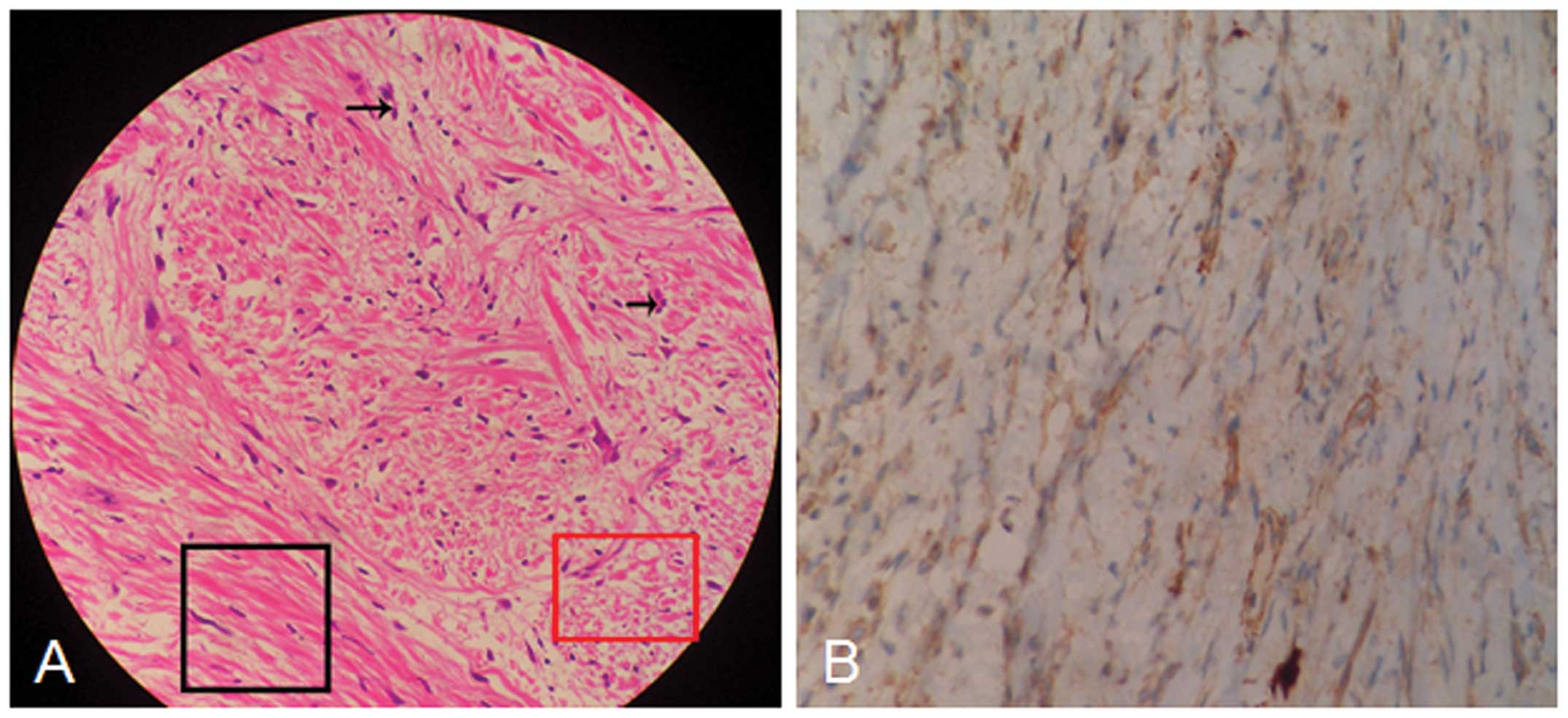
Histological examination (Fig. 3A) showed that the tumor was composed of abundant spindle-shaped cells, which locally invaded the surrounding fat tissues. Karyokinesis of the tumor cells could occasionally be observed. A primary diagnosis of a spindle cell tumor and neurofibroma was made. Immunohistochemical examination showed that the specimen was negative for S-100 (Fig. 3B). Based on the imaging findings and histopathological results, a final diagnosis of a schwannoma of low malignant potential was made. The patient was discharged 10 days after the surgery and no further treatment was required. No evidence of recurrence was found during the 3-month follow-up.

Figure 3

(A) Histopathological and (B) immunochemical appearance of the schwannoma (hematoxylin and eosin stainning; magnification, ×40). (A) Abundant spindle-shaped cells localized in the Antoni type A component area (black frame) and the Antoni type B component area (red frame). Sporadic karyokinesis (arrow) was occasionally observed (magnification, ×40). (B) Immunohistological assay showing cells negative for S-100 staining.

Discussion

Schwannomas or neurilemmomas are benign, encapsulated, slow-growing mesenchymal neoplasms that arise from Schwann cells and have a low malignant potential. These neoplasms can present at any age, and most commonly occur in adult females between the ages of 20 and 50 years old. Up to 20% of cases are associated with neurofibromatosis type 1 (4). Although sporadic cases of these tumors arising in the retroperitoneum, pelvis, perineum, adrenals, kidneys and inguen have been previously reported (1), their occurrence in the abdominal wall is extremely rare and only a few of these cases have been reported (1,4–6).

The majority of schwannomas arise from the nerve sheath of large peripheral nerves, occurring at the level of or below the subcutaneous fat layer (7). Thus, the clinical signs and symptoms may vary according to the tumor location. The tumors may be asymptomatic and only found incidentally during examination. However, when they grow larger, they can put pressure on the surrounding large nerves (1), as found in the patient of the present study.

Radiological examinations, CT or magnetic resonance imaging (MRI) can provide important information about these tumors, including the tumor site, its characteristics and its associations with other tissues. The imaging features of schwannomas are associated with its two components: Antoni type A (cellular component) and Antoni type B (myxoid component). The former consists of spindle-shaped cells that are arranged compactly in interlacing fascicles or short bundles. Antoni B schwannomas are loosely composed of reticular fibers, cysts, vascellum and the Schwann cells (8). Accordingly, schwannomas usually present as a well-defined, homogeneous soft-tissue mass on conventional CT scan (9,10). The tumors show minimal or mild heterogeneous enhancement following the injection of contrast agent (9,10). In the present case, marked peripheral enhancement was demonstrated during the artery and venous phases. The progressive centripetal fill-in was then observed, and homogeneous enhancement occurred in the lag period. The CT imaging observations of the schwannoma in this study were similar with that of a hemangioma. In general, schwannomas appear as hypointense on T1-weighted and hyperintense on T2-weighted MRI. The hypercellular (Antoni A) and hypocellular (Antoni B) components display hypointense and hyperintense signals, respectively, on T2-weighted images (4,9). With gadolinium administration, the enhancement portion corresponds to the solid component of the Antoni A. Loose cellularity with diffuse edematous change may result in minimal contrast enhancement. If there is degenerative change of the neurilemmoma, it often shows a poor blood supply, cyst formation, calcifications, hemorrhage and hyalinization (11). In the present case, no cyst formation, calcification or necrosis was observed.

Although MRI and CT are ideally suited to detect the tumor pathology and delineate the soft tissue and its components, the final diagnosis can only be confirmed by histopathological examination. Immunohistochemical examinations besides hematoxylin and eosin staining are required to form a differential diagnosis (12). Although a previous study has shown that the diffuse immunoreactivity for S-100 is almost universal in schwannomas (13), another study showed that 10–50% of cases were negative (5). In the present case, the schwannoma was also negative for S-100.

The ideal treatment for schwannoma is complete surgical excision, and the prognosis is good (11), although recurrence of schwannomas in situ or at a distant site, attributed to incomplete resection, has also been reported (2). The present patient recovered well and no evidence of recurrence was found on CT images during the 3-month follow-up.

In conclusion, the present study indicates that a diagnosis of schwannoma should be considered for certain patients with masses in the abdominal wall. The observations of gradual and heterogeneous enhancement during the artery and venous phases, progressive centripetal fill-in and homogeneous enhancement during the delayed phase may be useful information for the imaging diagnosis of schwannomas.

References

1 

Bhatia RK, Banerjea A, Ram M and Lovett BE: Benign ancient schwannoma of the abdominal wall: an unwanted birthday present. BMC Surg. 10:12010.

2 

Forthman CL and Blazar PE: Nerve tumors of the hand and upper extremity. Hand Clin. 20:233–242. 2004.

3 

Canda MŞ: Peripheral nerve sheath tumors. Türkiye Ekopatoloji Dergisi. 10:65–74. 2004.(In Turkish).

4 

Mishra A, Hamadto M, Azzabi M and Elfagieh M: Abdominal wall schwannoma: case report and review of the literature. Case Rep Radiol. 2013:4568632013.

5 

Khorgami Z, Nasiri S, Rezakhanlu F and Sodagari N: Malignant schwannoma of anterior abdominal wall: report of a case. J Clin Med Res. 1:233–236. 2009.

6 

Kataoka D, Nonaka M, Yamamoto S, Kawada T, Takaba T and Kunimura T: Multiple synchronous intrathoracic neurilemmomas who had a past history of neurilemmoma on the abdominal wall; report of a case. Kyobu Geka. 58:158–160. 2005.(In Japanese).

7 

Cho SB, Kim HS, Paik JH, Ryu DJ and Oh SH: Dome-shaped tumor with surface changes on the abdominal wall. Clin Exp Dermatol. 35:95–96. 2010.

8 

Qin S, Yang C and Ren H: Cerebral hernia caused by a thoracic surgery for multiple schwannomas in a patient with neurofibromatosis type 2. Turk Neurosurg. 23:245–248. 2013.

9 

Ogose A, Hotta T, Morita T, et al: Diagnosis of peripheral nerve sheath tumors around the pelvis. Jpn J Clin Oncol. 34:405–413. 2004.

10 

Hu S, Chen Y, Wang Y, Chen KM and Song Q: Clinical and CT manifestation of pleural schwannoma. Acta Radiol. 53:1137–1141. 2012.

11 

Lao WT, Yang SH, Chen CL and Chan WP: Mesentery neurilemmoma: CT, MRI and angiographic findings. Intern Med. 50:2579–2581. 2011.

12 

de Mesquita Neto JW, Lima Verde Leal RM, de Brito EV, Cordeiro DF and Costa ML: Solitary schwannoma of the cecum: case report and review of the literature. Case Rep Oncol. 6:62–65. 2013.

13 

Rodriguez FJ, Folpe AL, Giannini C and Perry A: Pathology of peripheral nerve sheath tumors: diagnostic overview and update on selected diagnostic problems. Acta Neuropathol. 123:295–319. 2012.

Related Articles

  • Abstract
  • View
  • Download
  • Twitter
Copy and paste a formatted citation
Spandidos Publications style
Liu Y, Chen X, Wang T and Wang Z: Imaging observations of a schwannoma of low malignant potential in the anterior abdominal wall: A case report. Oncol Lett 8: 1159-1162, 2014.
APA
Liu, Y., Chen, X., Wang, T., & Wang, Z. (2014). Imaging observations of a schwannoma of low malignant potential in the anterior abdominal wall: A case report. Oncology Letters, 8, 1159-1162. https://doi.org/10.3892/ol.2014.2305
MLA
Liu, Y., Chen, X., Wang, T., Wang, Z."Imaging observations of a schwannoma of low malignant potential in the anterior abdominal wall: A case report". Oncology Letters 8.3 (2014): 1159-1162.
Chicago
Liu, Y., Chen, X., Wang, T., Wang, Z."Imaging observations of a schwannoma of low malignant potential in the anterior abdominal wall: A case report". Oncology Letters 8, no. 3 (2014): 1159-1162. https://doi.org/10.3892/ol.2014.2305
Copy and paste a formatted citation
x
Spandidos Publications style
Liu Y, Chen X, Wang T and Wang Z: Imaging observations of a schwannoma of low malignant potential in the anterior abdominal wall: A case report. Oncol Lett 8: 1159-1162, 2014.
APA
Liu, Y., Chen, X., Wang, T., & Wang, Z. (2014). Imaging observations of a schwannoma of low malignant potential in the anterior abdominal wall: A case report. Oncology Letters, 8, 1159-1162. https://doi.org/10.3892/ol.2014.2305
MLA
Liu, Y., Chen, X., Wang, T., Wang, Z."Imaging observations of a schwannoma of low malignant potential in the anterior abdominal wall: A case report". Oncology Letters 8.3 (2014): 1159-1162.
Chicago
Liu, Y., Chen, X., Wang, T., Wang, Z."Imaging observations of a schwannoma of low malignant potential in the anterior abdominal wall: A case report". Oncology Letters 8, no. 3 (2014): 1159-1162. https://doi.org/10.3892/ol.2014.2305
Follow us
  • Twitter
  • LinkedIn
  • Facebook
About
  • Spandidos Publications
  • Careers
  • Cookie Policy
  • Privacy Policy
How can we help?
  • Help
  • Live Chat
  • Contact
  • Email to our Support Team