Imaging observations of a schwannoma of low malignant potential in the anterior abdominal wall: A case report

  • Authors:
    • Yongkang Liu
    • Xiao Chen
    • Tianyao Wang
    • Zhongqiu Wang
  • View Affiliations

  • Published online on: July 2, 2014     https://doi.org/10.3892/ol.2014.2305
  • Pages: 1159-1162
Metrics: Total Views: 0 (Spandidos Publications: | PMC Statistics: )
Total PDF Downloads: 0 (Spandidos Publications: | PMC Statistics: )


Abstract

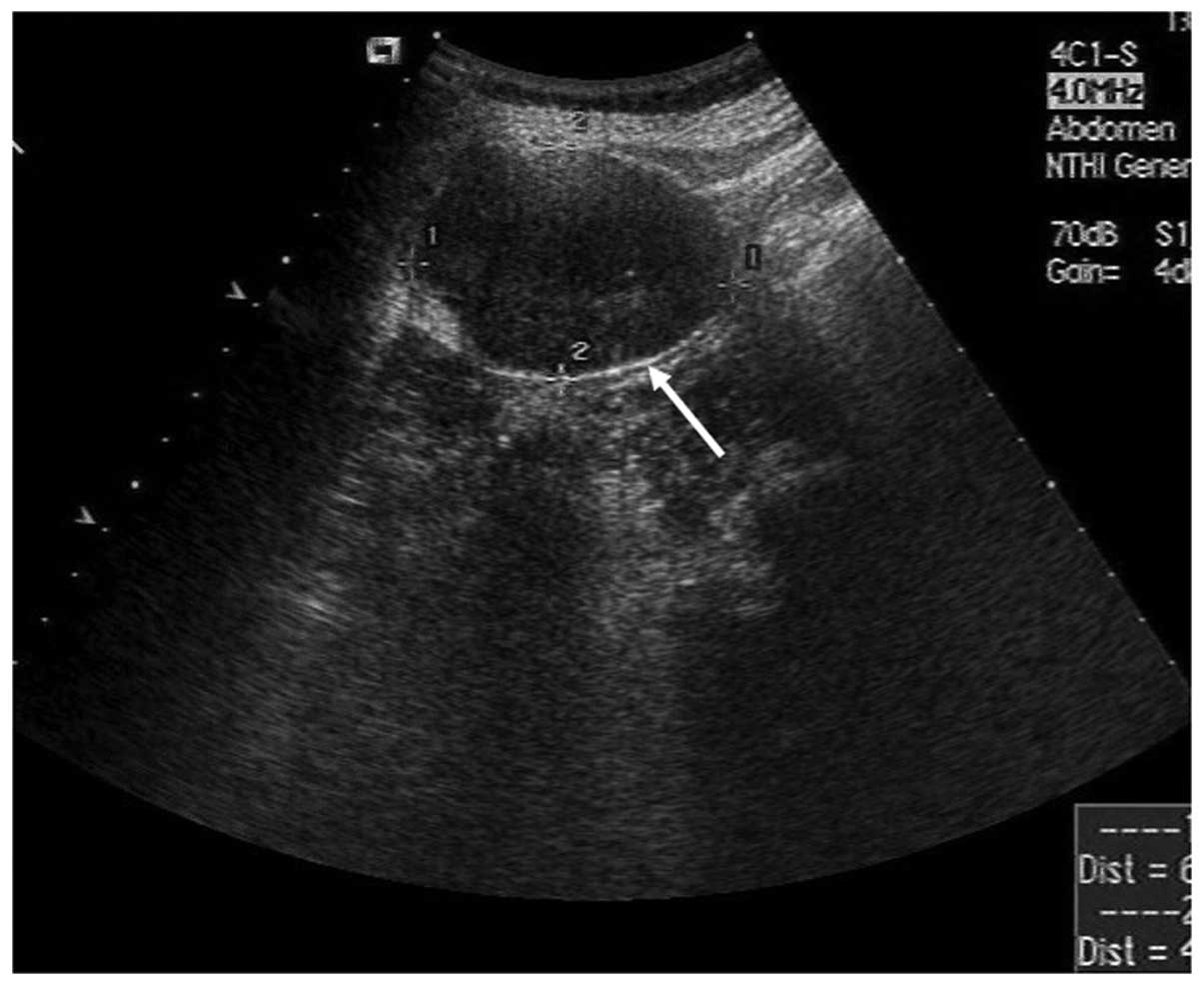
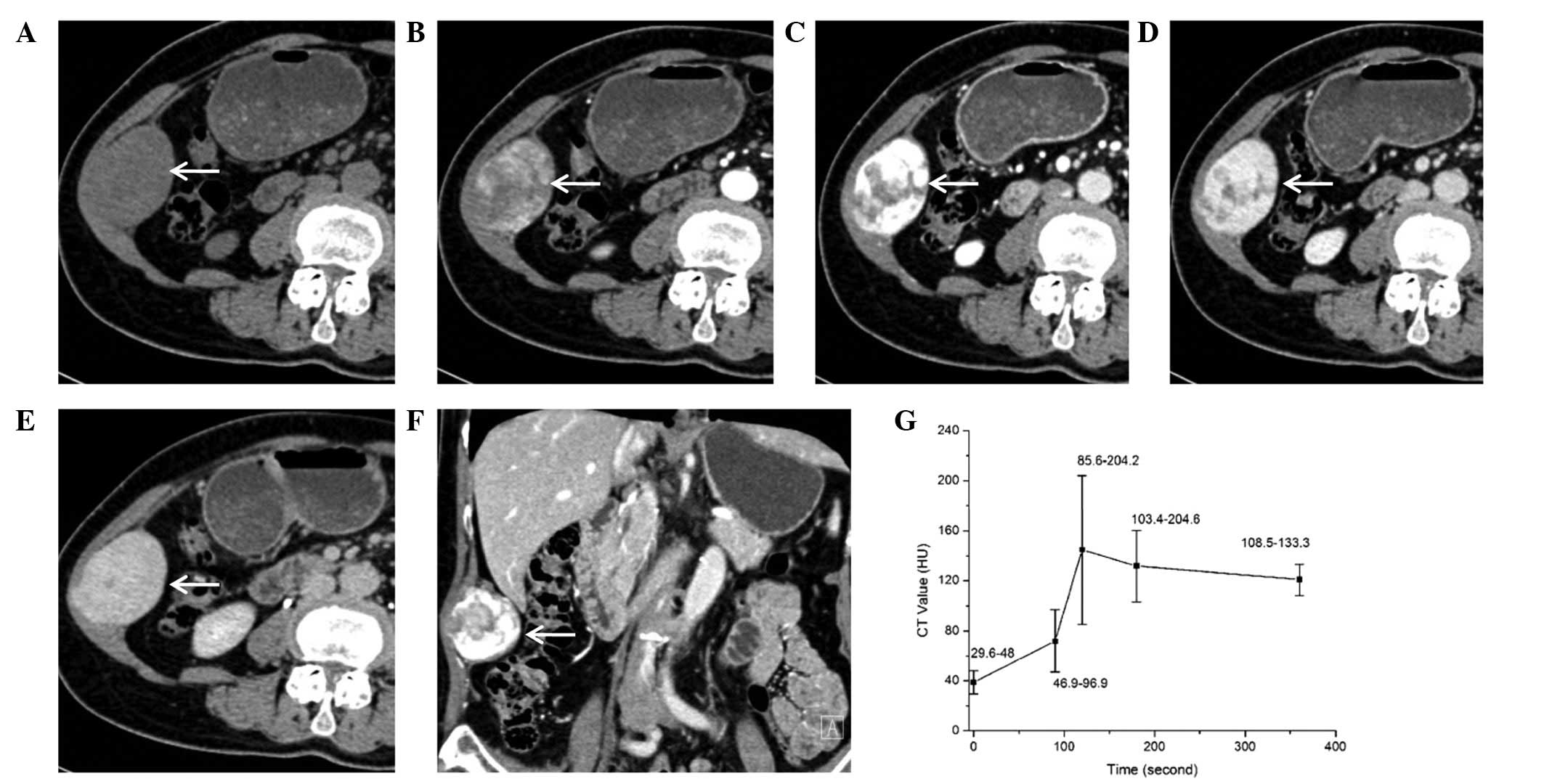
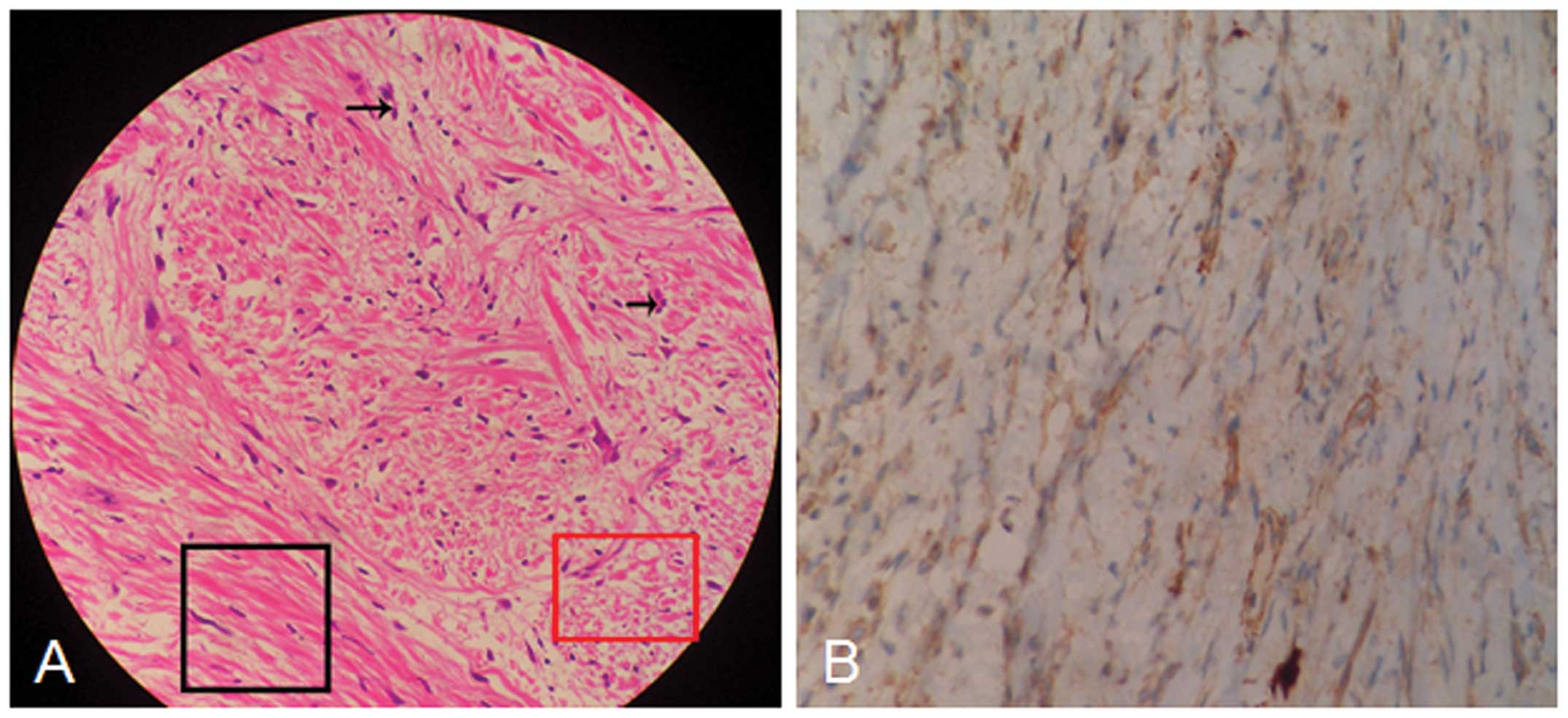
Neurilemmoma, also known as schwannoma, is an uncommon benign neoplasm that is most commonly found in the trunk and head and neck regions. The present study reports the case of a 67‑year‑old female with schwannoma localized in the anterior abdominal wall and analyzes the ultrasound and computed tomography (CT) imaging observations of the schwannoma. A dynamic time‑intensity curve was also recorded in the study. A well‑defined, elliptic low echo level, heterogeneous mass was observed during ultrasound examination. The CT scan revealed a solid, heterogeneous, low‑density mass in the abdominal wall. Contrast‑enhanced scans showed a heterogeneously enhanced mass during the arterial and venous phase. Centripetal fill‑in was demonstrated and the mass was markedly, homogenously enhanced relative to the muscles during the delayed phase. Peak enhancement was observed during the venous phase and then slowly declined. However, the mass was hyperattenuated during the delayed phase. The lesion was completely excised and no evidence of recurrence has been identified during the 3 months of follow‑up. The present study suggested that a diagnosis of schwannoma should be considered for certain patients with masses in the abdominal wall. Peripheral enhancement during the arterial and venous phases and homogeneous enhancement in the delayed phase are the significant imaging findings of a schwannoma.
View Figures
View References

Related Articles

Journal Cover

September-2014
Volume 8 Issue 3

Print ISSN: 1792-1074
Online ISSN:1792-1082

Sign up for eToc alerts

Recommend to Library

Copy and paste a formatted citation
x
Spandidos Publications style
Liu Y, Chen X, Wang T and Wang Z: Imaging observations of a schwannoma of low malignant potential in the anterior abdominal wall: A case report. Oncol Lett 8: 1159-1162, 2014
APA
Liu, Y., Chen, X., Wang, T., & Wang, Z. (2014). Imaging observations of a schwannoma of low malignant potential in the anterior abdominal wall: A case report. Oncology Letters, 8, 1159-1162. https://doi.org/10.3892/ol.2014.2305
MLA
Liu, Y., Chen, X., Wang, T., Wang, Z."Imaging observations of a schwannoma of low malignant potential in the anterior abdominal wall: A case report". Oncology Letters 8.3 (2014): 1159-1162.
Chicago
Liu, Y., Chen, X., Wang, T., Wang, Z."Imaging observations of a schwannoma of low malignant potential in the anterior abdominal wall: A case report". Oncology Letters 8, no. 3 (2014): 1159-1162. https://doi.org/10.3892/ol.2014.2305