Introduction
Horseshoe kidneys are the most common renal fusion
anomaly. Abnormal vasculature and the possibility of isthmusectomy
are the primary issues that require attention when surgery is
considered for renal cell carcinoma in horseshoe kidneys. To date,
there have been few reports of renal cell carcinomas excised from
horseshoe kidneys using the laparoscopic or hand-assisted
laparoscopic radical heminephrectomy approaches (1–4).
Therefore, the incidence, treatment and outcome of such remains
unclear. The present study describes the case of a pure
transperitoneal laparoscopic radical heminephrectomy for a large
renal tumor in a horseshoe kidney. Written informed consent was
obtained from the patient.
Case report
Case details
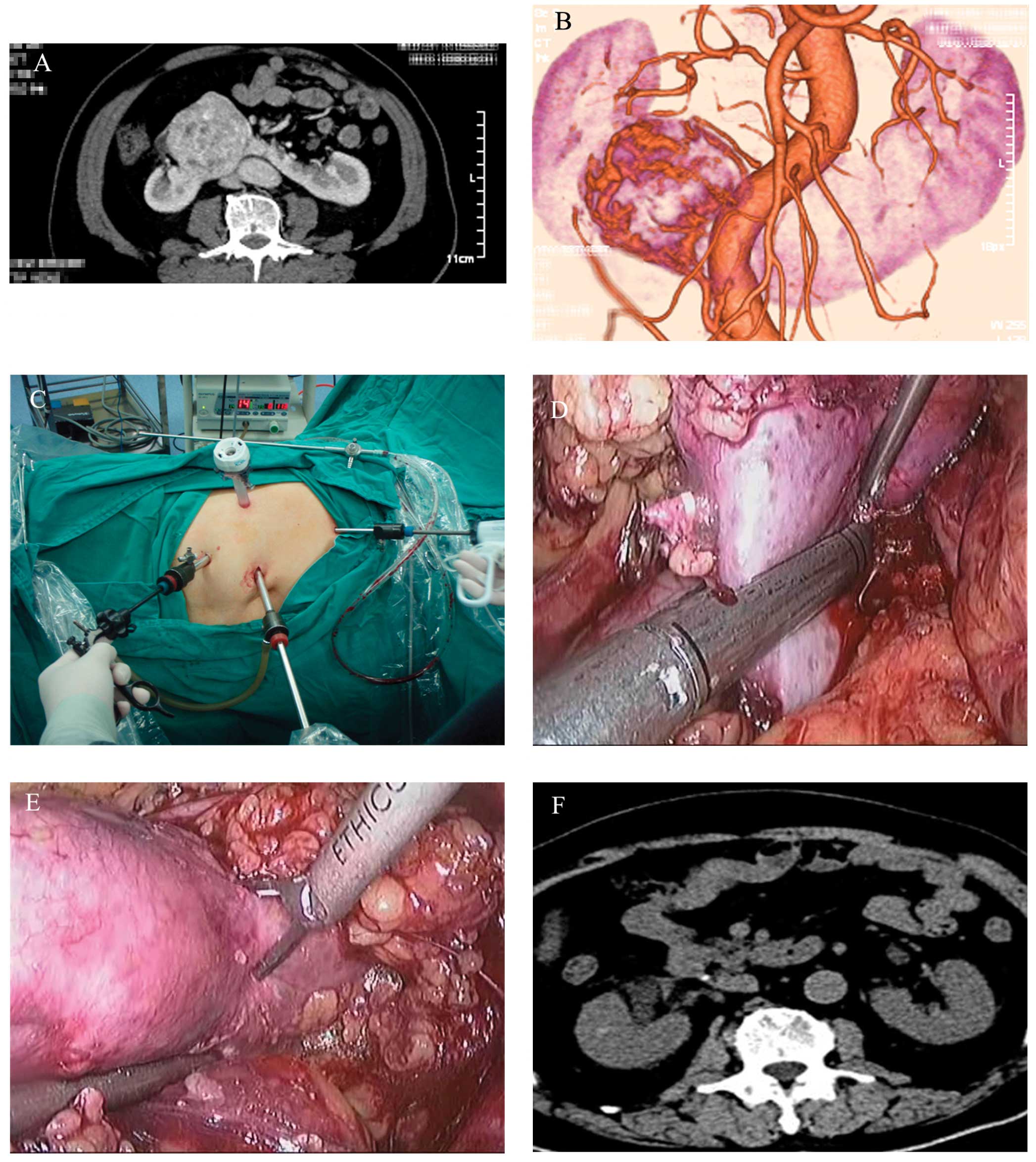
A solid renal tumor in the right moiety of a
horseshoe kidney was incidentally detected in a 72-year-old male
during a routine physical examination. Computed tomography
angiography (CTA) identified a 7-cm enhancing mass supplied by
three arteries in the right renal moiety (Fig. 1A and B). The results of the
evaluation for metastases were negative. Following a careful
explanation of the risks and benefits of alternative treatments,
the patient elected to undergo a laparoscopic radical
heminephrectomy.
Surgical technique
Under general anesthesia, a 16F urethral catheter
was inserted. The patient was placed in a 80° left lateral
decubitus position and a transperitoneal access approach was used.
A 14-mmHg pneumoperitoneum was established first. The positioning
and trocar placements are shown in Fig.
1C. Mobilization of the ascending and transverse sections of
the colon revealed the underlying kidney with a wide isthmus.
Mobilization of the right side of the horseshoe kidney extending to
the isthmus was carried out following the exposure of the inferior
vena cava. The tumor was identified in the inferoanterior section
of the right kidney. An Endo-GIA vascular stapler (Ref, 6TB45; 5-mm
staple line, 3.5-mm staple leg length; six rows; Ethicon
Endo-Surgery, Inc., Blue Ash, OH, USA) was used for the division of
the isthmus (Fig. 1D), while the
renal arteries and veins were secured with Hem-O-Lok clips
(Teleflex Medical, Research Triangle Park, NC, USA). The tumor
excision was performed using ultrasonic scissors (UltraCision;
Ethicon Endo-Surgery, Inc.) (Fig.
1E). The renal tissue defect was repaired using Vicryl 1
sutures (SutureVCP358; Ethicon Endo-Surgery, Inc.). The excised
tissue was removed with an Endo-bag (T Bag; Guangzhou TK Medical
Instrument Co., Ltd., Guangzhou, China). Frozen sections of the
tissue confirmed tumor-free borders. The surgical time was 153 min
and the estimated blood loss was 150 ml. There were no immediate or
delayed complications. Physical activity and oral intake were
resumed on the day after surgery. The patient was discharged on
post-operative day eight. Pathology revealed a pT2N0M0 grade 3
clear cell carcinoma of the kidney with free surgical margins.
Following an 18-month follow-up, there was no disease relapse
(Fig. 1F).
Discussion
Hayakawa et al (5) reported the first practical application
of laparoscopic partial nephrectomy using microwave coagulation for
the heminephrectomy of a horseshoe kidney. The treatment of a tumor
localized in a horseshoe kidney is challenging. The present study
describes a case treated by transperitoneal radical heminephrectomy
for the laparoscopic removal of a large tumor (7 cm; pT2) from a
horseshoe kidney. Horseshoe kidneys have unique anatomical features
that make surgery technically challenging. These include aberrant
vasculature, abnormal kidney location and the renal isthmus.
Pre-operative CTA is appropriate for determining the tumor size and
location, and the extrarenal anatomy of the renal vessels.
The treatment of horseshoe kidneys by minimally
invasive surgery using laparoscopy is rapidly becoming the leading
most suitable surgical option. The advancement in laparoscopic
instruments and techniques has led to the management of renal cell
carcinoma in horseshoe kidneys. Transperitoneal laparoscopic
heminephrectomy is an achievable and effective alternative to
conventional management of horseshoe kidney tumors.
In conclusion, in the management of horseshoe
kidneys with renal cell carcinoma, laparoscopic heminephrectomy has
been demonstrated to be a valuable alternative treatment. The
technique is a challenging approach and more experience is required
prior to it becoming the standard of care. The radical
heminephrectomy technique is useful in patients with horseshoe
kidneys, as it provides the surgeon with a safe and efficient
approach for the treatment of renal cell carcinoma in these
patients.
References
|
1
|
Kitamura H, Tanaka T, Miyamoto D and
Hatakeyama J: Retroperitoneoscopic nephrectomy of a horseshoe
kidney with renal-cell carcinoma. J Endourol. 17:907–908. 2003.
|
|
2
|
Molina WR and Gill IS: Laparoscopic
partial nephrectomy in a horseshoe kidney. J Endourol. 17:905–906.
2003.
|
|
3
|
Bhayani SB and Andriole GL: Pure
laparoscopic radical heminephrectomy and partial isthmusectomy for
renal cell carcinoma in a horseshoe kidney: case report and
technical considerations. Urology. 66:8802005.
|
|
4
|
Araki M, Link BA, Galati V and Wong C:
Case report: hand-assisted laparoscopic radical heminephrectomy for
renal-cell carcinoma in a horseshoe kidney. J Endourol.
21:1485–1487. 2007.
|
|
5
|
Hayakawa K, Baba S, Aoyagi T, et al:
Laparoscopic heminephrectomy of a horseshoe kidney using microwave
coagulator. J Urol. 161:15591999.
|