Introduction
Bladder cancer (BCa) is one of the most common types
of malignant tumor of the urinary system, worldwide. In Western
countries, BCa is the fifth most common type of cancer and is a
chronic disease with varying oncological outcomes. Patients require
frequent follow-up and repeated treatment, rendering the cost per
patient between diagnosis and mortality the highest of all types of
cancer (1). In China, the annual
incidence of BCa in 2002 was ~3.8/10 million in males and ~1.4/10
million in females (2). The most
commonly diagnosed histological type of BCa is urothelial carcinoma
(or transitional cell carcinoma), which accounts for ~95% cases. In
urothelial carcinoma, >70% cases are classified as
non-muscle-invasive bladder cancer (NMIBC). Remission is achieved
in the majority of NMIBC cases by transurethral resection of the
bladder tumor; however, after 3–5 years, the recurrence rate
reaches 60–90%. Postoperative intravesical chemotherapy and
Bacillus Calmette-Guerin (BCG) treatment may reduce the recurrence
and progression of the disease, but the optimal infusion doses and
treatment times have not been standardized. In addition, certain
patients suffer recurrence and progression following treatment
(1). Muscle-invasive BCa cases
require partial or total bladder resection. A number of clinicians
have expressed concern regarding how to inhibit the recurrence and
progression of BCa.
Recent studies indicate an association between the
occurrence and development of malignant tumors and immune escape.
Tumor immunological studies have suggested that the
T-cell-dependent immune response is the primary cellular antitumor
immune response (3–5). The activation of T cells requires two
signals. The first signal, termed the specific antigen stimulation
signal, is generated when the antigenic peptide-major
histocompatibility complex (MHC) on the antigen-presenting cells
(APCs) binds to the T-cell receptor (TCR)-CD3 complex on T cells.
The interaction between B7 family molecules on the APC and CD28
family molecules on the T cells provides the second signal, which
is known as the costimulation signal (3). The presence of coinhibitory molecules,
abnormally expressed in tumor cells, is one of the most important
immune escape mechanisms (6). B7-H4
is a recently identified member of the B7 family considered to be
predominantly expressed in full-time APCs, freshly isolated T
cells, B cells and monocytes. Subsequent to combining with the
corresponding receptor, B7-H4 is involved in tumor immune escape by
suppressing specific cellular and humoral immunity, and inducing
specific T-cell apoptosis (7–9). In
several types of tumor tissue, including those of ovarian cancer
(10), lung cancer (11), renal cell carcinoma (12), breast cancer (13) and prostate cancer (14), high levels of B7-H4 protein
expression have been identified and found to be closely associated
with tumor development, invasion and metastasis. Studies have shown
that tumor cells directly bind T-cell surface receptors through
expressing B7-H4 protein or secreting soluble B7-H4 (sB7-H4) to
inhibit the proliferation of CD4+ T cells, block the
T-cell division cycle, and inhibit the release of antitumor
cytokines and CD8+ T-cell cytotoxic activity against
tumor cells (15,16). However, whether B7-H4 is expressed
in BCa remains unclear, as, to the best of our knowledge, no
studies investigating this association have been reported.
The prognosis of patients with BCa treated with
infusion therapy using BCG or combined cytokines, such as
interleukin 2 (IL-2), has been shown to be superior to that of
patients with tumors treated with chemotherapeutic drugs.
Additionally, BCG remains the most effective treatment for the
prevention of moderate- and high-risk NMIBC recurrence and
progression following surgery (17–21).
Therefore, tumor immune escape may be involved in the incidence and
development, or recurrence and progression of BCa. The present
study focused on B7-H4 expression in bladder urothelial carcinoma
(part one) and investigated the association between B7-H4 and
immune escape mechanisms in this type of cancer (part two).
Materials and methods
Clinical data
Bladder urothelial carcinoma tissue specimens were
obtained by surgical resection or partial removal of the bladder
from 49 patients (42 males and 7 females; age range, 44–84 years;
30 newly diagnosed cases and 19 recurrent cases), between January
2009 and August 2011 at Fujian Medical University Union Hospital
(Fuzhou, China). These specimens were divided into 32 cases of
non-muscle-invasive tumor (Ta–T1) and 17
cases of muscle-invasive tumor (T2–T4) by the
2009 Union for International Cancer Control standards (22). Histological grading, conducted
according to the 2004 World Health Organization classification
criteria (23), divided the
specimens into 15 low-grade cases and 34 high-grade cases.
Urothelial cancer serum samples were selected from
45 clinically diagnosed cases from Fujian Medical University Union
Hospital, which were pathologically confirmed following surgery (33
males and 12 females; age range, 35–87 years; 36 initial onset
cases and 9 recurrent cases). The TNM staging standards and
histological grading criteria were defined as above; 32 non-muscle
invasive (Ta–T1) cases and 13 muscle invasive
(T2–T4 stage) cases, and 34 low-grade cases
and 11 high-grade cases were identified. In the control group, 45
healthy individuals with normal physical examination and no
identified tumors, 26 males and 19 females, aged 31 to 78 years,
were selected at Fujian Medical University Union Hospital. All
patients were confirmed as negative for autoimmune diseases and
other types of tumor, and had not undergone any preoperative
therapy. This study was approved by the ethics committee of Fujian
Medical University Union Hospital (Fuzhou, China). Written informed
consent was obtained from all patients.
Cell maintenance
BIU-87 human bladder urothelial carcinoma cells was
purchased from Wuhan Cell Bank (Wuhan, China) and maintained in
RPMI 1640 medium (Fuzhou DingGuo Biotechnology Co., Ltd., Fuzhou,
China) supplemented with 10% fetal bovine serum at 37°C in an
atmosphere of 5% CO2. T lymphocytes isolated from the
peripheral blood of a healthy 45 year-old male were maintained in
RPMI-1640 medium supplemented with 10% fetal calf serum (Gibco-BRL,
Carlsbad, CA, USA).
Part one
Immunohistochemistry
Immunostaining was performed on the BCa samples
using the Polink-2 Plus® horseradish peroxidase (HRP)
Polymer Detection system for monoclonal rabbit anti-human primary
antibody (PV-9001; GBI Labs, Mukilteo, WA, USA). Resected tissue
specimens were fixed in formalin, embedded in paraffin and cut into
5-μm serial sections. The slides were deparaffinized with xylene
and dehydrated in graded alcohol. Subsequent to retrieval of the
antigen by heating in a microwave oven for 1–2 min, the slides were
incubated with 0.3% H2O2 solution in methanol
for 20 min to block endogenous peroxidase activity. Following three
washes with phosphate-buffered saline (PBS), the slides were
incubated in 1.5% goat serum to block nonspecific background
staining. The sections were incubated with polyclonal rabbit
anti-B7-H4 (diluted 1:200; R&D Systems, Minneapolis, MN, USA)
in a humid chamber at 4°C overnight. Subsequent to washing with
PBS, the sections were incubated successively with polymer Helper
and poly-HRP goat anti-rabbit IgG (Beijing Zhongshan Golden Bridge
Biotechnology Co., Ltd., Beijing, China) at 37°C for 10–20 min, and
were then stained with 3,3′-diaminobenzidine solution. The controls
were incubated with PBS.
B7-H4 expression and
immunohistochemical analysis
B7-H4 expression was defined as the percentage of
the tumor cytoplasm or membrane with brown or tan particles. Five
horizons were randomly observed at a high magnification using an
optical microscope (×400; Ti-100, Nikon Corporation, Tokyo, Japan).
The specimens were classified into two groups, as determined by
staining intensity: Negative (<10% positive cells) and positive
(10–100% positive cells). The associations between B7-H4 expression
and clinical pathological parameters were analyzed using the
χ2 and Fisher’s exact tests.
ELISA
For serum sample collection and preservation, the
bladder urothelial carcinoma patient group and the healthy group
were physically examined in the morning, and 3–5 ml peripheral
fasting venous blood was siphoned, stood for 1 h and centrifuged
for 3–5 min at 300 × g. The upper serum was separated carefully
into a 1.5 ml Eppendorf tube using a micropipette, and
cryopreserved at −20°C.
The serum samples were removed from the −20°C
freezer (after <6 months) and placed in a 4°C refrigerator for 2
h. A human B7-H4 ELISA kit (Shanghai, BlueGene Biotech Co., Ltd.,
Shanghai, China) was removed from a 4°C refrigerator and maintained
at 20–25°C for 15–30 min. Following the manufacturer’s
instructions, 50 μl standard solution, 50 μl sample or 50 μl
distilled water serving as a blank control was added to each blank
micropore of the microtiter plates. Subsequently, 100 μl
enzyme-labeled solution was added to each well, excluding the blank
control wells. The microtiter plates were incubated for 1 h after
sealing with sealing compound at 37°C in the incubator to maintain
a stable temperature and humidity. The plates were fully cleaned
five times and adequate pressure was maintained in each well using
concentrated detergent solution diluted 1:100 in distilled water.
The plates were thoroughly washed and patted dry with absorbent
paper. A volume of 50 μl chromogenic agent A,B solution was added
to each well. Following 15 min reaction time in the dark at
20–25°C, 50 μl stop solution was added to each well to terminate
the reaction. Each sample was assayed in triplicate.
Calculation of sB7-H4 concentration
and statistical analysis
The optical density (OD) for each well was measured
using a microplate reader (Epoch, BioTek Instruments, Inc.,
Winooski, VT, USA) under a 450-nm wavelength for 30 min. A curve
was drawn as determined by the standard OD values, with ordinate
and standard concentrations as abscissae, and the concentration of
each of the samples was calculated according to the standard curve
(sensitivity, 0.1 ng/ml). The associations between sB7-H4
concentration and clinical pathological parameters were analyzed
using Student’s t-test. SPSS version 13.0 (SPSS, Inc., Chicago, IL,
USA) and Microsot Excel 2007 (Microsoft Corporation, Redmond, WA,
USA) were used for all statsitical analyses. P<0.05 was
considered to indicate a statistically significant difference.
Part two
Immunocytochemistry
The BIU-87 BCa cells were adhered on coverslips and
fixed with 4% paraformaldehyde. The coverslips were incubated in
1.5% goat serum to block nonspecific background staining. The
remaining steps performed and the antibodies used were the same as
for the tissue staining, as described above.
Isolation of peripheral blood
mononuclear cells (PBMCs) by density gradient centrifugation
A volume of 4 ml healthy human peripheral blood was
injected into sterile vials containing heparin, and the solution
was mixed thoroughly subsequent to the addition of an equal volume
of Hanks’ solution. Subsequently, 8 ml of this mixture was slowly
injected along the wall of a centrifuge tube containing 4 ml
lymphocyte separation medium, in order to overlap in the separation
medium (ratio 2:1). The tube was centrifuged at 200 × g for 15–20
min. The buffy coat cells were pipetted off carefully using a
capillary pipette.
Separation of lymphocytes by adherent
separation
The PBMCs, including monocytes and lymphocytes, were
seeded in a culture flask, which was well-agitated, and placed in
an incubator thermostat (37°C and 5% CO2) for 2 h, then
the cell suspension was carefully pipetted off.
Transformation of T lymphocytes
Designated lymphocyte densities
(1×10⁶–4×10⁶/ml), were added to different
concentrations of concanavalin A (ConA; 1, 2, 4 and 8 μg/ml) for 48
h, then IL-2 (100 IU/ml; Jiangsu Kingsley Pharmaceutical Co., Ltd.,
Jiangsu, China) was added to maintain activation. The T-lymphocyte
appreciation rate was detected using Cell Counting Kit-8 (CCK-8;
Dojindo, Kunamoto, Japan).
Proliferation rate calculation
The different lymphocyte densities stimulated with
the various concentrations of ConA were inoculated in 96-well
plates. OD450nm values were measured using the
microplate reader subsequent to the addition of 10 μl CCK-8 reagent
to each hole. The stimulation index (SI) was calculated using the
following equation: SI = (ConA group OD450nm value −
medium group OD450nm value)/(unstimulated group
OD450nmvalue − medium group OD450nm value).
Each condition was established in three wells. The cell density
with the average highest proliferation rate was selected for
subsequent experiments.
Identification of T lymphocytes by the
E-rosette test
A volume of 1 ml T lymphocytes (density
1×10⁶/ml) was centrifuged at 200 × g for 10 min, and
then the cell supernatant was discarded. Equal quantities of 10%
RPMI-1640 liquid were added to the T lymphocytes to resuspend the
cells. Subsequently, 0.2 ml 1% sheep red blood cells (SRBCs; Fuzhou
DingGuo Biotechnology Co., Ltd.) were mixed with the T-cell
suspension and the solution was maintained at 4°C overnight. On the
following day, 0.1 ml 0.8% glutaraldehyde was added dropwise to the
mixture of cells. Subsequent to fixing the cells with formalin,
Wright’s staining method was used for cell staining.
Cytotoxicity assay
BIU-87 cells in the logarithmic growth phase were
divided into experimental and control groups. Mouse anti-human
B7-H4 monoclonal antibodies (mAbs) for sterile environments
(R&D Systems) were added to the cells in the experimental group
at different concentrations (0, 5, 10, 15 and 20 μg/ml), and equal
quantities of monoclonal mouse anti-human IgG2b antibody
(eBioscience, Inc., San Diego, CA, USA) were added to the controls.
The two groups were cultured in an incubator at 37°C with 5%
CO2 for 1 h.
T lymphocytes stimulated by ConA and IL-2 were
designated effector cells, and BIU-87 cells were termed target
cells. The two groups of cells (100 μl each) were mixed together in
96-well plates according to different density ratios (effector to
target cells, 10:1, 20:1 and 30:1) in an incubator for 48 h at 37°C
and 5% CO2 (Table I).
The mixed cell solution was termed the experimental group. Equal
numbers of effector cell and target cell wells were simultaneously
independently set. Each experimental condition was repeatedly
analyzed in three wells. The OD450nm values were
measured using the microplate reader subsequent to the addition of
10 μl CCK-8 reagent to each well. Cytotoxic activity was calculated
using the following formula: [1−(experimental well
OD450nm value − effector cell OD450nm
value)/target cell OD450nm value] × 100%.
 | Table IDensity ratio of effector cells to
target cells. |
Table I
Density ratio of effector cells to
target cells.
| T cell density
(cells/ml) | BIU-87 cell density
(cells/ml) |
|---|
| 3×10⁵ | 1×10⁴ |
| 2×10⁵ | 1×10⁴ |
| 1×10⁵ | 1×10⁴ |
Statistical analysis of cytotoxicity
assay
The results are expressed as the mean ± standard
deviation and the groups were compared using Student’s t-test. SPSS
version 13.0 (SPSS, Inc.) and Microsoft Excel 2007 (Microsoft
Corporation) were used for all statistical analyses. P<0.05 was
considered to indicate a statistically significant difference.
Results
Part one
Immunochemistry
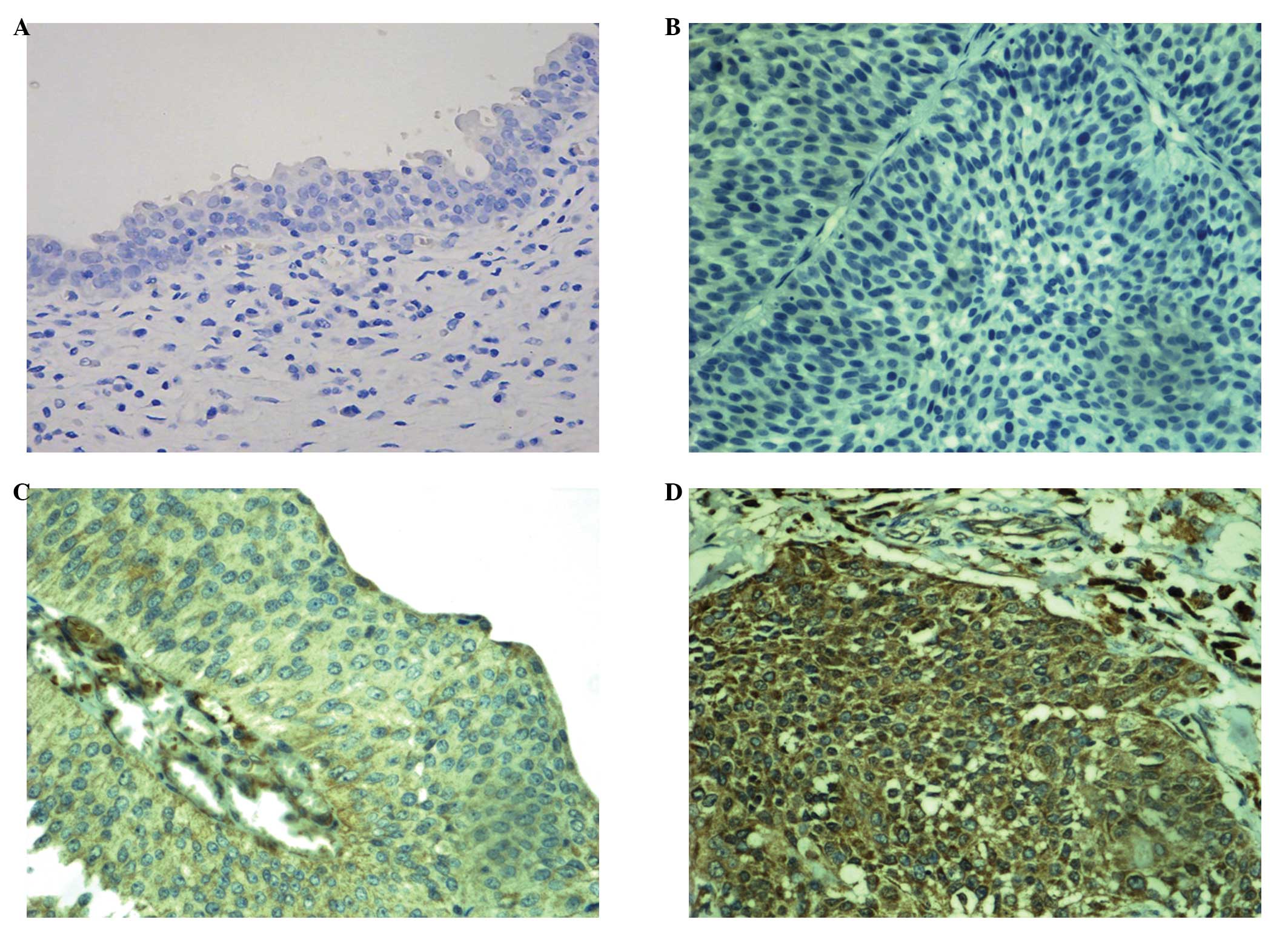
B7-H4 was not detected in the normal bladder tissue
samples. In the bladder urothelial carcinoma samples, the positive
rate of B7-H4 expression was 49.0% (24/49). Both weak (low-grade)
and strong (high-grade) immunostaining intensity was demonstrated
(Fig. 1). B7-H4 expression was
clearly associated with clinical stage and pathological grade, as
the positive rate of B7-H4 expression in patients at stage
T2–T4 was significantly higher than that in
patients at stage Ta–T1 (P<0.05).
Furthermore, the positive rate of B7-H4 expression in high-grade
cases was significantly higher than that in low-grade cases
(P<0.05). Although the positive expression rate in the
infiltration group was higher than that in the non-infiltration
group, the difference was not statistically significant
(P>0.05). No significant differences (P>0.05) were detected
between B7-H4 expression and the other pathological parameters
(Table II).
| Table IIAssociations between B7-H4 expression
and clinicopathological factors. |
Table II
Associations between B7-H4 expression
and clinicopathological factors.
| Clinicopathological
factors | Positive, n=24 | Negative, n=25 | Positive rate
(%) | χ2 | P-value |
|---|
| Gender |
| Male | 21 | 21 | 50.0 | 0.123 | 0.726 |
| Female | 3 | 4 | 42.9 | | |
| Age, years |
| <60 | 9 | 8 | 52.9 | 0.163 | 0.686 |
| ≥60 | 15 | 17 | 46.9 | | |
| TNM stage |
|
T0–T1 | 11 | 21 | 34.4 | 7.873 | 0.005 |
|
T2–T4 | 13 | 4 | 76.5 | | |
| Histological
gradea |
| Low-grade | 3 | 12 | 20.0 | 7.265 | 0.007 |
| High-grade | 21 | 13 | 61.8 | | |
| Tumor status |
| Initial group | 14 | 16 | 46.7 | 0.166 | 0.684 |
| Recurrent
group | 10 | 9 | 52.6 | | |
| Infiltration
degree |
| Non-infiltration
(Ta) | 5 | 11 | 31.3 | 2.988 | 0.084 |
| Infiltration | 19 | 14 | 57.6 | | |
ELISA
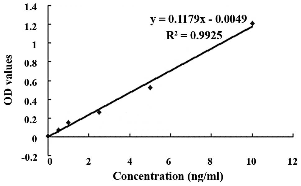
A standard curve was generated with standard B7-H4
concentrations (0, 0.5, 1.0, 2.5, 5.0 and 10.0 ng/ml) as the
abscissae and the corresponding OD values as the ordinates. This
was used to produce an equation: Y = 0.1179X − 0.0049,
R2=0.9925, to determine the sB7-H4 concentrations in
serum from the case and control patients (Fig. 2). The sB7-H4 concentrations in the
case group were significantly increased compared with those of the
healthy control group (P<0.05; Table III). Analysis of the differences
between groups with different urothelial carcinoma histological
grades revealed that patients with high-grade histology exhibited
significantly higher sB7-H4 concentrations than patients with
low-grade histology (P<0.05; Table
IV). However, no statistically significant differences in
sB7-H4 concentration were detected between groups classified by
other factors.
| Table IIIsB7-H4 concentrations (ng/ml) in the
case and control groups. |
Table III
sB7-H4 concentrations (ng/ml) in the
case and control groups.
| Group | n | Mean ± SD |
|---|
| Control | 45 | 1.664±1.316 |
| Case | 45 | 2.561±1.965a |
| Table IVsB7-H4 concentrations (ng/ml) in low-
and high-grade bladder urothelial carcioma groups. |
Table IV
sB7-H4 concentrations (ng/ml) in low-
and high-grade bladder urothelial carcioma groups.
| Histological
grade | n | Mean ± SD |
|---|
| Low | 34 | 2.178±1.881 |
| High | 11 | 3.745±1.811a |
Part two
Transformation of T lymphocytes
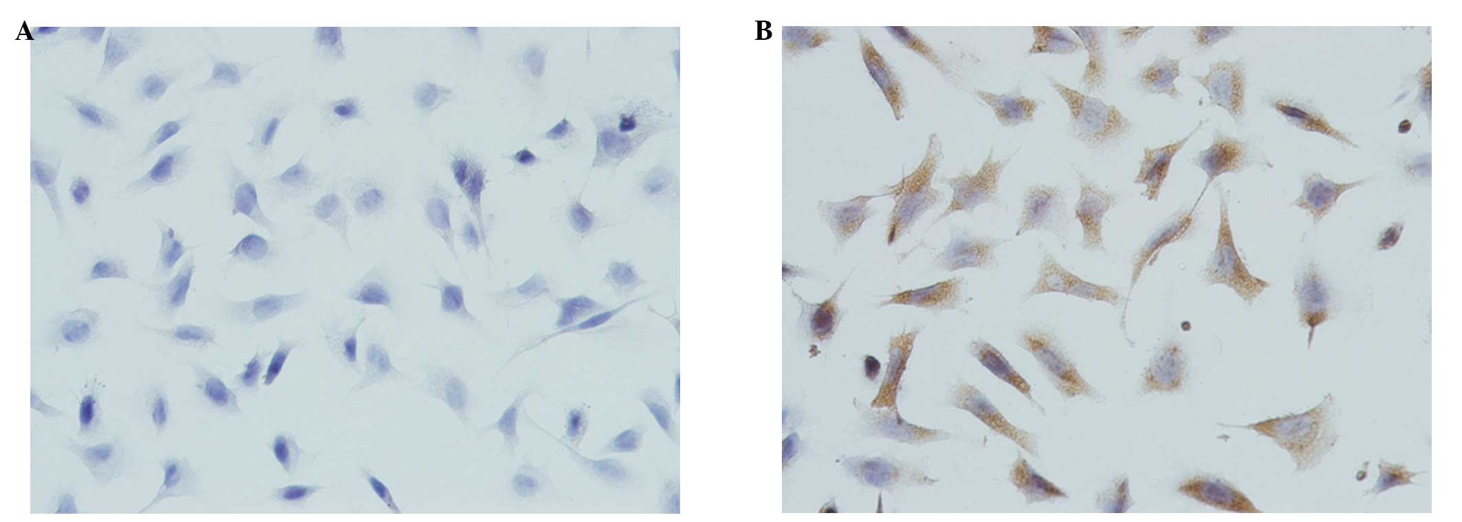
Immunocytochemical analysis revealed positive B7-H4
expression in the BIU-87 cells incubated with B7-H4 mAbs, and
negative expression in the control BIU-87 cells incubated with PBS
instead of primary antibody (Fig.
3). The SI results revealed that optimal lymphocyte
proliferative activity occurred when the cell density was set at
2×10⁶ cells/ml and the ConA concentration was set at 4
μg/ml (P<0.05) when compared with the other ConA concentrations
(1, 2 and 8 μg/ml). Thus, lymphocytes at a density of
2×10⁶ cells/ml were used as effector cells.
E-rosette test
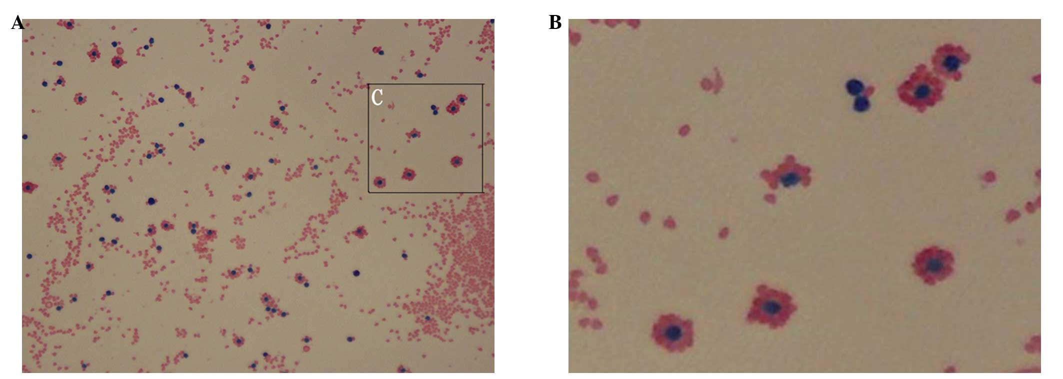
For the identification of T lymphocytes, Wright’s
staining method was used. SRBCs were stained red and lymphocytes
were stained blue. ‘Rosette cells’, with one lymphocyte surrounded
by ≥3 SRBCs were identified. The blue cells at the centers of these
rosette cells were demonstrated to be mature T lymphocytes
(Fig. 4). The calculation formula
for the rosette formation rate was as follows: Rosette formation
rate (%)= bound T lymphocytes (n)/[bound + unbound T lymphocytes
(n)]. The results demonstrated that activated T-lymphocyte purity
reached 80%, which was similar to the purity previously reported in
the literature (26).
Cytotoxicity assay
To examine activated T-lymphocyte cytotoxicity
against BIU-87 human bladder urothelial carcinoma cells following
blockade of the B7-H4 signaling pathway in vitro, control
and B7-H4 mAB-stimulated BIU-87 cells were mixed with the activated
T lymphocytes at different ratios. When the mouse anti-human B7-H4
mAb concentration used to block B7-H4 activity in the BIU-87 cells
was 10 μg/ml, cytotoxicity was significantly greater than that in
the normal BIU-87 cells (P<0.01 or P<0.05). The cytotoxic
effect was significantly enhanced and the T-lymphocyte
concentration was significantly increased following blockade
(P<0.01 and P<0.05; Table
V).
| Table VEffects of activated T-lymphocyte
cytotoxicity against BIU-87 human bladder urothelial carcinoma
cells following blockade of B7-H4 activity in vitro (%, mean
± SD). |
Table V
Effects of activated T-lymphocyte
cytotoxicity against BIU-87 human bladder urothelial carcinoma
cells following blockade of B7-H4 activity in vitro (%, mean
± SD).
| Density ratio
(effector:target cells) |
|---|
|
|
|---|
| Group | 30:1 | 20:1 | 10:1 |
|---|
| Control | 0.6621±0.0653 | 0.5402±0.0557 | 0.3341±0.0385 |
| Blocked |
0.7987±0.0717a,c |
0.6672±0.0454b,d |
0.4292±0.0634b |
Discussion
In part one, the study focused on B7-H4 expression
in 49 cases of human bladder urothelial carcinoma tissues and the
adjacent tissues. A total of 24 cases (49.0%) were found to exhibit
positive B7-H4 expression, whereas the adjacent bladder tissue did
not express B7-H4. In the cancer tissue specimens, B7-H4 was
expressed and located in the cytoplasm and plasma membranes of the
cancer cells, a finding consistent with studies examining B7-H4
expression in non-small-cell lung cancer (11) and breast cancer tissues (13). However, in renal clear cell
carcinoma, B7-H4 was shown to be expressed only on the cell
membrane (12). In the present
study, B7-H4 expression was shown to be closely associated with
increased TNM stage and histological grade in the bladder
urothelial carcinoma samples, as the positive expression rate in
the myometrial invasion group was significantly higher than that of
the non-muscle-invasive group, and the positive expression rate in
the high-grade group was significantly higher than that in the
low-grade group.
Bladder urothelial carcinoma cells may inhibit
T-cell activity and induce apoptosis in tumor antigen-specific
cells due to B7-H4 combining with the corresponding T-cell surface
receptor. These mechanisms may cause bladder tumor immune escape to
occur, and contribute to bladder urothelial carcinoma development
and progression. Thus, the detection of B7-H4 protein may reflect
the degree of malignancy, and the risk of recurrence and
progression in bladder urothelial carcinoma. One study by the
European Organization for Research and Treatment of Cancer observed
that the morphological and structural diversity of the tumor, tumor
size and short-term recurrence of BCa were important prognostic
factors for recurrence or secondary recurrence (1). Important prognostic factors for tumor
progression included tumor grade, stage and carcinoma in
situ, and cases with a history of recurrence and/or high-grade
histology were more likely to relapse and progress. In the present
study, no significant differences were identified between the rates
of B7-H4 positive expression in newly diagnosed and recurrent BCa
groups; however, B7-H4 expression was closely associated with TNM
stage and histological grade. Therefore, blocking B7-H4 protein
activity in bladder tumor tissues may become an effective
immunotherapeutic method to inhibit stage and grade progression in
bladder urothelial carcinoma.
B7-H4 protein is also present in the blood and body
fluids in a soluble form, sB7-H4. Tumor cells secrete sB7-H4, which
restrains T-cell proliferation through blocking the cell cycle at
the G0/G1 phase and further inhibits the T
cell immune response by inducing T cell apoptosis (21,24).
sB7-H4 levels detected in several genitourinary system tumors have
been shown to have important clinical significance at diagnosis,
and stage and prognosis assessment. Simon et al (10) found that sB7-H4 levels in serum and
ascites samples from ovarian cancer patients were significantly
higher than those in normal and benign lesions of the reproductive
system. Another study (26) found
that the combined detection of sB7-H4 and CA125 significantly
improved the diagnostic yield for ovarian cancer. Thompson et
al (24) found that the
sB7-H4-positive rate and the average sB7-H4 expression levels in
renal cell cancer (RCC) patients were significantly higher than
those in normal individuals, and were associated with the
development of lymph node and distant metastases. The authors
hypothesized that sB7-H4 may become a novel serum marker in RCC
diagnosis, and that sB7-H4 detection has significant value in the
prediction of tumor stage and prognosis. The present study found
that sB7-H4 concentrations in patients with bladder urothelial
carcinoma were significantly higher than those of normal
individuals. The sB7-H4 concentrations in cases with high-grade
histology were significantly higher than those in the low-grade
cases, but no significant differences in expression rates were
identified between gender, age, TNM stage, tumor size or the
initial issuance/relapse status groups. These results suggest that
sB7-H4 may be involved in the development of bladder urothelial
carcinoma, and that the detection of serum sB7-H4 concentrations
may provide certain value in the diagnosis and pathological grade
assessment of bladder urothelial cancer.
Host T lymphocytes are activated and proliferate
under the effects of polyclonal activators, anti-CD3 antibody and
antigen-MHC peptide complexes. T lymphocytes are involved in the
host adaptive immune response only when differentiated into
functional T cells (27,28). High initial T cell clonal expansion
may produce a large number of effective CD4+ and
CD8+ T cells, which is conducive to the rapid
elimination of pathogens and the formation of memory T cells. ConA
is a common polyclonal T cell activator, which activates T
lymphocyte proliferation by acting on the TCR-CD3 complex on the T
cell membrane (29). PBMCs include
T cells, B cells and monocytes. However, the largest proportion of
T cells are in PBMC, monocytes may proliferate and differentiate
into macrophages in vitro without stimulation. Due to the
adherent growth of monocytes and the suspended growth of
lymphocytes, in the present study, the adherent separation method
was used to remove mononuclear cells, to reduce background
interference and increase the reliability of the CCK-8
analysis.
B7-H4 expression in the bladder urothelial carcinoma
samples was demonstrated in part one of the present study. In order
to further analyze the association between B7-H4 and bladder
urothelial carcinoma immune escape mechanisms, cellular level
experiments in vitro were conducted. The results revealed
B7-H4 expression in BIU-87 BCa cells. High-purity activated T
lymphocytes were obtained; following B7-H4 antigen blockade in the
BIU-87 cells, the cytotoxic activity of these activated T cells
against BIU-87 cells was significantly enhanced compared with that
against normal BIU-87 cells, in a T-cell density-dependent and
blocking antibody dose-dependent manner. The results indicate that
the cytotoxic activity of the activated T cells against the BIU-87
cells was significantly enhanced following B7-H4 blockade. The
B7-H4-mediated immune escape effect in BCa was reversed when this
tumor antigen binding the T cell surface corresponding receptor was
blocked. However, Miyatake et al (25) hypothesized that B7-H4 tumor cells
mediate immune escape through inhibiting T-cell chemotaxis to the
tumor tissue, as B7-H4 expression intensity in breast cancer was
found to be inversely proportional to the number of infiltrating
CD3+ and CD8+ T cells. Therefore, altering
B7-H4 protein expression in bladder urothelial carcinoma cells may
enhance T-cell cytotoxicity to the cancer cells, and also promote
and maintain a functional T cell immune response; thus the rate of
BCa recurrence and progression may be reduced.
In conclusion, the results of the present study
revealed that B7-H4 was upregulated in bladder urothelial carcinoma
tissues and serum samples from patients, and was closely associated
with TNM stage and histological grade. Therefore, B7-H4 may be
involved in the occurrence and development of BCa, and may be
important in bladder urothelial carcinoma immune escape. B7-H4
expression in tissue and serum was analyzed, along with the
clinical significance. Therefore, simulations of the body
environment and the construction of relevant animal models may be
required in further studies.
Acknowledgements
The authors would like to thank all the medical
staff at the Department of Urology, Union Hospital of Fujian
Medical University, who aided in the collection of the tissue and
serum samples, and all other individuals involved in the study.
References
|
1
|
van Rhijn BW, Burger M, Lotan Y, et al:
Recurrence and progression of disease in non-muscle-invasive
bladder cancer: from epidemiology to treatment strategy. Eur Urol.
56:430–442. 2009.
|
|
2
|
Parkin MD, Bray F, Ferlay J and Pisani P:
Global cancer statistics, 2002. CA Cancer J Clin. 55:74–108.
2005.
|
|
3
|
Shin T, Kennedy G, Gorski K, et al:
Cooperative B7-1/2 (CD80/CD86) and B7-DC costimulation of
CD4+ T cells independent of the PD-1 receptor. J Exp
Med. 198:31–38. 2003.
|
|
4
|
Hsu FJ and Komarovskaya M: CTLA4 blockade
maximizes antitumor T-cell activation by dendritic cells presenting
idiotype protein or opsonized anti-CD20 antibody-coated lymphoma
cells. J Immunother. 25:455–468. 2002.
|
|
5
|
Nagamori M, Kawaguchi S, Murakami M, et
al: Intrinsic and extrinsic manipulation of B7/CTLA-4 interaction
for induction of anti-tumor immunity against osteosarcoma cells.
Anticancer Res. 22:3223–3227. 2002.
|
|
6
|
Liu X, Gao JX, Wen J, et al: B7DC/PDL2
promotes tumor immunity by a PD-1-independent mechanism. J Exp Med.
197:1721–1730. 2003.
|
|
7
|
Sica GL, Choi IH, Zhu G, et al: B7-H4, a
molecule of the B7 family, negatively regulates T cell immunity.
Immunity. 18:849–861. 2003.
|
|
8
|
Choi IH, Zhu G, Sica GL, et al: Genomic
organization and expression analysis of B7-H4, an immune inhibitory
molecule of the B7 family. J Immunol. 171:4650–4654. 2003.
|
|
9
|
Mao YX, Chen YJ, Ge Y, et al: Recombinant
human B7-H4 expressed in Escherichia coli inhibits T lymphocyte
proliferation and IL-2 secretion in vitro. Acta Pharmaco1 Sin.
27:741–746. 2006.
|
|
10
|
Simon I, Zhuo S, Corra1 L, et al: B7-H4 is
a novel membrane-bound protein and a candidate serum and tissue
biomarker for ovarian cancer. Cancer Res. 66:1570–1575. 2006.
|
|
11
|
Sun Y, Wang Y, Zhao J, et al: B7-H3 and
B7-H4 expression in non-small-cell lung cancer. Lung Cancer.
53:143–151. 2006.
|
|
12
|
Krambeck AE, Thompson RH, Dong H, et al:
B7-H4 expression in renal cell carcinoma and tumor vasculature:
associations with cancer progression and survival. Proc Natl Acad
Sci USA. 103:10391–10396. 2006.
|
|
13
|
Tringler B, Zhuo S, Pilkington G, et al:
B7-H4 is highly expressed in ductal and lobular breast cancer. Cli
Cancer Res. 11:1842–1848. 2005.
|
|
14
|
Zang X, Thompson RH, Al-Ahmadie HA, et al:
B7-H3 and B7x are highly expressed in human prostate cancer and
associated with disease spread and poor outcome. Proc Natl Acad Sci
USA. 104:19458–19463. 2007.
|
|
15
|
Kryczek I, Wei S, Zou L, et al: Cutting
edge: induction of B7-H4 on APCs through IL-10: novel suppressive
mode for regulatory T cells. J Immunol. 177:40–44. 2006.
|
|
16
|
Suh WK, Wang S, Duncan GS, et al:
Generation and characterization of B7-H4/B7Sl/B7x-deficient mice.
Mol Cell Biol. 26:6403–6411. 2006.
|
|
17
|
Margel D, Tal R, Golan S, et al: Long-term
follow-up of patients with Stage T1 high-grade transitional cell
carcinoma managed by Bacille Calmette-Guérin immunotherapy.
Urology. 69:78–82. 2007.
|
|
18
|
Porena M, Del Zingaro M, Lazzeri M, et al:
Bacillus Calmette-Guérin versus gemcitabine for intravesical
therapy in high-risk superficial bladder cancer: a randomised
prospective study. Urol Int. 84:23–27. 2010.
|
|
19
|
Hinotsu S, Akaza H, Naito S, et al:
Maintenance therapy with bacillus Calmette-Guérin Connaught strain
clearly prolongs recurrence-free survival following transurethral
resection of bladder tumor for non-muscle-invasive bladder cancer.
BJU Int. 108:187–195. 2011.
|
|
20
|
Järvinen R, Kaasinen E, Sankila A and
Rintala E; FinnBladder Group. Long-term efficacy of maintenance
bacillus Calmette-Guérin versus maintenance mitomycin C
instillation therapy in frequently recurrent TaT1 tumours without
carcinoma in situ: a subgroup analysis of the prospective,
randomised FinnBladder I study with a 20-year follow-up. Eur Urol.
56:260–265. 2009.
|
|
21
|
Duchek M, Johansson R, Jahnson S, et al:
Members of the Urothelial Cancer Group of the Nordic Association of
Urology: Bacillus Calmette-Guérin is superior to a combination of
epirubicin and interferon-alpha2b in the intravesical treatment of
patients with stage T1 urinary bladder cancer. A prospective,
randomized, Nordic study. Eur Urol. 57:25–31. 2010.
|
|
22
|
Sobin LH, Gospodariwicz M and Wittekind C:
TNM classification of malignant tumors. UICC International Union
Against Cancer. 7th edition. Wiley-Blackwell; Hoboken, USA: pp.
262–265. 2009
|
|
23
|
Sauter G, Algaba F, Amin M, et al: Tumour
of urinary system: non-invasive urothelial neoplasis. WHO
Classification of Tumours Pathology and Genetics of Tumors of the
Urinary System and Male Genital Organs. Eble JN, Sauter G, Epstein
JI and Sesterhenn IA: IARCC Press; Lyon: 2004
|
|
24
|
Thompson RH, Zang X, Lohse CM, et al:
Serum-soluble B7x is elevated in renal cell carcinoma patients and
is associated with advanced stage. Cancer Res. 68:6054–6058.
2008.
|
|
25
|
Miyatake T, Tringler B, Liu W, et al:
B7-H4 (DD-0110) is overexpressed in high risk uterine endometrioid
adenocarcinomas and inversely correlated with tumor T-cell
infiltration. Gynecol Oncol. 106:119–127. 2007.
|
|
26
|
Simon I, Katsaros D, Rigault de la
Longrais I, et al: B7-H4 is over-expressed in early-stage ovarian
cancer and is independent of CA125 expression. Gynecol Oncol.
106:334–341. 2007.
|
|
27
|
Acuto O and Michel F: CD28-mediated
co-stimulation: a quantitative support for TCR signaling. Nat Rev
Immunol. 3:939–951. 2003.
|
|
28
|
Alegre ML, Frauwirth KA and Thompson CB:
T-cell regulation by CD28 and CTLA-4. Nat Rev Immunol. 1:220–228.
2001.
|
|
29
|
Jiang J, Gross D, Elbaum P and Murasko DM:
Aging affects initiation and continuation of T cell proliferation.
Mech Ageing Dev. 128:332–339. 2007.
|