Introduction
Central neurocytoma is a rare grade II tumor of
neuronal origin, according to the World Health Organization staging
system (1). Young adults are most
commonly affected, with a similar incidence in males and females.
The tumors usually occur in the ventricular system of the brain
(2). Extraventricular neurocytomas
are extremely rare. Since the first case was reported by Hassoun
et al in 1982 (3), 271
studies regarding ventricular neurocytomas have been published,
while only 64 studies regarding extraventricular neurocytoma have
been published. By July 2012, extraventricular neurocytoma had been
reported in the cerebrum, including the frontal (4), temporal (5,6),
parietal (7) and occipital
(7) lobes, the pons (8), the skull base (9,10), the
vermis of the cerebellum (11), the
cerebellum (12), the sellar region
(13), the cauda equina (14), the thalamus (15) and the spinal cord (16–25).
To date, 17 cases of neurocytoma involving the spinal cord have
been reported; nine cases located in the cervical spinal cord
(16,17,19,21–23,25)
and eight cases located in the thoracic spinal cord (18,20,22,23).
The majority of spinal cord neurocytomas do not recur following
complete resection and radiation therapy. In the current study, two
cases of intramedullary neurocytomas in the craniocervical spinal
cord are reported and the clinical features, radiological
observations, histopathological presentation and two-year follow-up
results are presented, together with a review of the literature.
Written informed consent was obtained from both patients.
Case report
Case one
A 26-year-old male presented to Yuquan Hospital,
Tsinghua University (Beijing, China) with a nine-month history of
numbness in the right lower limb and a five-month history of
progressive weakness of the left upper limb. The patient’s general
health was good and no relevant family history was reported. Upon
neurological examination, muscle power in the left upper limb was
rated as grade 4/5, according to the Medical Research Council scale
(26), with decreased pinprick
sensation in the sole of the right foot. Myoatrophy was identified
in the right lower limb. A physical examination of the spine did
not reveal any abnormalities and perineal sensation was normal.
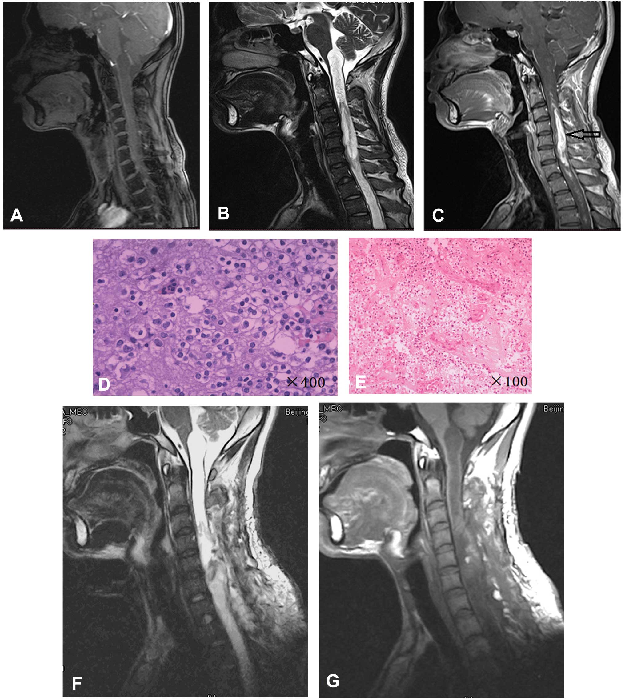
A magnetic resonance imaging (MRI) scan of the
craniocervical region revealed an expansile intramedullary mass
extending from the medulla oblongata to the T4 segment of the
spine. The mass was isointense on T1-weighted images, hyperintense
with partially cystic mass on T2-weighted images and showed intense
heterogeneous enhancement of solid tumor following the injection of
gadolinium diethylenetriamine pentaacetic acid (Gd-DTPA) (Fig. 1A–C).
A C3-7 laminectomy was performed and an
intramedullary solid mass extending from C3 to C7 was exposed. The
tumor was gray-purple and exhibited features of infiltrative
growth. A partial tumor resection was performed using a
micro-neurosurgery technique, resulting in 85% of the tumor being
resected. Hematoxylin and eosin staining revealed a neoplasm
composed of uniform, round cells (Fig.
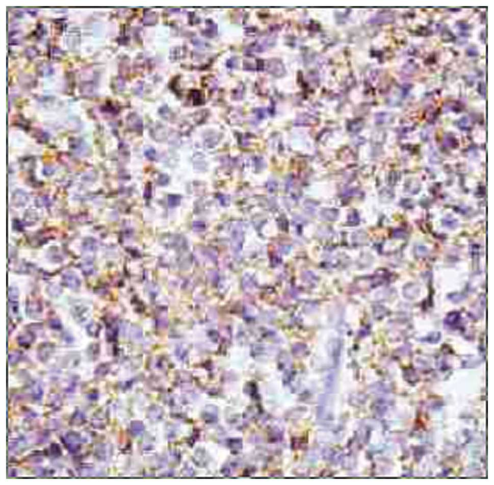
1D and E). Immunohistochemical staining revealed positivity for
glial fibrillary acidic protein (GFAP), neuronal nuclear antigen,
vimentin, neuron-specific enolase (NSE), S-100 protein,
synaptophysin (SYN; Fig. 2) and
oligo2. Post-operative radiotherapy (56 Gy) was administered for
three months. Two years after surgery, the patient’s symptoms were
in remission and post-operative MRI revealed no tumor recurrence
(Fig. 1F and G).
Case two
A 48-year-old female presented to Yuaquan Hospital,
Tsinghua University, with a seven-year history of pain in the right
lower limb and a five-year history of progressive numbness in all
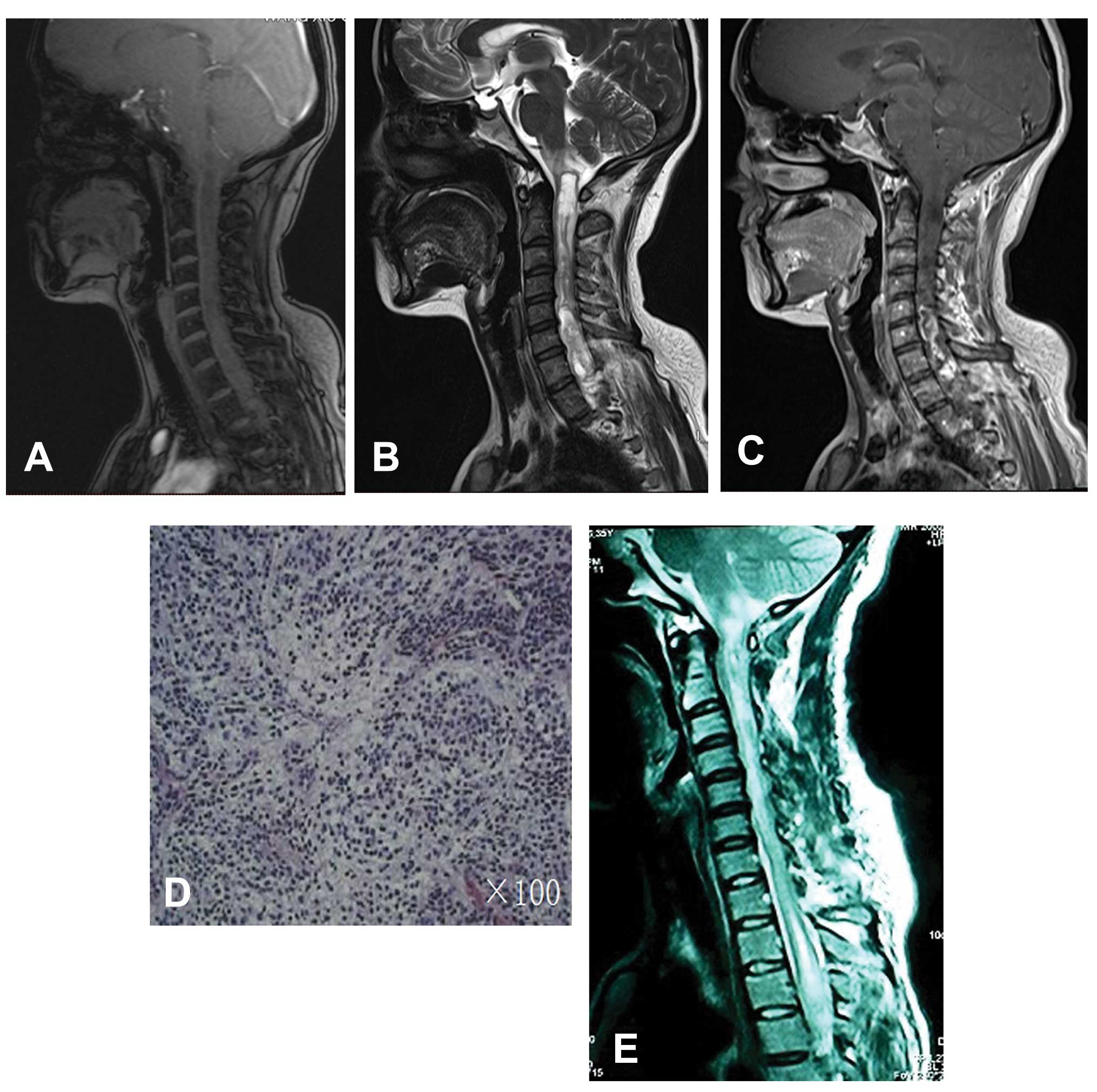
four limbs and lumbar zone anesthesia. MRI revealed a mass
extending from the medulla oblongata to the T2 level, with an
isointense T1-weighted signal and a hyperintense T2-weighted signal
with a partially cystic mass. Intense heterogeneous enhancement of
a solid tumor was shown following injection of Gd-DTPA (Fig. 3A–C). The tumor capsule could not be
clearly observed around the whole of the tumor, so a subtotal
resection of the mass was performed. Hematoxylin and eosin staining
revealed a lesion composed of uniformly small, round cells
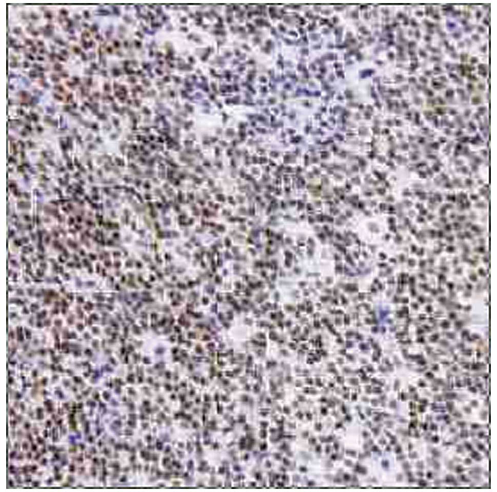
(Fig. 3D). Immunohistochemical
staining revealed positivity for GFAP, NSE, vimentin and SYN
(Fig. 4). Post-operative
radiotherapy (56 Gy) was administered for three months and no
deterioration was identified. Following radiotherapy, residual
tumor was observed by MRI, however, the patient’s condition had not
deteriorated on a follow-up MRI performed after two years (Fig. 3E).
Discussion
Neurocytomas are tumors of the central nervous
system that are derived from the neuronal precursor cells
surrounding the central canal region in the developing fetus
(2). The spinal cord is an
extremely rare site for extraventricular neurocytomas. To date, the
majority of cases of spinal neurocytomas cited in the literature
have occurred in the cervico-thoracic region. Neurocytomas
occurring in the cervical region of the spinal cord region were
first documented in 1994 (16).
Since then, only eight cases have been reported in the cervical
spinal cord (Table I). In the
present study, two cases of neurocytoma occurring in the
craniocervical region were reported. These are the first cases to
be reported in China.
 | Table ISummary of the literature review of
neurocytomas in the cervical spinal cord. |
Table I
Summary of the literature review of
neurocytomas in the cervical spinal cord.
| First author, year
(ref.) | Age, years | Location | MRI enhancement | Surgery | Radiotherapy | Recurrence (follow-up
time) |
|---|
| Tatter et al,
1994 (14) | 65 | C2-C6 | Homogenous | Biopsy | Yes | No (10 years) |
| 49 | C3-C4 | Homogenous | Total resection | Yes | Yes (30 months) |
| Stapleton et
al, 1997 (15) | 12 | C4-T1 | Heterogeneous | Total resection | No | No (24 months) |
| Ashkan et al,
2000 (20) | 12 | C6-T1 | Homogenous | Subtotal
resection | No | No (33 months) |
| Sharma et al,
2006 (1) | 24 | C5-T1 | Homogenous | Total resection | No | Yes (10 months) |
| Gokham, 2008
(20) | 49 | C3-C5 | Homogenous | Subtotal
resection | No | Unknown |
| Polli et al,
2009 (21) | 15 | C1-T11 | Heterogeneous | Subtotal
resection | No | Succumbed |
| 6 | C1-C7 | Heterogeneous | Subtotal
resection | No | No (12 months) |
| Gepp Rde et
al, 2012 (23) | 15 | Cervical spinal
cord | Unknown | Subtotal
resection | No | Unknown |
According to the literature review, six cases of
cervical spinal neurocytomas occurred in young adults while three
cases were reported in adults. In the current study, one patient
was 24 years old and the other was 48 years old. The clinical
presentation of neurocytoma depends on the location and size of the
tumor. Upon neurological examination of case one, the patient was
found to have abnormal sensation and loss of proximal muscle power
in the upper limbs. MRI revealed a mass with low to intermediate
signal intensity on T1-weighted images and intermediate to high
signal intensity on T2-weighted images. These tumors involved
multiple spinal segments and contrast-enhanced MRI revealed either
homogenous or heterogeneous enhancement of the tumor mass. The
tumors did not exhibit any characteristic manifestations and their
pre-operative diagnosis was relatively difficult, which is typical
of spinal neurocytoma. The differential diagnosis of spinal
neurocytomas include ependymoma and oligodendroglioma (19).
Neurocytomas are benign and slow growing, and the
majority of patients with ventricular neurocytomas may be treated
effectively by surgery. However, total resection of spinal cord
neurocytomas is relatively difficult, particularly in tumors
involving the upper cervical spinal cord and in those involving
multiple segments. Giant cervical cord neurocytomas may be treated
with subtotal resection followed by radiotherapy, however, in the
present literature review, only the two cases reported by Tatter
et al (16) received
radiotherapy. By contrast, Stapleton et al (17) reported that post-operative
radiotherapy should be avoided, and the remaining six cases did not
receive radiotherapy. Of these six cases, one patient succumbed to
the tumor and one patient presented with recurrence during
follow-up (19,21–23,25).
In the present study, for each case, the tumors involved the
craniocervical region and were difficult to remove intact. As a
consequence, partial and subtotal resections were performed in case
one and case two, respectively. Following surgery, the two patients
received radiotherapy for three months and no tumor recurrence was
observed at the end of the two-year follow-up period. Overall, the
number of reported cases of upper cervical neurocytoma is so small
that it is unclear whether post-operative radiotherapy is
beneficial in preventing tumor recurrence. However, the present
study indicates that radiotherapy for a period of three months
following surgical resection may prove useful in the prevention of
tumor recurrence.
In the present study, the cases of two patients with
craniocervical neurocytomas were reported, including the clinical
presentation, radiological observations and histopathological
features. The successful treatment of neurocytomas may be dependent
on an early diagnosis and the total surgical resection of the
tumors, however, it is unclear whether post-operative radiotherapy
is required to prevent tumor recurrence. Although craniocervical
neurocytomas are rare and difficult to diagnose, they must be
considered as a presurgical differential diagnosis for
craniocervical tumors. In addition, a long-term follow-up period is
required for craniocervical neurocytomas due to the possibility of
local recurrence.
References
|
1
|
Louis DN, Ohgaki H, Wiestler OD, et al:
The 2007 WHO classification of tumours of the central nervous
system. Acta Neuropathol. 114:97–109. 2007. View Article : Google Scholar : PubMed/NCBI
|
|
2
|
Sharma MC, Deb P, Sharma S and Sarkar C:
Neurocytoma: a comprehensive review. Neurosurg Rev. 29:270–285.
2006. View Article : Google Scholar : PubMed/NCBI
|
|
3
|
Hassoun J, Gambarelli D, Grisoli F, et al:
Central neurocytoma. An electon-microscopic study of two cases.
Acta Neuropathol. 56:151–156. 1982. View Article : Google Scholar
|
|
4
|
Harada M, Morioka T, Nishio S and Fukui M:
Neurocytoma in the left frontal lobe. No Shinkei Geka. 19:89–92.
1991.PubMed/NCBI
|
|
5
|
Treier M, Doostkam S and Meckel S:
Extraventricular neurocytoma: a rare cause of temporal lobe
epilepsy. Rofo. 183:1065–1066. 2011. View Article : Google Scholar : PubMed/NCBI
|
|
6
|
Giulioni M, Martinoni M, Rubboli G, et al:
Temporo-mesial extraventricular neurocytoma and cortical dysplasia
in focal temporal lobe epilepsy. J Clin Neurosci. 18:147–148. 2011.
View Article : Google Scholar
|
|
7
|
Liebert W, Szymas J, Majewski T and
Paprzycki W: Central neurocytoma of the right parietal and
occipital lobe, Case report. Neurol Neurochir Pol. 32:191–199.
1998.PubMed/NCBI
|
|
8
|
Swinson BM, Friedman WA and Yachnis AT:
Pontine atypical neurocytoma: case report. Neurosurgery.
58:E9902006. View Article : Google Scholar : PubMed/NCBI
|
|
9
|
Kowalski RJ, Prayson RA and Lee JH: Skull
base neurocytoma: case report and review of the literature of
extraventricular neurocytomas. Skull Base. 12:59–65. 2002.
View Article : Google Scholar
|
|
10
|
Shidoh S, Yoshida K, Saitoh K, et al:
Extraaxial neurocytoma in the skull base. Brain Tumor Pathol.
28:273–277. 2011. View Article : Google Scholar : PubMed/NCBI
|
|
11
|
Kapoor N, Gandhi A and Chaurasia AK:
Central neurocytoma in the vermis of the cerebellum. Indian J
Pathol Microbiol. 52:108–109. 2009. View Article : Google Scholar : PubMed/NCBI
|
|
12
|
Ogiwara H, Dubner S, Bigio E and Chandler
J: Neurocytoma of the cerebellum. Surg Neurol Int. 2:362011.
View Article : Google Scholar : PubMed/NCBI
|
|
13
|
Wang YY, Kearney T, du Plessis D and
Gnanalingham KK: Extraventricular neurocytoma of the sellar region.
Br J Neurosurg. 26:420–422. 2012. View Article : Google Scholar
|
|
14
|
Stephan CL, Kepes JJ, Arnold P, Green KD
and Chamberlin F: Neurocytoma of the cauda equina. Case report. J
Neurosurg. 90:247–251. 1992.
|
|
15
|
Cheung YK: Central neurocytoma occurring
in the thalamus: CT and MRI findings. Australas Radiol. 40:182–184.
1996. View Article : Google Scholar : PubMed/NCBI
|
|
16
|
Tatter SB, Borges LF and Louis DN: Central
neurocytomas of the cervical spinal cord. Report of two cases. J
Neurosurg. 81:288–293. 1994. View Article : Google Scholar : PubMed/NCBI
|
|
17
|
Stapleton SR, David KM, Harkness WF and
Harding B: Central neurocytoma of the cervical spinal cord. J
Neurol Neurosurg Psychiatry. 63:1191997. View Article : Google Scholar : PubMed/NCBI
|
|
18
|
Martin AJ, Sharr MM and Teddy PJ:
Neurocytoma of the thoracic spinal cord. Acta Neurochir (Wien).
144:823–828. 2002. View Article : Google Scholar
|
|
19
|
Sharma S, Sarkar C, Gaikwad S, Suri A and
Sharma MC: Primary neurocytoma of the spinal cord: a case report
and review of literature. J Neurooncol. 74:47–52. 2005. View Article : Google Scholar : PubMed/NCBI
|
|
20
|
Singh A, Chand K, Singh H, Sarkar C and
Sharma MC: Atypical neurocytoma of the spinal cord in a young
child. Childs Nerv Syst. 23:207–211. 2007. View Article : Google Scholar
|
|
21
|
Gokhan GA, Gurer IE and Akyuz: A case of
extraventricular neurocytoma of the spinal cord. Neuropathology.
28:322–325. 2008. View Article : Google Scholar : PubMed/NCBI
|
|
22
|
Ashkan K, Casey AT and D’Arrigo C: Benign
central neurocytoma. Cancer. 89:1111–1120. 2000. View Article : Google Scholar : PubMed/NCBI
|
|
23
|
Polli FM, Salvati M and Miscusi M:
Neurocytoma of the spinal cord: report of three cases and review of
the literature. Acta Neurochir (Wien). 151:569–574. 2009.
View Article : Google Scholar
|
|
24
|
Tsai CY, Tsai TH and Lin CH: Unusual
exophytic neurocytoma of thoracic spine mimicking meningioma: a
case report and review of the literature. Eur Spine. 20(Suppl 2):
S239–S242. 2011. View Article : Google Scholar
|
|
25
|
de Gepp RA, Sacco RC and Brandão IC:
Central neurocytoma of spinal cord. Arq Neuropsiquiatr. 70:234–235.
2012. View Article : Google Scholar
|
|
26
|
Medical Research Council. Aids to the
examination of the peripheral nervous system. Memorandum no. 45.
Her Majesty’s Stationery Office; London: 1981
|