Introduction
Oral squamous cell carcinoma (OSCC) is the eighth
most prevalent malignant neoplasm worldwide, and mortality rates
are high in the majority of countries, with an overall five-year
survival rate of <50% (1). OSCCs
are usually diagnosed at an advanced stage; however, invasive
diseases are often preceded by the presence of clinically
identifiable potentially malignant lesions, including leukoplakias,
erythroplakias and oral submucous fibrosis (2). Although the presence and severity of
epithelial dysplasia has been associated with transformation into
carcinoma, the mechanisms of progression are poorly understood, and
the transformation rates are extremely variable (2,3).
Previous studies have indicated that specific
alterations in the underlying connective tissue are associated with
tumor development and progression (4,5). Tumor
and stromal cells exchange cytokines, extracellular matrix proteins
and enzymes that promote growth directly via stimulation of
proliferation and survival, as well as invasion via local
proteolysis of the extracellular matrix (6–8).
Notably, previously, an increased density of myofibroblasts, also
termed carcinoma-associated fibroblasts, in the stroma of OSCCs was
found to correlate with lymph node metastasis and higher mortality
(9–12). However, only a small number of
studies have assessed the involvement of the subjadjacent
connective tissue, particularly of myofibroblasts, on malignant
transformation of potentially malignant oral lesions. In the
present study, the density of myofibroblasts in potentially
malignant and malignant lesions of the oral cavity was investigated
to provide novel insights with regard to the involvement of
myofibroblast cells in the development of OSCCs.
Material and methods
Tissue samples
This study included 29 cases of oral fibrous
hyperplasia, 69 oral leukoplakias (of which 24 cases were
histologically classified as mild epithelial dysplasia, 26 as
moderate dysplasia and 19 as severe dysplasia), 90 OSCCs (35
well-differentiated, 39 moderately differentiated and 16 poorly
differentiated) and eight oral verrucous carcinomas. All samples
were obtained from patients aged >45 years. Samples were
obtained from the Department of Oral Diagnosis, School of
Dentistry, State University of Campinas (Piracicaba, Brazil), and
new sections were cut from the paraffin blocks and stained with
hematoxylin and eosin. Oral epithelial dysplasias and OSCCs were
classified according to the World Health Organization grading
system (13). This study was
approved by the ethics committee of the School of Dentistry, State
University of Campinas.
Immunohistochemistry
Immunohistochemical staining was performed on 3-μm
tissue sections using the avidin-biotin-peroxidase complex method.
Briefly, sections were deparaffinized and dehydrated using a graded
series of ethanol. Sections were then subjected to antigen
retrieval with 0.01 M citrate buffer, pH 6.0 in an electric
pressure cooker and incubated with 3% aqueous hydrogen peroxide for
15 min to quench endogenous peroxidase. Next, the sections were
incubated with monoclonal mouse anti-human α-smooth muscle actin
(α-SMA) antibody (1:400; Clone 1A4; Dako, Carpenteria, CA, USA),
followed by the LSAB detection system (Dako). Reactions were
developed by incubating the sections with 0.6 mg/ml
3,3′-diaminobenzidine tetrahydrochloride (Sigma-Aldrich, St. Louis,
MO, USA) containing 0.01% H2O2 and
counterstained with Mayer’s hematoxylin (Sigma-Aldrich). The
exclusion of the primary antibody served as the negative control,
whereas normal blood vessels were used as internal positive
controls. The density of myofibroblasts was classified as negative,
scanty or abundant as described by Kellermann et al
(9), where tumors with 0%
α-SMA-positive cells were classified as negative, if >1–<50%
of the stromal myofibroblasts were α-SMA-positive tumors were
classified as scanty, and abundant if >50% of the stromal cells
were α-SMA-positive cells.
Statistical analysis
Differences in the density of myofibroblasts between
groups were analyzed using cross-tabulation and χ2
tests, and P<0.05 was considered to indicate a statistically
significant difference (GraphPad Prism software, version 5.0;
GraphPad Software, Inc., La Jolla, CA, USA.).
Results
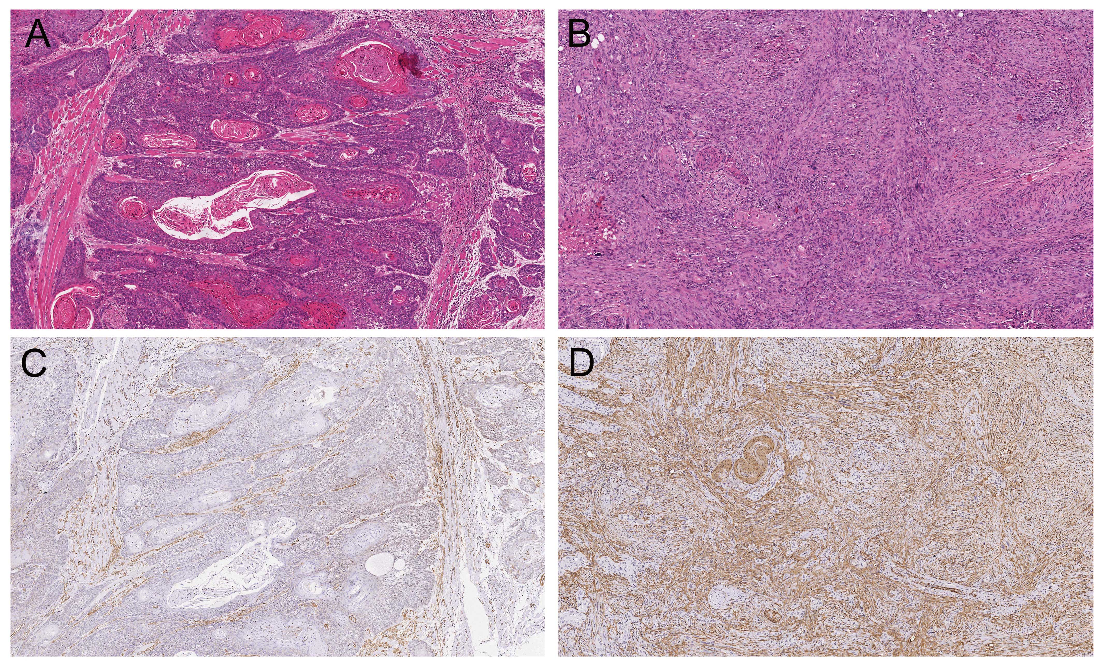
The density of myofibroblasts in the different oral
lesions is shown in Table I.
Myofibroblasts were not detected in oral fibrous hyperplasias or
any of the 69 oral leukoplakias. Oral cancers exhibited positivity
for myofibroblasts in 59.8% of the samples (Fig. 1). In well-differentiated OSCCs, the
presence of myofibroblasts was classified as negative in 17 (48.6%)
cases, scanty in 13 (37.1%) and abundant in five (14.3%). In
moderately differentiated OSCCs nine (23.1%) cases were negative,
17 (43.6%) scanty and 13 (33.3%) abundant; whereas in poorly
differentiated OSCCs, three (18.8%) cases were classified as
negative, four (25.0%) as scanty and nine (56.2%) as abundant
(Table I). In the samples of oral
verrucous carcinomas, the presence of myofibroblasts was classified
as negative in all cases (Table I).
According to the histopathological differentiation, the density of
myofibroblasts was significantly higher in moderately
differentiated and poorly differentiated OSCCs, when compared with
well-differentiated OSCCs (P=0.0402 and P=0.007, respectively). No
significant differences were observed between moderately and poorly
differentiated OSCCs (P=0.271).
 | Table IDensity of myofibroblasts in fibrous
hyperplasias, potentially malignant oral lesions (leukoplakias with
histologically confirmed dysplasia), OSCCs and oral verrucous
carcinomas. |
Table I
Density of myofibroblasts in fibrous
hyperplasias, potentially malignant oral lesions (leukoplakias with
histologically confirmed dysplasia), OSCCs and oral verrucous
carcinomas.
| Density of
myofibroblast |
|---|
|
|
|---|
| Negative, n (%) | Scanty, n (%) | Abundant, n (%) |
|---|
| Fibrous
hyperplasia | 29 (100) | 0 (0) | 0 (0) |
| Leukoplakia with mild
dysplasia | 24 (100) | 0 (0) | 0 (0) |
| Leukoplakia with
moderate dysplasia | 26 (100) | 0 (0) | 0 (0) |
| Leukoplakia with
severe dysplasia | 19 (100) | 0 (0) | 0 (0) |
| Well-differentiated
OSCCa,b | 17 (48.6) | 13 (37.1) | 5 (14.3) |
| Moderately
differentiated OSCCc | 9 (23.1) | 17 (43.6) | 13 (33.3) |
| Poorly differentiated
OSCC | 3 (18.8) | 4 (25.0) | 9 (56.2) |
| Oral verrucous
carcinoma | 8 (100) | 0 (0) | 0 (0) |
Discussion
Although a number of studies have indicated that the
stroma is important in the development and progression of malignant
tumors, the specific mechanisms associated with its activation and
effects on regulation of the tumorigenesis remain unclear (4). One of the most evident processes in
tumor stroma is the acquisition of myofibroblasts. The
transdifferentiation and emergence of myofibroblasts has been
considered a crucial event in tumorigenesis, and is mediated by
growth factors and cytokines released by tumor cells (10,12,14,15).
Myofibroblasts were originally identified in granulation tissues as
modified fibroblasts with prominent rough endoplasmic reticulum and
Golgi apparatus producing collagen, abundant myofilaments
characterized by the presence of α-smooth muscle actin and
fibronexus junctions (16). Later,
it was shown that myofibroblasts may control a number of
physiological and pathological events via the secretion of an
extensive range of cytokines, growth factors, chemokines, hormones,
neurotransmitters, inflammatory mediators, adhesion proteins and
extracellular matrix proteins (7,17).
Numerous studies have detected myofibroblasts in oral cancers
(9–12,18–26);
however, few studies have evaluated myofibroblasts in potentially
malignant oral lesions (9,20,21,24–26)
and in oral verrucous carcinomas (25).
In the present study, myofibroblasts were not found
in oral fibrous hyperplasias or any of the 69 potentially malignant
oral lesions. Previous studies did not identify myofibroblasts in
oral normal mucosa or oral leukoplakias with dysplasia, which
highlights the importance of the molecular crosstalk between
stromal elements and tumor cells during invasion of the connective
tissue for the emergence of myofibroblasts (9,21,26).
However, a small number of studies identified myofibroblasts in
oral dysplasias (20,24,25).
Vered et al (20) analyzed
11 samples classified as mild dysplasia and 12 as moderate/severe
dysplasia by directly counting α-SMA-stained cells, and identified
sparse stromal myofibroblasts in the premalignant lesions with a
mean percentage of ~1%, although the number of positive samples was
not reported. Seifi et al (24) used the same scoring system that was
applied in the present study and identified myofibroblasts in 4/18
(22.2%) epithelial dysplasia samples; however, this study did not
grade the epithelial dysplasias. Chaudhary et al (25) reported that 7/15 (46.6%) cases of
high-risk epithelial dysplasia (moderate and severe) exhibited
myofibroblasts in various intensities, whereas all low risk
dysplasias (hyperplasia and mild) were negative for myofibroblasts.
However, in all three studies (20,24,25)
the immunohistochemical images provided by the authors were not
sufficient to distinguish smooth muscle cells from blood vessels of
myofibroblasts or stromal reaction due to superficially invasive
OSCC. The present study, which had the largest sample size when
compared with previous studies, indicated that myofibroblasts are
not associated with the transformation process of potentially
malignant lesions of the oral cavity.
The present study also demonstrated that
myofibroblasts were frequently identified in the stroma of invasive
OSCCs. However, myofibroblasts were not detected in oral verrucous
carcinoma, which is consistent with the hypothesis that oral
verrucous carcinoma is a form of well-differentiated squamous cell
carcinoma with specific clinical and histological features,
including slow growth and no invasive potential, which is unlikely
to metastasize (31). This
reinforces the hypothesis that interactions between tumor cells and
tumor microenvironment are important for oral carcinogenesis.
Myofibroblasts in OSCCs may exert an active role in disease
progression via autocrine effects on tumor stroma and paracrine
effects on malignant epithelial cells through tumor-stromal
interactions (10,27–29).
The neoplastic changes that occur in the epithelium are followed by
changes in the stroma surrounding tumor cells, which promote the
differentiation of fibroblasts into myofibroblasts (30). Furthermore, a previous study
demonstrated that transforming growth factor-β1, which is released
by OSCC cells, was responsible for oral fibroblast to myofibroblast
transdifferentiation in an experimental model (10). The presence of myofibroblasts was
significantly higher in moderately and poorly differentiated OSCCs
when compared with that of well-differentiated OSCCs. These results
may indicate that the loss of cellular differentiation affects the
number of myofibroblasts in the tumor stroma (22,25).
In conclusion, the results of this study indicated
that myofibroblasts are not associated with potentially malignant
oral lesions; however, moderately and poorly differentiated OSCCs
exhibited a higher density of myofibroblasts when compared with
well-differentiated tumors. The prognostic value of tumor
differentiation in OSCC is unclear and, thus, further studies with
larger patient cohorts are required to confirm the association
between increased myofibroblast density, loss of differentiation
and the biological behavior of tumors.
Acknowledgements
This study was supported by grants from the State of
São Paulo Research Foundation-FAPESP (São Paulo, Brazil), the
National Council for Scientific and Technological Development-CNPq
(Brasília, Brazil) and the Procad/Casadinho-CNPq/CAPES (Brasília,
Brazil). Dr Miguel was supported by the National Council for
Scientific and Technological Development-CNPq (Brasília,
Brazil).
References
|
1
|
Warnakulasuriya S: Living with oral
cancer: Epidemiology with particular reference to prevalence and
life-style changes that influence survival. Oral Oncol. 46:407–410.
2010. View Article : Google Scholar : PubMed/NCBI
|
|
2
|
Napier SS and Speight PM: Natural history
of potentially malignant oral lesions and conditions: an overview
of the literature. J Oral Pathol Med. 37:1–10. 2008. View Article : Google Scholar
|
|
3
|
Hsue SS, Wang WC, Chen CH, et al:
Malignant transformation in 1458 patients with potentially
malignant oral mucosal disorders: a follow-up study based in a
Taiwanese hospital. J Oral Pathol Med. 36:25–29. 2007. View Article : Google Scholar
|
|
4
|
Hanahan D and Coussens LM: Accessories to
the crime: functions of cells recruited to the tumor
microenvironment. Cancer Cell. 21:309–322. 2012. View Article : Google Scholar : PubMed/NCBI
|
|
5
|
Alitalo A and Detmar M: Interaction of
tumor cells and lymphatic vessels in cancer progression. Oncogene.
31:4499–4508. 2012. View Article : Google Scholar
|
|
6
|
De Wever O and Mareel M: Role of tissue
stroma in cancer cell invasion. J Pathol. 200:429–447. 2003.
View Article : Google Scholar : PubMed/NCBI
|
|
7
|
Desmouliere A, Guyot C and Gabbiani G: The
stroma reaction myofibroblasts: a key player in the control of
tumor cell behavior. Int J Dev Biol. 48:509–517. 2004. View Article : Google Scholar
|
|
8
|
De Wever O, Demetter P, Mareel M and
Bracke M: Stromal myofibroblasts are drivers of invasive cancer
growth. Int J Cancer. 123:2229–2238. 2008. View Article : Google Scholar : PubMed/NCBI
|
|
9
|
Kellermann MG, Sobral LM, da Silva SD, et
al: Myofibroblasts in the stroma of oral squamous cell carcinoma
are associated with poor prognosis. Histopathology. 51:849–853.
2007. View Article : Google Scholar : PubMed/NCBI
|
|
10
|
Kellermann MG, Sobral LM, da Silva SD, et
al: Mutual paracrine effects of oral squamous cell carcinoma cells
and normal oral fibroblasts: Induction of fibroblast to
myofibroblast transdifferentiation and modulation of tumor cell
proliferation. Oral Oncol. 44:509–517. 2008. View Article : Google Scholar
|
|
11
|
Bello IO, Vered M, Dayan D, et al:
Cancer-associated fibroblasts, a parameter of the tumor
microenvironment, overcomes carcinoma-associated parameters in the
prognosis of patients with mobile tongue cancer. Oral Oncol.
47:33–38. 2011. View Article : Google Scholar
|
|
12
|
Marsh D, Suchak K, Moutasim KA, et al:
Stromal features are predictive of disease mortality in oral cancer
patients. J Pathol. 223:470–548. 2011. View Article : Google Scholar : PubMed/NCBI
|
|
13
|
Gale N, Pilch BZ, Sidransky D, El-Naggar
AK, Westra W, Califano J, Johnson N and MacDonald DG: Epithelial
precursor lesions (Oral cavity and oropharynx). World Health
Organization Classification of Tumours. Pathology and Genetics of
Head and Neck Tumours. Barnes L, Eveson JW, Reichart P and
Sidransky D: IARC Press; Lyon: pp. 177–179. 2005
|
|
14
|
Baglole CJ, Ray DM, Bernstein SH, et al:
More than structural cells, fibroblasts create and orchestrate the
tumor microenvironment. Immunol Invest. 35:297–325. 2006.
View Article : Google Scholar : PubMed/NCBI
|
|
15
|
Thode C, Jørgensen TG, Dabelsteen E,
Mackenzie I and Dabelsteen S: Significance of myofibroblasts in
oral squamous cell carcinoma. J Oral Pathol Med. 40:201–207. 2011.
View Article : Google Scholar : PubMed/NCBI
|
|
16
|
Gabbiani G, Ryan GB and Majno G: Presence
of modified fibroblasts in granulation tissue and their possible
role in wound contraction. Experientia. 27:549–550. 1971.
View Article : Google Scholar : PubMed/NCBI
|
|
17
|
Powell DW, Adegboyega PA, Di Mari JF and
Mifflin RC: Epithelial cells and their neighbors I. Role of
intestinal myofibroblasts in development, repair, and cancer. Am J
Physiol Gastrointest Liver Physiol. 289:G2–G7. 2005. View Article : Google Scholar : PubMed/NCBI
|
|
18
|
Barth PJ, Schenck ZU, Schweinsberg T,
Ramaswamy A and Moll R: CD34+ fibrocytes, alpha-smooth muscle
antigen positive myofibroblasts and CD117 expression in the stroma
of invasive squamous cell carcinoma of the oral cavity, pharynx,
and larynx. Virchows Arch. 444:231–234. 2004. View Article : Google Scholar : PubMed/NCBI
|
|
19
|
Kojc N, Zidar N, Vodopivec B and Gale N:
Expression of CD34, alpha-smooth muscle actin, and transforming
growth factor beta1 in squamous intraepithelial lesions and
squamous cell carcinoma of the larynx and hypopharynx. Hum Pathol.
36:16–21. 2005. View Article : Google Scholar : PubMed/NCBI
|
|
20
|
Vered M, Allon I, Buchner A and Dayan D:
Stromal myofibroblasts accompany modifications in the epithelial
phenotype of tongue dysplastic and malignant lesions. Cancer
Microenviron. 2:49–57. 2009. View Article : Google Scholar : PubMed/NCBI
|
|
21
|
Etemad-Moghadam S, Khalili M, Tirgary F
and Alaeddini M: Evaluation of myofibroblasts in oral epithelial
dysplasia and squamous cell carcinoma. J Oral Pathol Med.
38:639–643. 2009. View Article : Google Scholar : PubMed/NCBI
|
|
22
|
Kawashiri S, Tanaka A, Noguchi N, et al:
Significance of stromal desmoplasia and myofibroblast appearance at
the invasive front in squamous cell carcinoma of the oral cavity.
Head Neck. 31:1346–1353. 2009. View Article : Google Scholar : PubMed/NCBI
|
|
23
|
Vered M, Dobriyan A, Dayan D, et al:
Tumor-host histopathologic variables, stromal myofibroblasts and
risk score, are significantly associated with recurrent disease in
tongue cancer. Cancer Sci. 101:274–280. 2010. View Article : Google Scholar
|
|
24
|
Seifi S, Shafaei S, Shafigh E, Sahabi SM
and Ghasemi H: Myofibroblast stromal presence and distribution in
squamous epithelial carcinomas, oral dysplasia and hyperkeratosis.
Asian Pac J Cancer Prev. 11:359–364. 2010.PubMed/NCBI
|
|
25
|
Chaudhary M, Gadbail AR, Vidhale G, et al:
Comparison of myofibroblasts expression in oral squamous cell
carcinoma, verrucous carcinoma, high risk epithelial dysplasia, low
risk epithelial dysplasia and normal oral mucosa. Head Neck Pathol.
6:305–313. 2012. View Article : Google Scholar : PubMed/NCBI
|
|
26
|
De-Assis EM, Pimenta LGGS, Costa-e-Silva
E, Souza PEA and Horta MCR: Stromal myofibroblasts in oral
leukoplakia and oral squamous cell carcinoma. Med Oral Patol Oral
Cir Bucal. 17:e733–e738. 2012. View Article : Google Scholar : PubMed/NCBI
|
|
27
|
Lewis MP, Lygoe KA, Nystrom ML, et al:
Tumour-derived TGF-beta1 modulates myofibroblast differentiation
and promotes HGF/SF-dependent invasion of squamous carcinoma cells.
Br J Cancer. 90:822–832. 2004. View Article : Google Scholar : PubMed/NCBI
|
|
28
|
Sobral LM, Bufalino A, Lopes MA, et al:
Myofibroblasts in the stroma of oral cancer promote tumorigenesis
via secretion of activin A. Oral Oncol. 47:840–846. 2011.
View Article : Google Scholar : PubMed/NCBI
|
|
29
|
Hinsley EE, Kumar S, Hunter KD, Whawell SA
and Lambert DW: Endothelin-1 stimulates oral fibroblasts to promote
oral cancer invasion. Life Sci. 91:557–561. 2012. View Article : Google Scholar : PubMed/NCBI
|
|
30
|
Bremnes RM, Dønnem T, Al-Saad S, et al:
The role of tumor stroma in cancer progression and prognosis:
emphasis on carcinoma-associated fibroblasts and non-small cell
lung cancer. J Thorac Oncol. 6:209–217. 2011. View Article : Google Scholar
|
|
31
|
Alkan A, Bulut E, Gunhan O and Ozden B:
Oral verrucous carcinoma: a study of 12 cases. Eur J Dent.
4:202–207. 2010.PubMed/NCBI
|