Introduction
Sarcomas are tumors that rarely occur in the breast,
accounting for 0.5–1% of all breast cancers (1). Sarcomas arise from the stromal tissue
of the mammary gland and if angiosarcoma, which has a more severe
prognosis, is excluded, the course of the disease is not
significantly different compared with tumors arising from other
sites (2). Chondrosarcoma is a rare
type of sarcoma that, to the best of our knowledge, was reported in
only five cases in the literature prior to 2001 (3–6) and 11
cases in the literature between 2002 and 2013 (7–17), and
mainly occurs in females >50 years old. Macroscopically,
chondrosarcoma appears as a whitish-gray, round mass with regular
margins and necrotic, hemorrhagic or cystic areas that is
characterized by rapid growth, although the sarcoma rarely invades
locally or metastasizes to lymph nodes (9–13,18,19).
Chondrosarcoma is microscopically characterized by chondroid
lacunae, in which numerous chondroblasts exhibit hyperchromatic
nuclei and the absence of epithelial areas or other stromal
components, with a high mitotic division rate. There is also an
absence of estrogen, androgen or HER2 receptors, with a strong
expression of the S100 protein, cytokeratin and the membrane
epithelial antigen (1).
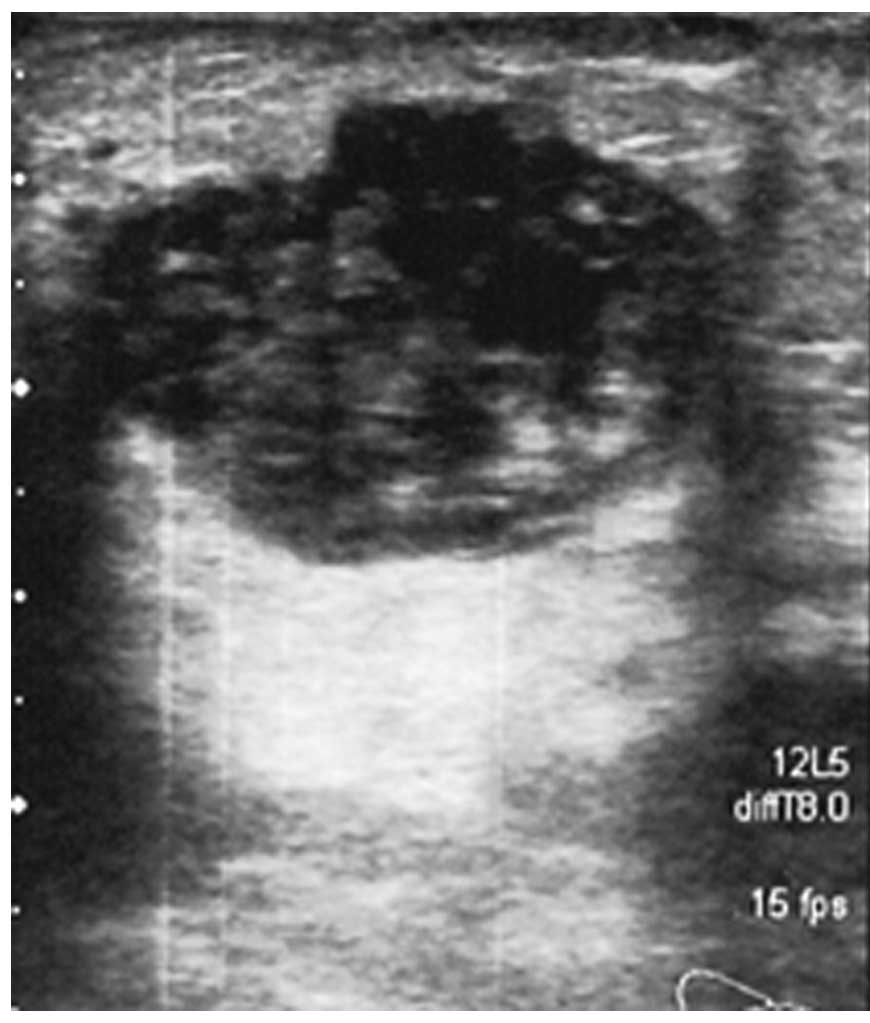
Ultrasonography reveals a hyperechogenic formation, with a
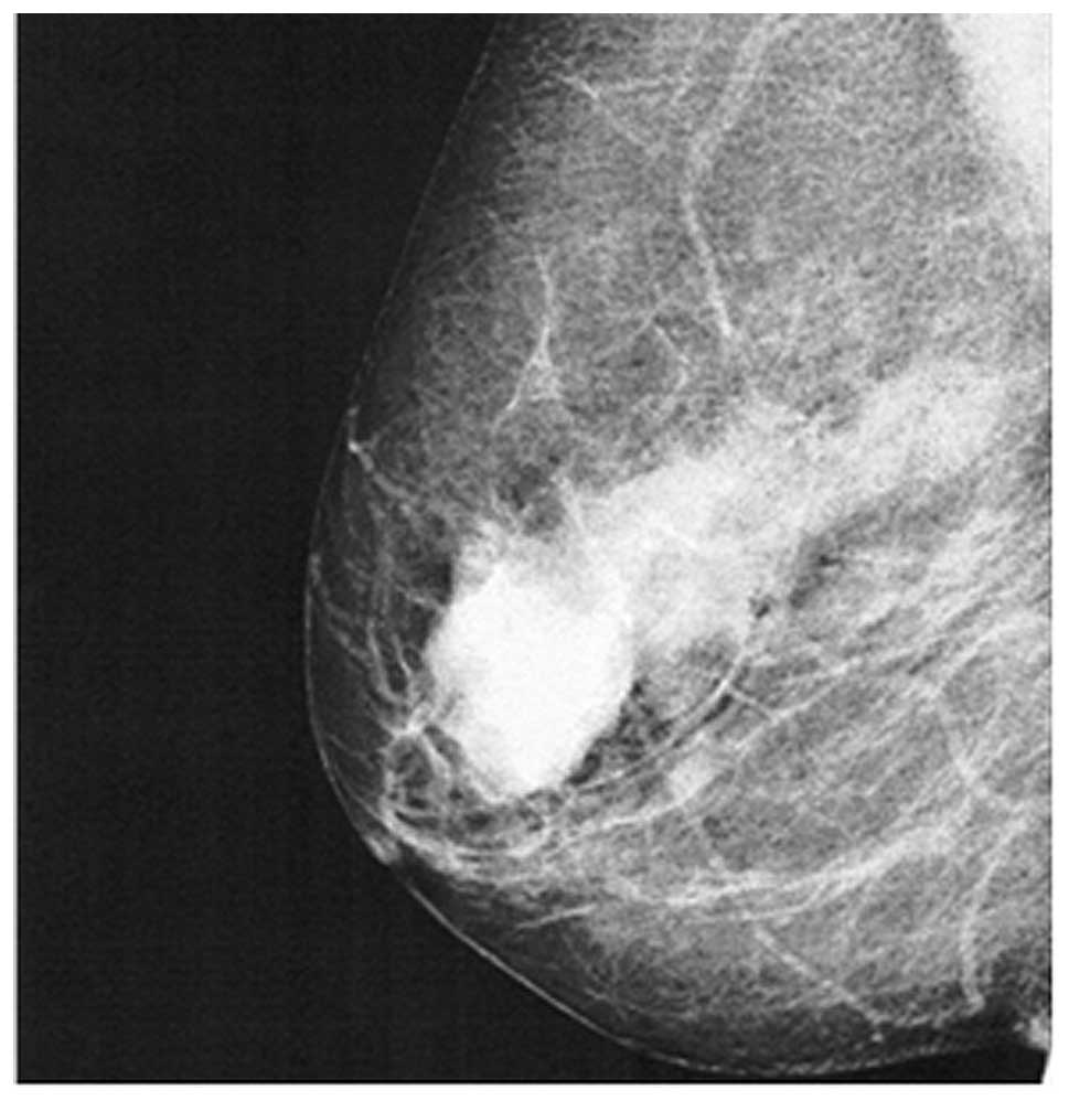
polylobated shape, while mammography exhibits a round hyperdense
mass with regular margins, which may simulate a benign lesion
(20). The differential diagnosis
of chondrosarcoma is usually between malignant phyllodes tumor
(PT), metaplastic carcinoma (MC), in which a significant percentage
of metaplastic elements (>10%) are present, and matrix-producing
metaplastic carcinoma (MP-MC), a rare cancer of the breast
characterized by osteoid and chondral matrices and the presence of
carcinomatous features (20,21).
The mainstay of treatment for sarcoma of the breast, as with
sarcomas at other sites, is a surgical procedure combined with
mastectomy (20). The present study
describes a novel case of extraskeletal chondrosarcoma of the
breast that was treated with a more conservative surgical treatment
instead of mastectomy, as an innovative surgical approach. Written
informed consent was obtained from the patient.
Case report
A 63-year-old female presented to Umberto I
Hospital, Sapienza University (Rome, Italy) with a neoplasm
localized in the upper-outer quadrant of the right breast (RUO).
The palpable lesion was a firm parenchymatous mass that was mobile
on the superficial and deep planes, exhibited sharp margins and a
poor homogeneous surface and was not painful. There was no evident
axillary lymphadenopathy. An X-ray mammogram revealed the
enhancement of a previously observed opacity in the superolateral
quadrant, with partial sharp margins. Using core biopsy, the mass
was determined to be a mammary parenchyma in continuity with
hypercellular condroid tissue, with the presence of atypical,
occasionally polynuclear elements, and rare mitosis.
Ultrasonography revealed the presence of a hypoechoic, solid
neoformation with jagged edges and a maximum diameter of ~3 cm
(Fig. 1). Mammography revealed an
upper-outer para-areolar oval opacity with partially shaded
contours that demonstrated the presence of an adequate margin of
healthy perilesional tissue (Fig.
2). The formation was delimited and split by fibrous stroma, in
which ductal structures and ectatic vessels were present. The tumor
appeared to be a malignant mesenchymal neoplasm with chondroid
differentiation, although it could not be excluded that the
formation was a component of a biphasic lesion similar to phyllodes
tumors. Considering the size of the lesion, the overall dimensions
of the breast and the lack of literature reporting a genuine
benefit of mastectomy compared with more conservative surgery
(Table I), it was decided to refer
the patient for a wide RUO quadrantectomy, to include any skip
metastasis. For an improved examination, tissue samples of the
resection margins were separately analyzed by a pathologist.
 | Table IReview of the cases of chondrosarcoma
of the breast reported in the literature between 2001 and 2013. |
Table I
Review of the cases of chondrosarcoma
of the breast reported in the literature between 2001 and 2013.
| First author, year
(reference) | Gender | Age, years | Tumor site,
breast | Method used for
diagnosis | Therapy | Follow-up |
|---|
| Errarhay et
al, 2013 (7) | Female | 24 | Right | Mammography and
tumorectomy | Mastectomy | NR |
| Mujtaba et al,
2013 (13) | Female | 40 | Right | Mammography and
CT | Mastectomy | |
| Badyal et al,
2012 (8) | Male | 80 | Right | FNAC | Mastectomy and
axillary lymphadenectomy | Yes |
| Patterson et
al, 2011 (12) | Female | 52 | Left | FNAC and core
biopsy | Mastectomy and
RT | Yes |
| Lakshmikant et
al, 2010 (14) | Female | 42 | Left | Core biopsy | Mastectomy | NR |
| Bhosale et al,
2010 (15) | Female | 45 | Right | Tumorectomy and
axillary lymph node FNAC | Mastectomy and
axillary lymphadenectomy, RT-CHT | Yes |
| De Padua et
al, 2009 (11) | Female | 56 | Right | FNAC | Mastectomy and
RT | NR |
| Gurleyik et
al, 2009 (16) | Female | 52 | Right | Ultrasonography,
mammography, FNAC and tumorectomy | Mastectomy and
axillary lymphadenectomy | NR |
| Gupta et al,
2006 (10) | Female | 46 | Left | FNAC | Mastectomy | NR |
| Verfaille et
al, 2005 (17) | Female | 77 | Right | Ultrasonography,
mammography and Tru-cut needle biopsy | Mastectomy | NR |
| Gupta et al,
2003 (9) | Female | 46 | Left | FNAC | Mastectomy,
neoadjuvant CHT, RT and lymphadenectomy | NR |
Histopathological results
The gross post-surgical examination revealed a
mammary-parenchymal section, 6.5×4.5×5 cm in size, covered by 4.1
cm of skin. Following the cut, a round-shaped neoplasm was
observed, possessing a maximum diameter of 3 cm and exhibiting
gelatinous and tense-elastic regions. The biopsy specimen from the
neoplasm revealed the proliferation of mesenchymal cells with round
nuclei, eosinophilic cytoplasm and ill-defined boundaries. The
neoplastic elements were arranged in groups of varying sizes, in
the context of a layer with a basophilic myxoid appearance. A
chondroid-type layer was present at the periphery of the lesion,
with a star-shaped appearance and pleomorphic nuclei. Certain
figures were suggestive of invasion, however, no perilesional
venous vessel invasion was detected. The mitotic index was seven
out of 10 fields of high magnification. The tumor possessed large
areas of necrosis. The neoplastic elements exhibited 50% of the
proliferation index, as evaluated by Ki-67 staining. The remaining
parenchyma demonstrated a nodular lesion with a diameter of 1.2 cm
attributable to stromal fibrosis, in the absence of further
significant atypia. All resection margins were separately analyzed
by a pathologist and were found to be free from cancer-associated
elements. A diagnosis of chondrosarcoma of the breast was finally
made.
Post-surgical treatments
Subsequent to surgery there was a discussion between
the various consulted oncologists on the necessity of further
treatments, as the role of chemotherapy and radiotherapy in primary
breast chondrosarcoma is unresolved (1). Despite the possibility that adjuvant
therapy may decrease the rates of local and systematic recurrence,
the literature is lacking in significant information regarding the
benefit of post-surgery chemo- or radiotherapy in the face of the
side-effects of those therapies, due to the rarity of this disease
and the small number of cases reported (1). It was decided, with the consent of the
patient, to administer five cycles of standard radiotherapy on the
residual breast tissue (50 Gy) and operative site (10 Gy), used
only as a complement to radical surgery, and six cycles of
precautionary chemotherapy for six weeks, with epirubicin (120 mg
per cycle) and ifosfamide (2,950 mg per cycle), as previously
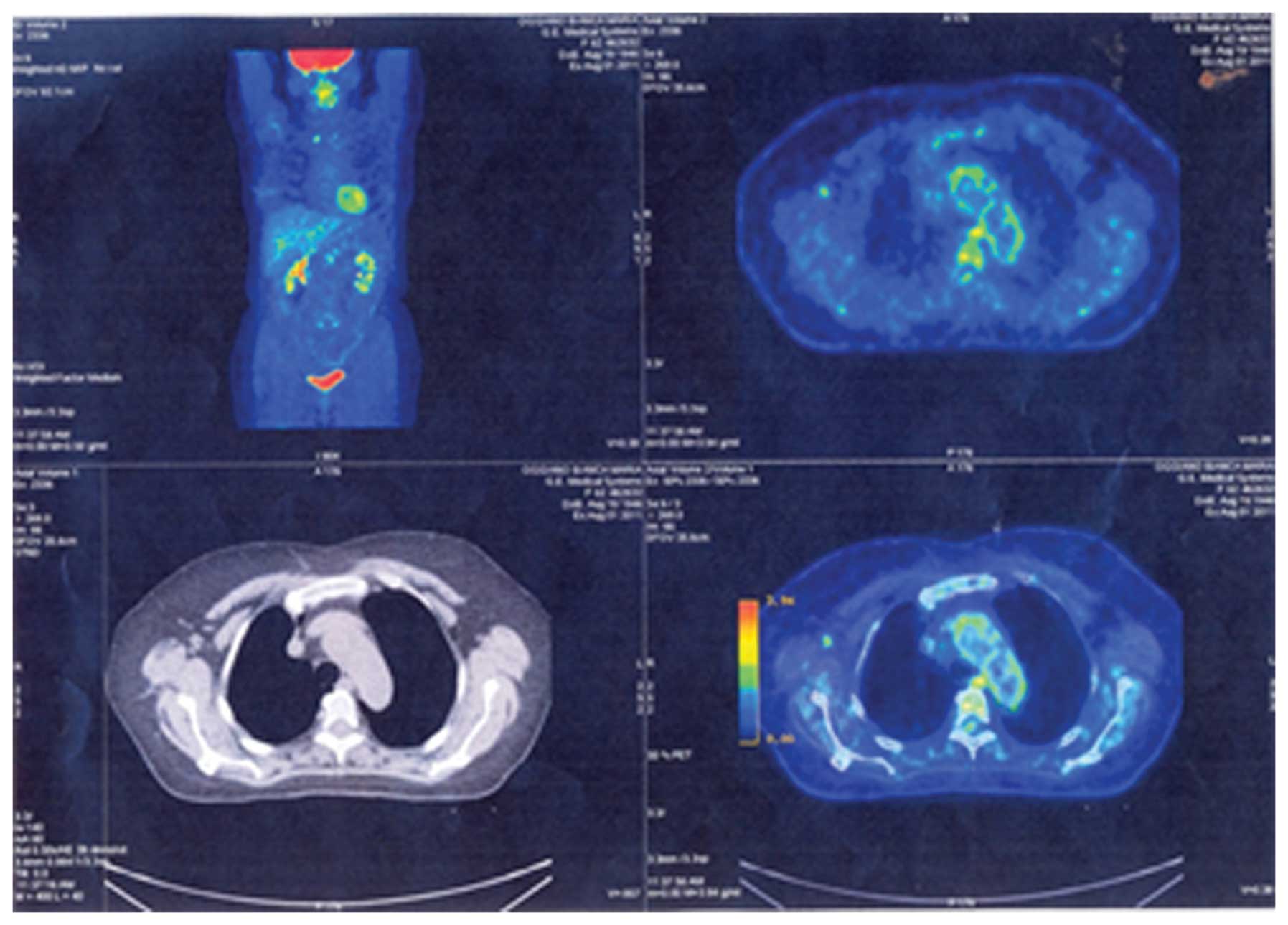
reported (1). A positron emission
tomography scan performed five months after the surgery excluded
the presence of remnants of the disease (Fig. 3). Therefore, whilst the patient
continues to undergo a strict clinical and instrumental follow-up
two and half years after surgery, there are no signs of recurrent
disease.
Discussion
The present study describes a novel case of
extraskeletal chondrosarcoma of the breast that was treated with a
more conservative quadrantectomy instead of mastectomy, as an
innovative surgical approach. The benefit of performing
quadrantectomy instead of mastectomy is not addressed in the
literature (1,2). Between 1967 and 2001, only five cases
of chondrosarcoma of the breast were reported in the literature,
and all were treated with mastectomy (3–6). The
present review revealed that between 2001 and 2013 only 11 other
cases were described, as summarized in Table I. All the 16 studies, consisting of
15 female patients and one male patient, reported mastectomy as the
surgical treatment choice. In four cases mastectomy was associated
with lymphadenectomy and in one case mastectomy was preceded by
neoadjuvant chemotherapy (Table I).
The choice of mastectomy was dictated not only by a strictly local
situation of the lesion, but also by the lack of previous studies,
due to the rarity of the lesion, and by an old interpretation of
the surgical approach to sarcoma at this particular site.
Although surgery remains the gold standard for the
treatment of breast sarcoma, and chondrosarcoma in particular
(1), there is no uniformity of view
on the most effective type of surgery that may justify the
requirement for radical intervention, such as mastectomy, compared
to surgery with wide tumor-free margins (20). According to a study performed by
Zelek et al (22), and
another previous study (23),
mastectomy for sarcomatous malignancy should not be associated with
axillary lymphadenectomy, as the sarcoma does not exhibit a
tendency to spread through lymphatic system, but mainly through the
haematogenous route. Furthermore, the lymphectomy exposes the
patient to greater morbidity without a real benefit in terms of
disease-free and overall survival (20). Thus, lymphectomy should be indicated
only if the lesion is associated with an epithelial component or
when all breast quadrants are involved, in particular the
upper-outer (superolateral) quadrants (20).
In previous years, the surgical approach to all
sarcomas, and in particular to those of the breast, has been
reevaluated as it has been found that an extensive surgical
procedure may be considered adequate, providing that the curative
wide margins of healthy peritumoral tissue are sufficiently
respected, to ensure that the margins include any skip metastasis
with the excision (2,24,25).
At present, only two factors have been demonstrated to affect the
outcome of surgery, the extent of the tumor and the margins of
excision. Tumors >5 cm in size result in a poorer prognosis
compared with those of smaller dimensions (20). In addition, the majority of authors
agree that a resection margin of 1 cm is sufficient for small and
localized sarcomas, and this approach is compatible with a
conservative surgery. Therefore, in the present study, it was
decided to treat the chondrosarcoma of the breast with surgery, in
consideration of the size of the lesion, and, in particular, to use
a more conservative quadrantectomy instead of mastectomy, as a
novel surgical approach. This approach was chosen with the
consideration that a greater efficacy has not been proven or
demonstrated in patients treated with mastectomy in terms of
overall and disease-free survival compared with patients treated by
adequate quadrantectomy.
The present treatment strategy was in agreement with
a previous retrospective study revealing that, for sarcomas of the
breast, the radical treatment of mastectomy did not offer
significant survival benefits compared with the wide excision
option of quadrantectomy (20). By
contrast, the study revealed a more severe prognosis for the
patients that underwent simple lumpectomy. Notably, at the time of
writing, a novel case report was published in the literature that
used a similar conservative breast surgery approach (26), supporting the present approach of
performing quadrantectomy instead of mastectomy.
Acknowledgements
The authors would like to thank the patient who
participated in this study.
References
|
1
|
Lewin J, Puri A, Quek R, Ngan R, Alcasabas
AP, Wood D and Thomas D: Management of sarcoma in the Asia-Pacific
region: resource-stratified guidelines. Lancet Oncol. 14:e562–e570.
2013. View Article : Google Scholar : PubMed/NCBI
|
|
2
|
Demetri GD, Antonia S, Benjamin RS, et al:
National Comprehensive Cancer Network Soft Tissue Sarcoma Panel:
Soft tissue sarcoma. J Natl Compr Canc Netw. 8:630–674.
2010.PubMed/NCBI
|
|
3
|
Kennedy T and Biggart JD: Sarcoma of the
breast. Br J Cancer. 21:635–644. 1967. View Article : Google Scholar : PubMed/NCBI
|
|
4
|
Beltaos E and Banerjee TK: Chondrosarcoma
of the breast: Report of two cases. Am J Clin Pathol. 71:345–349.
1979.PubMed/NCBI
|
|
5
|
Thilagavathi G, Subramanian S, Samuel AV,
Rani U and Somasundaram C: Primary chondrosarcoma of the breast. J
Indian Med Assoc. 90:16–17. 1992.PubMed/NCBI
|
|
6
|
Guymar S, Ferlicot S, Genestie C, Gelberg
JJ, Blondon J, Le Charpentier Y and Zafrani B: Breast
chondrosarcoma: a case report and review. Ann Pathol. 21:168–171.
2001.(In French). PubMed/NCBI
|
|
7
|
Errarhay S, Fetohi M, Mahmoud S, et al:
Primary chondrosarcoma of the breast: a case presentation and
review of the literature. World J Surg. 11:2082013. View Article : Google Scholar
|
|
8
|
Badyal RK, Kataria AS and Kaur M: Primary
chondrosarcoma of male breast: a rare case. Indian J Surg.
74:418–419. 2012. View Article : Google Scholar :
|
|
9
|
Gupta S, Gupta V, Aggarwal PN, Kant R,
Khurana N and Mandal AK: Primary chondrosarcoma of the breast: a
case report. Indian J Cancer. 40:77–79. 2003.
|
|
10
|
Gupta S, Gupta V, Aggarwal PN, Kant R,
Dass PM, Khurana N and Mandal AK: Primary chondrosarcoma of the
breast. J Indian Med Assoc. 104:99–100. 2006.PubMed/NCBI
|
|
11
|
De Padua M and Bhandari TP: Primary
mesenchymal chondrosarcoma of the breast. Indian J Pathol
Microbiol. 52:129–130. 2009. View Article : Google Scholar : PubMed/NCBI
|
|
12
|
Patterson JD, Wilson JE, Dim D and Talboy
GE: Primary chondrosarcoma of the breast: report of a case and
review of the literature. Breast Dis. 33:189–191. 2011.PubMed/NCBI
|
|
13
|
Mujtaba SS, Haroon S and Faridi N: Primary
chondrosarcoma of breast. J Coll Physicians Surg Pak. 23:754–755.
2013.PubMed/NCBI
|
|
14
|
Lakshmikantha A, Kawatra V, Varma D and
Khurana N: Primary breast chondrosarcoma. Breast J. 16:553–554.
2010. View Article : Google Scholar : PubMed/NCBI
|
|
15
|
Bhosale SJ, Kshirsagar AY, Sulhyan SR, et
al: Metaplastic carcinoma with predominant chondrosarcoma of the
right breast. Case Rep Oncol. 3:277–281. 2010. View Article : Google Scholar
|
|
16
|
Gurleyik E, Yildirim U, Gunai O and
Pehlivan M: Malignant mesenchymal tumor of the breast: primary
chondrosarcoma. Breast Care (Basel). 4:101–103. 2009. View Article : Google Scholar
|
|
17
|
Verfaillie G, Breucq C, Perdaens C, et al:
Chondrosarcoma of the breast. Breast J. 11:147–148. 2005.
View Article : Google Scholar : PubMed/NCBI
|
|
18
|
Rao L, Kudva R, Rao RV and Kumar B:
Extraskeletal myxoid chondrosarcoma of the chest wall masquerading
as a breast tumor. A case report. Acta Cytol. 46:417–421. 2002.
View Article : Google Scholar : PubMed/NCBI
|
|
19
|
Vera-Sempere F and García-Martínez A:
Malignant phyllodes tumor of the breast with predominant
chondrosarcomatous differentiation. Pathol Res Pract. 199:841–845.
2003. View Article : Google Scholar
|
|
20
|
Pencavel TD and Hayes A: Breast sarcoma -
a review of diagnosis and management. Int J Surg. 7:20–23. 2009.
View Article : Google Scholar
|
|
21
|
Rakha EA, Tan PH, Shaaban A, et al: Do
primary mammary osteosarcoma and chondrosarcoma exist? A review of
a large multi-institutional series of malignant matrix-producing
breast tumours. Breast. 22:13–18. 2013. View Article : Google Scholar
|
|
22
|
Zelek L, Llombart-Cussac A, Terrier P, et
al: Prognostic factors in primary breast sarcomas: a series of
patients with long-term follow-up. J Clin Oncol. 21:2583–2588.
2003. View Article : Google Scholar : PubMed/NCBI
|
|
23
|
Pasta V, Monti M, Antonucci D, Di Matteo
FM, Boccaccini F and Brescia A: Primary sarcoma of the breast:
criteria for radical surgery. G Chir. 18:703–706. 1997.(In
Italian).
|
|
24
|
Baldini EH, Goldberg J, Jenner C, Manola
JB, Demetri GD, Fletcher CD and Singer S: Long-term outcomes after
function-sparing surgery without radiotherapy for soft tissue
sarcoma of the extremities and trunk. J Clin Oncol. 17:3252–3259.
1999.PubMed/NCBI
|
|
25
|
Stojadinovic A, Leung DH, Hoos A, Jaques
DP, Lewis JJ and Brennan MF: Analysis of the prognostic
significance of microscopic margins in 2,084 localized primary
adult soft tissue sarcomas. Ann Surg. 235:424–434. 2002. View Article : Google Scholar : PubMed/NCBI
|
|
26
|
Faraht A, Magdy N and Elaffandi A: Primary
myxoid chondrosarcoma of the breast. Ann R Coll Surg Engl.
96:112E–411E. 2014. View Article : Google Scholar
|