Spandidos Publications Logo
  • About
    • About Spandidos
    • Aims and Scopes
    • Abstracting and Indexing
    • Editorial Policies
    • Reprints and Permissions
    • Job Opportunities
    • Terms and Conditions
    • Contact
  • Journals
    • All Journals
    • Oncology Letters
      • Oncology Letters
      • Information for Authors
      • Editorial Policies
      • Editorial Board
      • Aims and Scope
      • Abstracting and Indexing
      • Bibliographic Information
      • Archive
    • International Journal of Oncology
      • International Journal of Oncology
      • Information for Authors
      • Editorial Policies
      • Editorial Board
      • Aims and Scope
      • Abstracting and Indexing
      • Bibliographic Information
      • Archive
    • Molecular and Clinical Oncology
      • Molecular and Clinical Oncology
      • Information for Authors
      • Editorial Policies
      • Editorial Board
      • Aims and Scope
      • Abstracting and Indexing
      • Bibliographic Information
      • Archive
    • Experimental and Therapeutic Medicine
      • Experimental and Therapeutic Medicine
      • Information for Authors
      • Editorial Policies
      • Editorial Board
      • Aims and Scope
      • Abstracting and Indexing
      • Bibliographic Information
      • Archive
    • International Journal of Molecular Medicine
      • International Journal of Molecular Medicine
      • Information for Authors
      • Editorial Policies
      • Editorial Board
      • Aims and Scope
      • Abstracting and Indexing
      • Bibliographic Information
      • Archive
    • Biomedical Reports
      • Biomedical Reports
      • Information for Authors
      • Editorial Policies
      • Editorial Board
      • Aims and Scope
      • Abstracting and Indexing
      • Bibliographic Information
      • Archive
    • Oncology Reports
      • Oncology Reports
      • Information for Authors
      • Editorial Policies
      • Editorial Board
      • Aims and Scope
      • Abstracting and Indexing
      • Bibliographic Information
      • Archive
    • Molecular Medicine Reports
      • Molecular Medicine Reports
      • Information for Authors
      • Editorial Policies
      • Editorial Board
      • Aims and Scope
      • Abstracting and Indexing
      • Bibliographic Information
      • Archive
    • World Academy of Sciences Journal
      • World Academy of Sciences Journal
      • Information for Authors
      • Editorial Policies
      • Editorial Board
      • Aims and Scope
      • Abstracting and Indexing
      • Bibliographic Information
      • Archive
    • International Journal of Functional Nutrition
      • International Journal of Functional Nutrition
      • Information for Authors
      • Editorial Policies
      • Editorial Board
      • Aims and Scope
      • Abstracting and Indexing
      • Bibliographic Information
      • Archive
    • International Journal of Epigenetics
      • International Journal of Epigenetics
      • Information for Authors
      • Editorial Policies
      • Editorial Board
      • Aims and Scope
      • Abstracting and Indexing
      • Bibliographic Information
      • Archive
    • Medicine International
      • Medicine International
      • Information for Authors
      • Editorial Policies
      • Editorial Board
      • Aims and Scope
      • Abstracting and Indexing
      • Bibliographic Information
      • Archive
  • Articles
  • Information
    • Information for Authors
    • Information for Reviewers
    • Information for Librarians
    • Information for Advertisers
    • Conferences
  • Language Editing
Spandidos Publications Logo
  • About
    • About Spandidos
    • Aims and Scopes
    • Abstracting and Indexing
    • Editorial Policies
    • Reprints and Permissions
    • Job Opportunities
    • Terms and Conditions
    • Contact
  • Journals
    • All Journals
    • Biomedical Reports
      • Information for Authors
      • Editorial Policies
      • Editorial Board
      • Aims and Scope
      • Abstracting and Indexing
      • Bibliographic Information
      • Archive
    • Experimental and Therapeutic Medicine
      • Information for Authors
      • Editorial Policies
      • Editorial Board
      • Aims and Scope
      • Abstracting and Indexing
      • Bibliographic Information
      • Archive
    • International Journal of Epigenetics
      • Information for Authors
      • Editorial Policies
      • Editorial Board
      • Aims and Scope
      • Abstracting and Indexing
      • Bibliographic Information
      • Archive
    • International Journal of Functional Nutrition
      • Information for Authors
      • Editorial Policies
      • Editorial Board
      • Aims and Scope
      • Abstracting and Indexing
      • Bibliographic Information
      • Archive
    • International Journal of Molecular Medicine
      • Information for Authors
      • Editorial Policies
      • Editorial Board
      • Aims and Scope
      • Abstracting and Indexing
      • Bibliographic Information
      • Archive
    • International Journal of Oncology
      • Information for Authors
      • Editorial Policies
      • Editorial Board
      • Aims and Scope
      • Abstracting and Indexing
      • Bibliographic Information
      • Archive
    • Medicine International
      • Information for Authors
      • Editorial Policies
      • Editorial Board
      • Aims and Scope
      • Abstracting and Indexing
      • Bibliographic Information
      • Archive
    • Molecular and Clinical Oncology
      • Information for Authors
      • Editorial Policies
      • Editorial Board
      • Aims and Scope
      • Abstracting and Indexing
      • Bibliographic Information
      • Archive
    • Molecular Medicine Reports
      • Information for Authors
      • Editorial Policies
      • Editorial Board
      • Aims and Scope
      • Abstracting and Indexing
      • Bibliographic Information
      • Archive
    • Oncology Letters
      • Information for Authors
      • Editorial Policies
      • Editorial Board
      • Aims and Scope
      • Abstracting and Indexing
      • Bibliographic Information
      • Archive
    • Oncology Reports
      • Information for Authors
      • Editorial Policies
      • Editorial Board
      • Aims and Scope
      • Abstracting and Indexing
      • Bibliographic Information
      • Archive
    • World Academy of Sciences Journal
      • Information for Authors
      • Editorial Policies
      • Editorial Board
      • Aims and Scope
      • Abstracting and Indexing
      • Bibliographic Information
      • Archive
  • Articles
  • Information
    • For Authors
    • For Reviewers
    • For Librarians
    • For Advertisers
    • Conferences
  • Language Editing
Login Register Submit
  • This site uses cookies
  • You can change your cookie settings at any time by following the instructions in our Cookie Policy. To find out more, you may read our Privacy Policy.

    I agree
Search articles by DOI, keyword, author or affiliation
Search
Advanced Search
presentation
Oncology Letters
Join Editorial Board Propose a Special Issue
Print ISSN: 1792-1074 Online ISSN: 1792-1082
Journal Cover
March-2015 Volume 9 Issue 3

Full Size Image

Sign up for eToc alerts
Recommend to Library

Journals

International Journal of Molecular Medicine

International Journal of Molecular Medicine

International Journal of Molecular Medicine is an international journal devoted to molecular mechanisms of human disease.

International Journal of Oncology

International Journal of Oncology

International Journal of Oncology is an international journal devoted to oncology research and cancer treatment.

Molecular Medicine Reports

Molecular Medicine Reports

Covers molecular medicine topics such as pharmacology, pathology, genetics, neuroscience, infectious diseases, molecular cardiology, and molecular surgery.

Oncology Reports

Oncology Reports

Oncology Reports is an international journal devoted to fundamental and applied research in Oncology.

Experimental and Therapeutic Medicine

Experimental and Therapeutic Medicine

Experimental and Therapeutic Medicine is an international journal devoted to laboratory and clinical medicine.

Oncology Letters

Oncology Letters

Oncology Letters is an international journal devoted to Experimental and Clinical Oncology.

Biomedical Reports

Biomedical Reports

Explores a wide range of biological and medical fields, including pharmacology, genetics, microbiology, neuroscience, and molecular cardiology.

Molecular and Clinical Oncology

Molecular and Clinical Oncology

International journal addressing all aspects of oncology research, from tumorigenesis and oncogenes to chemotherapy and metastasis.

World Academy of Sciences Journal

World Academy of Sciences Journal

Multidisciplinary open-access journal spanning biochemistry, genetics, neuroscience, environmental health, and synthetic biology.

International Journal of Functional Nutrition

International Journal of Functional Nutrition

Open-access journal combining biochemistry, pharmacology, immunology, and genetics to advance health through functional nutrition.

International Journal of Epigenetics

International Journal of Epigenetics

Publishes open-access research on using epigenetics to advance understanding and treatment of human disease.

Medicine International

Medicine International

An International Open Access Journal Devoted to General Medicine.

Journal Cover
March-2015 Volume 9 Issue 3

Full Size Image

Sign up for eToc alerts
Recommend to Library

  • Article
  • Citations
    • Cite This Article
    • Download Citation
    • Create Citation Alert
    • Remove Citation Alert
    • Cited By
  • Similar Articles
    • Related Articles (in Spandidos Publications)
    • Similar Articles (Google Scholar)
    • Similar Articles (PubMed)
  • Download PDF
  • Download XML
  • View XML
Article

Hepatic hemangioma masquerading as a tumor originating from the stomach

  • Authors:
    • Xingmao Zhang
    • Zhixiang Zhou
  • View Affiliations / Copyright

    Affiliations: Department of Gastrointestinal Surgery, Cancer Hospital, Chinese Academy of Medical Sciences, Peking Union Medical College, Beijing 100021, P.R. China
  • Pages: 1406-1408
    |
    Published online on: January 12, 2015
       https://doi.org/10.3892/ol.2015.2863
  • Expand metrics +
Metrics: Total Views: 0 (Spandidos Publications: | PMC Statistics: )
Metrics: Total PDF Downloads: 0 (Spandidos Publications: | PMC Statistics: )
Cited By (CrossRef): 0 citations Loading Articles...

This article is mentioned in:


Abstract

Hemangioma is the most common benign hepatic neoplasm. The majority of cases are asymptomatic and can be confirmed by imaging examinations, including enhanced computed tomography and magnetic resonance imaging. Exophytic growth is not common and pedunculated cases are extremely rare. The present study reports a case that was pre‑operatively misdiagnosed as a stomach‑originating tumor. Laparoscopic exploration confirmed that this tumor was a hepatic hemangioma with a long peduncle originating from the left edge of the liver. The final diagnosis of cavernous hemangioma was confirmed by postoperative pathology. This indicates that hepatic hemangioma with a long peduncle has the possibility to be inaccurately diagnosed. Laparoscopic examination is required for such cases.

Introduction

Hepatic cavernous hemangioma is the most common benign tumor of the liver and occurs more frequently in females (1). The majority of lesions are >3 cm in size and a significant proportion of patients are asymptomatic (2). Large lesions may cause a variety of symptoms, including an abdominal mass, pain, hemorrhage, jaundice, nausea and vomiting (3,4). Cavernous hemangioma usually presents as subcapsular, disclosed and solitary well-delineated nodules, these distinctive structures exhibit a characteristic hemodynamic pattern on enhanced computed tomography (CT) (5). Exophytic hemangioma, particularly in a pedunculated form, is extremely rare (6,7).

A patient possessing a pedunculated hepatic hemangioma that was misdiagnosed as a tumor originating from the stomach due to atypical imaging findings was recently treated at the Cancer Hospital (Chinese Academy of Medical Sciences, Peking Union Medical College, Beijing, China). In the present study, the case is reported together with a review of the literature. Written informed consent was obtained from the patient.

Case report

A 52-year-old male was referred to the Cancer Hospital for ascending colon cancer, which had been confirmed by colonoscopy with biopsy. The patient had visited Chengde Central Hospital (Chengde, China) one week prior to attending the Cancer Hospital, and the chief complaint of the patient was blood-stained stool. Colonoscopy with biopsy was performed at the local hospital, and a lesion with a diameter of ~5 cm was identified in the ascending colon. The surface of the lesion was rough, and a diagnosis of ascending colon adenocarcinoma was confirmed by biopsy. No neoadjuvant therapy was delivered to the patient. With the aim of receiving laparoscopic surgery and better comprehensive treatment, the patient was referred to the Cancer Hospital, where further examinations were performed prior to surgery. The further examinations included enhanced abdominal and pelvic CT scanning; examination of tumor markers, liver and renal function and coagulation function; chest X-ray; abdominal ultrasound examination; and re-examination of the pathological results of the local hospital by two pathologists.

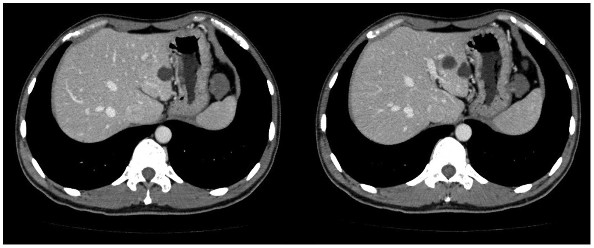
Enhanced CT revealed a mass in the left upper quadrant, with a maximum diameter of ~2.8 cm and a CT value of ~36 Hounsfield units (Fig. 1). This low-density mass was suspected to be a tumor originating from the stomach, as CT showed that the mass was adhered to the greater curvature of body of stomach and a peduncle appeared to originate from stomach wall. Hepatic ultrasonography did not reveal the mass.

Figure 1

A mass with a maximum diameter of ~2.8 cm and a CT value of ~36 Hounsfield units was identified in the left upper quadrant (left). It appeared that the mass was originating from the body of the stomach (right), and a gastric stromal tumor was suspected due to the CT scans.

The laboratory data were as follows: White blood cell count, 4,800/μl (normal range, 4,000–10,000/μl); red blood cell count, 5.3×10¹²/l (normal range, 3.5–5.5×10¹²/l); and hemoglobin, 82 g/μl (normal range, 120–160 g/μl). Tumor markers were as follows: α-fetoprotein, 3.2 ng/ml (normal levels, <20 ng/ml); carcinoembryonic antigen, 1.7 ng/ml (normal range, 0–5.0 ng/ml); and carbohydrate antigen 19–9, 16 U/ml (0–37 U/ml). Tests for hepatitis B surface antibody (HBsAb) provided a positive result, but the samples were negative for HBeAb, HBcAb, HB s antigen (Ag) and HBeAg; therefore, the patient in the present case did not have hepatitis B.

Based on preoperative examinations, including CT and blood tests, primary ascending colon cancer and cancer-related anemia were confirmed, and a left upper quadrant mass was also diagnosed. The patient was transfused with 800 ml blood and, two days after the transfusion, laparoscopic-assisted right 
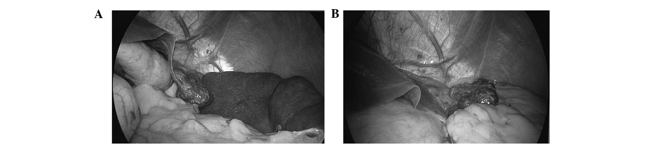
hemicolectomy was performed. A dark red mass with a smooth surface and long peduncle originating from the left edge of the liver was identified in left upper quadrant. The mass, which had previously been suspected to be a tumor originating in the stomach, was confirmed as hepatic hemangioma by the findings of laparoscopic exploration. This tumor was isolated from the liver and extended 
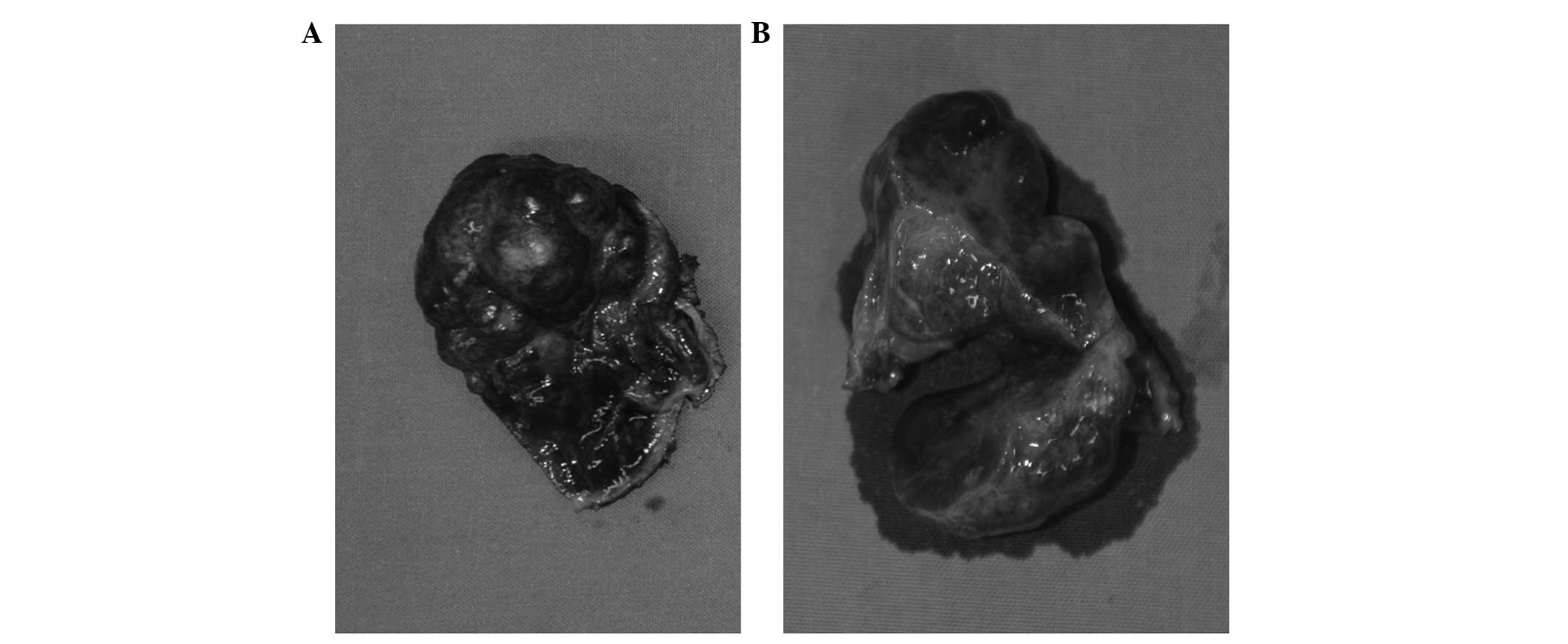
down to the left side of the stomach (Fig. 2) and, macroscopically, the tumor size was 3.0×2.5×1.5 cm (Fig. 3). A resection of the hepatic hemangioma was successfully performed under laparoscopy.

Figure 2

Laparoscopic exploration. (A) Dark red mass with a diameter of 3 cm, a smooth surface and a long peduncle was isolated from the liver extended down to the left side of the stomach. (B) The mass had a long peduncle originated from the left edge of the liver and was found in the left upper quadrant.

Figure 3

(A) The resected tumor was dark red in color with a complete capsule, soft texture and smooth surface; at its widest the diameter was ~3 cm. The diameter was ~3 cm. (B) On cutting the mass, the cross section was honeycomb-like, the inner surface was observed to be red and wet, and blood leaked from the section.

For the ascending colon lesion, a diagnosis of stage T4aN0M0 adenocarcinoma was confirmed by postoperative pathology, and chemotherapy was recommended for this patient. This patient was administered chemotherapy four weeks following surgery. Oxaliplatin was administered intravenously in addition to an oral capecitabine regimen (oxaliplatin 235 mg over 2 h on day 1, capecitabine 3250 mg on days 1–14) for 2 consecutive weeks followed by 1 week of rest, for a total of 8 cycles (24 weeks). Four cycles had been completed when this report was started. During the follow-up period, examinations including clinical examination, abdominal ultrasonography, abdominal CT scanning, chest radiography and carcinoembryonic antigen assessment should be delivered for patients once every 3 months in the first 2 years after surgery, biannually in the next 3 years and then annually (8,9). For the hepatic hemangioma, a diagnosis of cavernous hemangioma was confirmed by pathology, and no other additional treatments were required for this hemangioma following surgery. At the time of writing, the patient had been followed-up for approximately three months following surgery.

Discussion

Hemangioma is the most common benign tumor of the liver, and the incidence of hepatic cavernous hemangioma among liver tumors is as high as 20%. A diagnosis of typical hemangioma is straightforward when using a combination of various imaging techniques (6). Typical hemangioma presents early peripheral enhancement of the tumor on dynamic contrast CT, followed by centripetal fill-in of the contrast medium with persistently enhancement on delayed-phase images (10). In the present study, the tumor was revealed as low density in enhanced and delayed-phase images. Hemangioma is usually solitary and it is frequently in a subcapsular location, more commonly in the right lobe, particularly the posterior segment (1). Exophytic growth is not common and pedunculated cases are extremely rare (11–13). Hemangioma with a peduncle possesses the ability to isolate and migrate to the outside of the liver. For example, Moon et al (1) reported a case of pedunculated hepatic hemangioma that was misdiagnosed as a submucosal tumor of the stomach due to the atypical position of the tumor.

The majority of hemangiomas are asymptomatic, but larger hemangiomas can produce various symptoms, including an abdominal mass, pain, nausea, vomiting, jaundice, hemorrhage and even rupture. The patient in the present study did not experience any abdominal discomfort or an abdominal mass. No treatment is required for the majority of hemangiomas, with the exception of symptomatic treatment, which includes the treatment of a palpable mass, pain, increasing size or complications, including consumptive coagulopathy and rupture (14).

In conclusion, hepatic hemangioma with a long peduncle originating from the left edge of the liver may be inaccurately diagnosed. Laparoscopic examination is required for accurate diagnosis in such cases.

References

1 

Moon HK, Kim HS, Heo GM, et al: A case of pedunculated hepatic hemangioma mimicking submucosal tumor of the stomach. Korean J Hepatol. 17:66–70. 2011. View Article : Google Scholar : PubMed/NCBI

2 

Chui AK, Vass J, McCaughan GW and Sheil AG: Giant cavernous haemangioma: a rare indication for liver transplantation. Aust N Z J Surg. 66:122–124. 1996. View Article : Google Scholar : PubMed/NCBI

3 

Bengisun U, Ozbas S, Gürel M and Ensari A: Laparoscopic hepatic wedge resection of hemangioma: report of two cases. Langenbecks Arch Surg. 385:363–365. 2000. View Article : Google Scholar : PubMed/NCBI

4 

Kochar R, Atiq M, Lee JH, et al: Giant hepatic hemangioma masquerading as a gastric subepithelial tumor. Gastroenterol Hepatol (NY). 9:396–398. 2013.

5 

Yamada S, Shimada M, Utsunomiya T, et al: Hepatic screlosed hemangioma which was misdiagnosed as metastasis of gastric cancer: report of a case. J Med Invest. 59:270–274. 2012. View Article : Google Scholar : PubMed/NCBI

6 

Masui T, Katayama M, Nakagawara M, Shimizu S and Kojima K: Exophytic giant cavernous hemangioma of the liver with growing tendency. Radiat Med. 23:121–124. 2005.PubMed/NCBI

7 

Maèkawa S, Mizutani Y, Terachi T, Okada Y and Yoshida O: A case of exophytic hepatic hemangioma mimicking adrenal tumor. Hinyokika Kiyo. 43:123–126. 1997.PubMed/NCBI

8 

Desch CE, Benson AB III, Somerfield MR, et al: American Society of Clinical Oncology: Colorectal cancer surveillance: 2005 update of an American Society of Clinical Oncology practice guideline. J Clin Oncol. 23:8512–8519. 2005. View Article : Google Scholar : PubMed/NCBI

9 

Locker GY, Hamilton S, Harris J, et al: ASCO: ASCO 2006 update of recommendations for the use of tumor markers in gastrointestinal cancer. J Clin Oncol. 24:5313–5327. 2006. View Article : Google Scholar : PubMed/NCBI

10 

Kim SJ, Yoon CM, Park IC, et al: Giant cavernous hemangiomas of the liver. Korean J Gastroenterol. 18:351–357. 1986.

11 

Brancatelli G, Federle MP, Blachar A and Grazioli L: Hemangioma in the cirrhotic liver: diagnosis and natural history. Radiology. 219:69–74. 2001. View Article : Google Scholar : PubMed/NCBI

12 

Hosokawa A, Maeda T, Tateishi U, et al: Hepatic hemangioma presenting atypical radiologic findings: a case report. Radiat Med. 23:371–375. 2005.PubMed/NCBI

13 

Leone N, Saettone S, De Paolis P, et al: Ectopic livers and related pathology: report of three cases of benign lesions. Dig Dis Sci. 50:1818–1822. 2005. View Article : Google Scholar : PubMed/NCBI

14 

Yoon SS, Charny CK, Fong Y, et al: Diagnosis, management and outcomes of 115 patients with hepatic hemangioma. J Am Coll Surg. 197:392–402. 2003. View Article : Google Scholar : PubMed/NCBI

Related Articles

  • Abstract
  • View
  • Download
  • Twitter
Copy and paste a formatted citation
Spandidos Publications style
Zhang X and Zhou Z: Hepatic hemangioma masquerading as a tumor originating from the stomach. Oncol Lett 9: 1406-1408, 2015.
APA
Zhang, X., & Zhou, Z. (2015). Hepatic hemangioma masquerading as a tumor originating from the stomach. Oncology Letters, 9, 1406-1408. https://doi.org/10.3892/ol.2015.2863
MLA
Zhang, X., Zhou, Z."Hepatic hemangioma masquerading as a tumor originating from the stomach". Oncology Letters 9.3 (2015): 1406-1408.
Chicago
Zhang, X., Zhou, Z."Hepatic hemangioma masquerading as a tumor originating from the stomach". Oncology Letters 9, no. 3 (2015): 1406-1408. https://doi.org/10.3892/ol.2015.2863
Copy and paste a formatted citation
x
Spandidos Publications style
Zhang X and Zhou Z: Hepatic hemangioma masquerading as a tumor originating from the stomach. Oncol Lett 9: 1406-1408, 2015.
APA
Zhang, X., & Zhou, Z. (2015). Hepatic hemangioma masquerading as a tumor originating from the stomach. Oncology Letters, 9, 1406-1408. https://doi.org/10.3892/ol.2015.2863
MLA
Zhang, X., Zhou, Z."Hepatic hemangioma masquerading as a tumor originating from the stomach". Oncology Letters 9.3 (2015): 1406-1408.
Chicago
Zhang, X., Zhou, Z."Hepatic hemangioma masquerading as a tumor originating from the stomach". Oncology Letters 9, no. 3 (2015): 1406-1408. https://doi.org/10.3892/ol.2015.2863
Follow us
  • Twitter
  • LinkedIn
  • Facebook
About
  • Spandidos Publications
  • Careers
  • Cookie Policy
  • Privacy Policy
How can we help?
  • Help
  • Live Chat
  • Contact
  • Email to our Support Team