Spandidos Publications Logo
  • About
    • About Spandidos
    • Aims and Scopes
    • Abstracting and Indexing
    • Editorial Policies
    • Reprints and Permissions
    • Job Opportunities
    • Terms and Conditions
    • Contact
  • Journals
    • All Journals
    • Oncology Letters
      • Oncology Letters
      • Information for Authors
      • Editorial Policies
      • Editorial Board
      • Aims and Scope
      • Abstracting and Indexing
      • Bibliographic Information
      • Archive
    • International Journal of Oncology
      • International Journal of Oncology
      • Information for Authors
      • Editorial Policies
      • Editorial Board
      • Aims and Scope
      • Abstracting and Indexing
      • Bibliographic Information
      • Archive
    • Molecular and Clinical Oncology
      • Molecular and Clinical Oncology
      • Information for Authors
      • Editorial Policies
      • Editorial Board
      • Aims and Scope
      • Abstracting and Indexing
      • Bibliographic Information
      • Archive
    • Experimental and Therapeutic Medicine
      • Experimental and Therapeutic Medicine
      • Information for Authors
      • Editorial Policies
      • Editorial Board
      • Aims and Scope
      • Abstracting and Indexing
      • Bibliographic Information
      • Archive
    • International Journal of Molecular Medicine
      • International Journal of Molecular Medicine
      • Information for Authors
      • Editorial Policies
      • Editorial Board
      • Aims and Scope
      • Abstracting and Indexing
      • Bibliographic Information
      • Archive
    • Biomedical Reports
      • Biomedical Reports
      • Information for Authors
      • Editorial Policies
      • Editorial Board
      • Aims and Scope
      • Abstracting and Indexing
      • Bibliographic Information
      • Archive
    • Oncology Reports
      • Oncology Reports
      • Information for Authors
      • Editorial Policies
      • Editorial Board
      • Aims and Scope
      • Abstracting and Indexing
      • Bibliographic Information
      • Archive
    • Molecular Medicine Reports
      • Molecular Medicine Reports
      • Information for Authors
      • Editorial Policies
      • Editorial Board
      • Aims and Scope
      • Abstracting and Indexing
      • Bibliographic Information
      • Archive
    • World Academy of Sciences Journal
      • World Academy of Sciences Journal
      • Information for Authors
      • Editorial Policies
      • Editorial Board
      • Aims and Scope
      • Abstracting and Indexing
      • Bibliographic Information
      • Archive
    • International Journal of Functional Nutrition
      • International Journal of Functional Nutrition
      • Information for Authors
      • Editorial Policies
      • Editorial Board
      • Aims and Scope
      • Abstracting and Indexing
      • Bibliographic Information
      • Archive
    • International Journal of Epigenetics
      • International Journal of Epigenetics
      • Information for Authors
      • Editorial Policies
      • Editorial Board
      • Aims and Scope
      • Abstracting and Indexing
      • Bibliographic Information
      • Archive
    • Medicine International
      • Medicine International
      • Information for Authors
      • Editorial Policies
      • Editorial Board
      • Aims and Scope
      • Abstracting and Indexing
      • Bibliographic Information
      • Archive
  • Articles
  • Information
    • Information for Authors
    • Information for Reviewers
    • Information for Librarians
    • Information for Advertisers
    • Conferences
  • Language Editing
Spandidos Publications Logo
  • About
    • About Spandidos
    • Aims and Scopes
    • Abstracting and Indexing
    • Editorial Policies
    • Reprints and Permissions
    • Job Opportunities
    • Terms and Conditions
    • Contact
  • Journals
    • All Journals
    • Biomedical Reports
      • Information for Authors
      • Editorial Policies
      • Editorial Board
      • Aims and Scope
      • Abstracting and Indexing
      • Bibliographic Information
      • Archive
    • Experimental and Therapeutic Medicine
      • Information for Authors
      • Editorial Policies
      • Editorial Board
      • Aims and Scope
      • Abstracting and Indexing
      • Bibliographic Information
      • Archive
    • International Journal of Epigenetics
      • Information for Authors
      • Editorial Policies
      • Editorial Board
      • Aims and Scope
      • Abstracting and Indexing
      • Bibliographic Information
      • Archive
    • International Journal of Functional Nutrition
      • Information for Authors
      • Editorial Policies
      • Editorial Board
      • Aims and Scope
      • Abstracting and Indexing
      • Bibliographic Information
      • Archive
    • International Journal of Molecular Medicine
      • Information for Authors
      • Editorial Policies
      • Editorial Board
      • Aims and Scope
      • Abstracting and Indexing
      • Bibliographic Information
      • Archive
    • International Journal of Oncology
      • Information for Authors
      • Editorial Policies
      • Editorial Board
      • Aims and Scope
      • Abstracting and Indexing
      • Bibliographic Information
      • Archive
    • Medicine International
      • Information for Authors
      • Editorial Policies
      • Editorial Board
      • Aims and Scope
      • Abstracting and Indexing
      • Bibliographic Information
      • Archive
    • Molecular and Clinical Oncology
      • Information for Authors
      • Editorial Policies
      • Editorial Board
      • Aims and Scope
      • Abstracting and Indexing
      • Bibliographic Information
      • Archive
    • Molecular Medicine Reports
      • Information for Authors
      • Editorial Policies
      • Editorial Board
      • Aims and Scope
      • Abstracting and Indexing
      • Bibliographic Information
      • Archive
    • Oncology Letters
      • Information for Authors
      • Editorial Policies
      • Editorial Board
      • Aims and Scope
      • Abstracting and Indexing
      • Bibliographic Information
      • Archive
    • Oncology Reports
      • Information for Authors
      • Editorial Policies
      • Editorial Board
      • Aims and Scope
      • Abstracting and Indexing
      • Bibliographic Information
      • Archive
    • World Academy of Sciences Journal
      • Information for Authors
      • Editorial Policies
      • Editorial Board
      • Aims and Scope
      • Abstracting and Indexing
      • Bibliographic Information
      • Archive
  • Articles
  • Information
    • For Authors
    • For Reviewers
    • For Librarians
    • For Advertisers
    • Conferences
  • Language Editing
Login Register Submit
  • This site uses cookies
  • You can change your cookie settings at any time by following the instructions in our Cookie Policy. To find out more, you may read our Privacy Policy.

    I agree
Search articles by DOI, keyword, author or affiliation
Search
Advanced Search
presentation
Oncology Letters
Join Editorial Board Propose a Special Issue
Print ISSN: 1792-1074 Online ISSN: 1792-1082
Journal Cover
April-2015 Volume 9 Issue 4

Full Size Image

Sign up for eToc alerts
Recommend to Library

Journals

International Journal of Molecular Medicine

International Journal of Molecular Medicine

International Journal of Molecular Medicine is an international journal devoted to molecular mechanisms of human disease.

International Journal of Oncology

International Journal of Oncology

International Journal of Oncology is an international journal devoted to oncology research and cancer treatment.

Molecular Medicine Reports

Molecular Medicine Reports

Covers molecular medicine topics such as pharmacology, pathology, genetics, neuroscience, infectious diseases, molecular cardiology, and molecular surgery.

Oncology Reports

Oncology Reports

Oncology Reports is an international journal devoted to fundamental and applied research in Oncology.

Experimental and Therapeutic Medicine

Experimental and Therapeutic Medicine

Experimental and Therapeutic Medicine is an international journal devoted to laboratory and clinical medicine.

Oncology Letters

Oncology Letters

Oncology Letters is an international journal devoted to Experimental and Clinical Oncology.

Biomedical Reports

Biomedical Reports

Explores a wide range of biological and medical fields, including pharmacology, genetics, microbiology, neuroscience, and molecular cardiology.

Molecular and Clinical Oncology

Molecular and Clinical Oncology

International journal addressing all aspects of oncology research, from tumorigenesis and oncogenes to chemotherapy and metastasis.

World Academy of Sciences Journal

World Academy of Sciences Journal

Multidisciplinary open-access journal spanning biochemistry, genetics, neuroscience, environmental health, and synthetic biology.

International Journal of Functional Nutrition

International Journal of Functional Nutrition

Open-access journal combining biochemistry, pharmacology, immunology, and genetics to advance health through functional nutrition.

International Journal of Epigenetics

International Journal of Epigenetics

Publishes open-access research on using epigenetics to advance understanding and treatment of human disease.

Medicine International

Medicine International

An International Open Access Journal Devoted to General Medicine.

Journal Cover
April-2015 Volume 9 Issue 4

Full Size Image

Sign up for eToc alerts
Recommend to Library

  • Article
  • Citations
    • Cite This Article
    • Download Citation
    • Create Citation Alert
    • Remove Citation Alert
    • Cited By
  • Similar Articles
    • Related Articles (in Spandidos Publications)
    • Similar Articles (Google Scholar)
    • Similar Articles (PubMed)
  • Download PDF
  • Download XML
  • View XML
Case Report

Unicentric mesenteric Castleman's disease with littoral cell angioma, anemia, growth retardation and amenorrhea: A case report

  • Authors:
    • Ling Yang
    • Shuang Zhao
    • Rong‑Bo Liu
  • View Affiliations / Copyright

    Affiliations: Department of Radiology, West China Hospital, Sichuan University, Chengdu, Sichuan 610000, P.R. China
  • Pages: 1779-1781
    |
    Published online on: February 5, 2015
       https://doi.org/10.3892/ol.2015.2933
  • Expand metrics +
Metrics: Total Views: 0 (Spandidos Publications: | PMC Statistics: )
Metrics: Total PDF Downloads: 0 (Spandidos Publications: | PMC Statistics: )
Cited By (CrossRef): 0 citations Loading Articles...

This article is mentioned in:


Abstract

Castleman's disease (CD) is a rare lymphoproliferative disorder of unknown origin, and littoral cell angioma (LCA) is a rare vascular tumor of the spleen with an unknown etiology. The current study reports the case of a 28‑year‑old female who presented with anemia, growth retardation and amenorrhea. Physical examination revealed a mass in the mesentery, splenomegaly with multiple small nodules, hepatomegaly and an infantile uterus. Histopathological analysis of the resected mass and spleen confirmed the diagnosis of hyaline‑vascular CD and LCA. The patient's anemia resolved, and menstruation and breast development also commenced following surgery. To the best of our knowledge, this is the first report of CD accompanied by littoral cell angioma, anemia, growth retardation and amenorrhea.

Introduction

Castleman’s disease (CD) is a rare lymphoproliferative disorder with unknown origin. It is not frequently identified in the abdomen, however, it can occur in any region containing lymph nodes (1,2). Two principal histological subtypes of CD have been reported: Hyaline-vascular and plasma cell variants; additionally, there are two clinicoradiological entities, denoted unicentric and multicentric CD (1). CD is exclusively diagnosed histologically and immunohistochemically following biopsy or resectioning of the lesion. Furthermore, the majority of unicentric cases are cured by resection of the involved lymph node(s). Radiation or chemotherapy may be administered, however, these treatments are not curative (1,3). The five-year survival rate for unicentric CD is ~100% (3).

Littoral cell angioma (LCA) is also a rare disease, and refers to a benign, vascular tumor of the spleen (4), although malignant variants have been described (5,6). Although no age predilection has been identified, the median age for patients with LCA is 50 years (range, 1–77 years) and it usually occurs in adults and appears to be extremely rare in children. Currently, a final diagnosis is only possible by histopathological examination. LCA may be cured by with a splenectomy (5,6).

The current study describes the clinical, laboratory, radiological and histological findings, and the successful treatment of a patient with concurrent CD and LCA. Written informed consent was obtained from the patient. To date, no cases of CD accompanied by LCA have been previously reported.

Case report

A 28-year-old female presented to the Department of Radiology, West China Hospital (Chengdu, China) with growth retardation, primary amenorrhea and a 10 year history of microcytic hypochromic anemia that was resistant to iron therapy. The patient had long-term paleness and gradually reduced physical strength. On physical examination, the patient was 145 cm tall and exhibited no secondary gender characteristics. In addition, moderate hepatomegaly, splenomegaly and a nontender mass in the hypogastrium were observed.

Laboratory test results indicated microcytic hypochromic anemia [hemoglobin, 14 g/l (normal range, 110–150 g/l); hematocrit, 7% (normal range, 35–45%); and mean corpuscular volume, 70.0 fl (normal range, 80–98 fl)], and thrombocytosis (396×109/l; normal range, 100–300×109/l), however, leukocyte count and hemoglobin electrophoresis were normal. A bone marrow smear revealed increased erythrocytic series (41%; normal range, 20–25%) and decreased granulocytic series (46.5%; normal range, 50–60%). Hormone, tumor marker, hepatic and renal function test results were all within normal ranges. An infantile uterus and a solid mass with central calcification were 
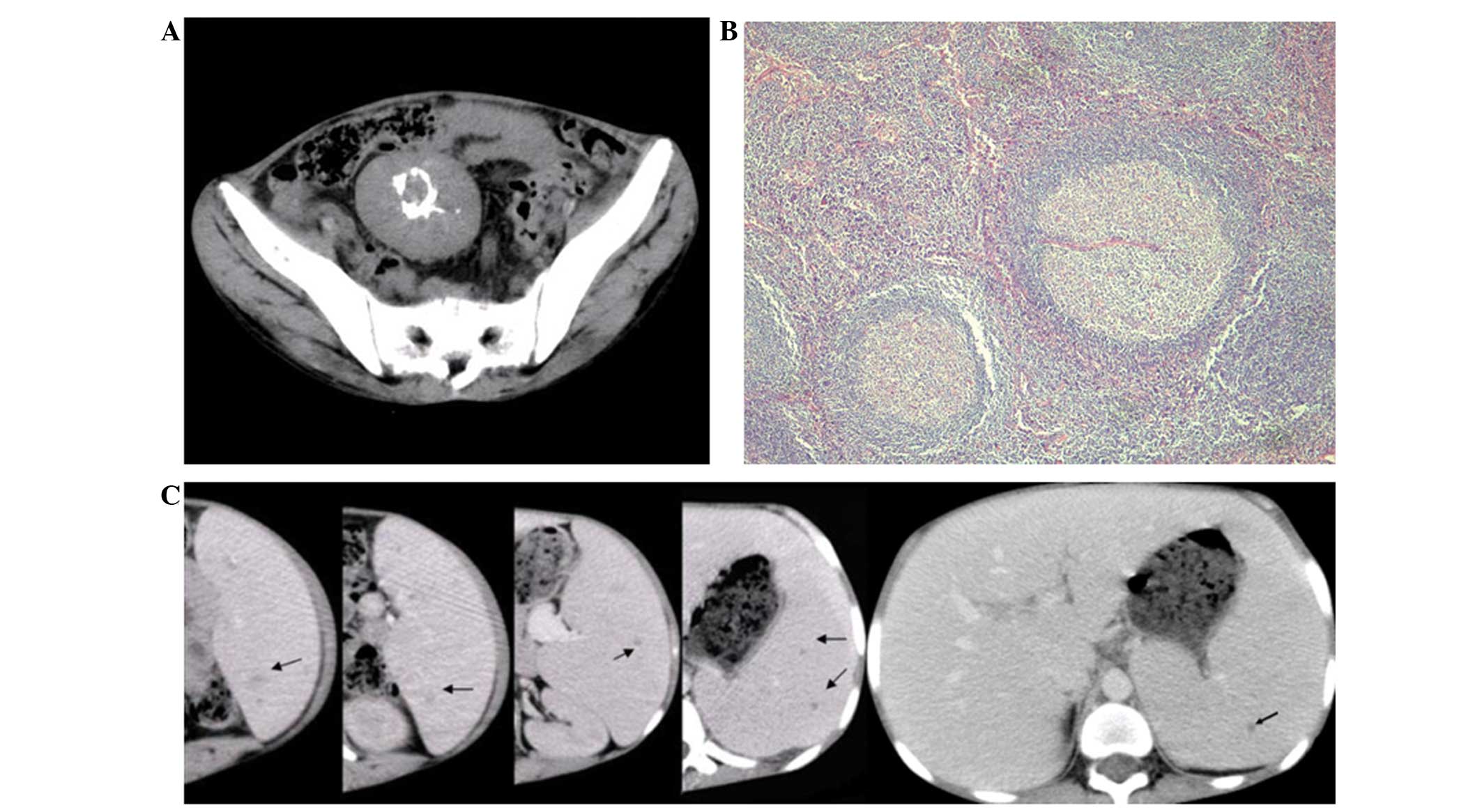
detected by pelvic ultrasonic examination. A moderately enhanced, well-defined mass of 4.8×5.6 cm in size was detected at the mesentery on computed tomography (CT) imaging; centrally located ring-shaped calcification could also be observed. In addition, hepatomegaly and splenomegaly were detected. Scattered small hypo-attenuated nodules were identified in the spleen on the arterial and portal venous phases of enhanced CT images, however, these were isoattenuated on normal CT images (Fig. 1A and C). No abnormalities were observed on liver parenchyma, chest or cervical CT imaging.

Figure 1

(A) A well-demarcated, moderately enhanced mass with ring-shaped calcification in the center was detected in the mesentery. (B) Hematoxylin and eosin-stained section of the mesentery mass showed involuting germinal center surrounded by concentric rings of small lymphocytes penetrated by hyalinized vessel (original magnification, ×200). (C) Enhanced abdominal computed scan on portal venous phase showed hepatomegaly and splenomegaly with scattered small low-attenuated lesions (arrows showing the lesions of littoral cell angioma in the spleen).

As the underlying nature of the mass, nodules of the spleen and hepatomegaly could not be determined in this patient, exploratory laparotomy was performed to remove the mass and spleen, with additional liver biopsy. Subsequent histopathological analysis of the mass was consistent with hyaline-vascular CD, revealing involuting germinal centers surrounded by concentric rings of small lymphocytes penetrated by hyalinized vessels (Fig. 1B). Immunohistochemically, the mass was reactive for CD3±, CD20±, bcl-2 (+) and Ki-67. In addition, pathological and immunohistochemical examination of the spleen showed a hybrid endothelial-histiocytic phenotype, which confirmed a diagnosis of littoral-cell angioma. Fatty degeneration was identified in the specimen of the liver. Following surgery, the patient’s symptoms resolved and laboratory tests normalized. Follow-up was performed once a month, and menstruation and breast development commenced three months following the surgery. At the time of writing, no recurrence had been identified.

Discussion

CD was initially first described in 1954 and is also known as angiofollicular hyperplasia or giant lymph node hyperplasia; it is a rare disorder of lymphoid tissue (7). Little is known about the cause of this disorder, however, it is thought to be infectious, inflammatory, or hamartomatous in nature (1). CD may occur in any area in which lymphoid tissue is normally found, however, it most commonly occurs in the mediastinum (70%). Additionally, extrathoracic sites have been reported in the neck, axilla, mesentery, pelvis, pancreas, adrenal, and retroperitoneum (2).

Clinically, the hyaline-vascular type of CD is typically asymptomatic and tends to be localized, whereas the less common plasma cell variant is occasionally associated with symptoms including fever, anemia, weight loss, night sweats, and polyclonal hypergammaglobulinemia, and is typically multicentric (2). The present case exhibited unique clinical features including amenorrhea, growth retardation and anemia. The mechanism for growth retardation in CD is unclear, and multiple factors may be related to this, with IL-6 being one of the important contributing factors (8). We hypothesize that amenorrhea, which is extremely rare in CD (9), may be associated with the infantile uterus resulting from growth retardation. Anemia is also extremely rare in the hyaline-vascular CD, and Yang et al (8) proposed that elevated IL-6 acts on the IL-6 receptors in liver and tumor lymph node cells, resulting in the enhanced expression of hepcidin, consistent with the role of the IL-6/hepcidin pathway in anemia of chronic disease in CD (9).

LCA, initially described in 1991, is a rare vascular tumor, originating from the splenic littoral cells (10). Due to the limited number of cases, the etiology of LCA remains unclear. The histopathological characteristics of LCA include the presence of anastomosing vascular channels lined with flat and tall endothelial cells, with focal papillary fronds extended into the vascular channels and normal splenic sinuses at the periphery of the lesions in the splenic red pulp. Immunohistochemical analysis also exhibits specific staining patterns, as the tumor shows immunoreactivity with factor 8, CD31 and CD68, which indicates the presence of endothelial and histocytic cells (4).

Clinically, patients with LCA may present with an abdominal mass from splenomegaly or as an incidental finding. Symptoms of hypersplenism (thrombocytopenia and anemia), portal hypertension and pyrexia of unknown origin have also been described, but are rare (5). An association between LCA and malignancy, chronic infection or autoimmune diseases has been proposed (6), however, the coexistence of LCA and CD has not been previously reported in the literature. The association between CD and LCA in the present case was unclear, however, they have previously exhibited an association with altered immune host response (6) thus, we hypothesized that the coexistence of these two rare diseases in one patient may be due to immune disorder.

Unicentric CD exhibits a number of important features on imaging. It is usually shown to be a single, well circumscribed, soft-tissue mass with moderate to intense enhancement on CT, and may be homogeneous or heterogeneous dependent on its size; additionally, low attenuation areas in the mass has been shown to correspond to necrosis (11,12). Intratumoral calcification, found in approximately 5–10% of CD cases, is considered to be another characteristic CT feature of this disease (11). CD typically displays a variety of patterns in the abdomen and pelvis, including punctuate, coarse, flocculent, peripheral and arborizing pattern, which radiates from the center of the mass (2,12); however, the central ring-shaped pattern observed in the present case has not been previously described.

Conversely, the CT features of LCA have been well described, with the majority being isoattenuating to slightly hypoattenuating on non-contrast examinations. On contrast enhancement, early hypoattenuation on arterial and most early portal phase scans may be revealed. Heterogeneous to homogeneous enhancement is present on the late portal phase and delayed phase. However, a number of delayed scans have revealed a complete contrast washout with return to isoattenuation (13). In the current study, the patient exhibited a significantly enlarged spleen with scattered small hypoattenuated nodules on enhanced CT, which was imperceptible on plain CT images.

Surgical resection of the mass is usually curative in localized CD, with dissipation of the symptoms, and splenectomy is curative in littoral-cell angioma. However, local recurrence following subtotal resection has also been described in localized CD (1), as well as long-term complications with malignancy, which has been reported in localized CD and LCA (1,6), therefore, close clinical follow-up is required.

In conclusion, the present study reported an unusual case of unicentric CD accompanied by LCA, anemia, growth retardation and amenorrhea, which was treated with surgery. However, the relationship between unicentric CD and LCA requires further investigation.

References

1 

Jongsma TE, Verburg RJ and Geelhoed-Duijvestijn PH: Castleman’s disease: A rare lymphoproliferative disorder. Eur J Intern Med. 18:87–97. 2007. View Article : Google Scholar : PubMed/NCBI

2 

Kim TJ, Han JK, Kim YH, Kim TK and Choi BI: Castleman disease of the abdomen: imaging spectrum and clinicopathologic correlations. J Comput Assist Tomogr. 25:207–214. 2001. View Article : Google Scholar : PubMed/NCBI

3 

Chronowski GM, Ha CS, Wilder RB, Cabanillas F, Manning J and Cox JD: Treatment of unicentric and multicentric Castleman disease and the role of radiotherapy. Cancer. 92:670–676. 2001. View Article : Google Scholar : PubMed/NCBI

4 

Ertan G, Tekes A, Mitchell S, Keefer J and Huisman TA: Pediatric littoral cell angioma of the spleen: multimodality imaging including diffusion-weighted imaging. Pediatr Radiol. 39:1105–1109. 2009. View Article : Google Scholar : PubMed/NCBI

5 

Tan YM, Chuah KL and Wong WK: Littoral cell angioma of the spleen. Ann Acad Med Singapore. 33:524–526. 2004.PubMed/NCBI

6 

Harmon RL, Cerruto CA and Scheckner A: Littoral cell angioma: a case report and review. Curr Surg. 63:345–350. 2006. View Article : Google Scholar : PubMed/NCBI

7 

Castleman B and Towne VW: CASE records of the Massachusetts General Hospital Weekly Clinicopathological Exercises: Case 40011. N Engl J Med. 250:26–30. 1954. View Article : Google Scholar

8 

Yang M, Wang FD, Han B, et al: Castleman’s disease presenting as prolonged anemia and growth retardation: a case report and literature review. Acta Haematol. 125:125–129. 2011. View Article : Google Scholar

9 

Cammisuli E, Catania V, Santuccio A and Pennisi S: Castleman’s disease: a case report. Ann Ital Chir. 74:713–716. 2003.(In Italian).

10 

Falk S, Stutte HJ and Frizzera G: Littoral cell angioma: A novel splenic vascular lesion demonstrating histiocytic differentiation. Am J Surg Pathol. 15:1023–1033. 1991. View Article : Google Scholar : PubMed/NCBI

11 

Zheng X, Pan K, Cheng J, Dong L, Yang K and Wu E: Localized Castleman disease in retroperitoneum: newly discovered features by multi-detector helical CT. Abdom Imaging. 33:489–492. 2008. View Article : Google Scholar

12 

Meador TL and McLarney JK: CT features of Castleman’s disease of the abdomen and pelvis. AJR Am J Roentgenol. 175:115–118. 2000. View Article : Google Scholar : PubMed/NCBI

13 

Johnson C, Goyal M, Kim B, Wasdahl D and Nazinitsky K: Littoral cell angioma. Clin Imaging. 31:27–31. 2007. View Article : Google Scholar

Related Articles

  • Abstract
  • View
  • Download
  • Twitter
Copy and paste a formatted citation
Spandidos Publications style
Yang L, Zhao S and Liu RB: Unicentric mesenteric Castleman's disease with littoral cell angioma, anemia, growth retardation and amenorrhea: A case report. Oncol Lett 9: 1779-1781, 2015.
APA
Yang, L., Zhao, S., & Liu, R. (2015). Unicentric mesenteric Castleman's disease with littoral cell angioma, anemia, growth retardation and amenorrhea: A case report. Oncology Letters, 9, 1779-1781. https://doi.org/10.3892/ol.2015.2933
MLA
Yang, L., Zhao, S., Liu, R."Unicentric mesenteric Castleman's disease with littoral cell angioma, anemia, growth retardation and amenorrhea: A case report". Oncology Letters 9.4 (2015): 1779-1781.
Chicago
Yang, L., Zhao, S., Liu, R."Unicentric mesenteric Castleman's disease with littoral cell angioma, anemia, growth retardation and amenorrhea: A case report". Oncology Letters 9, no. 4 (2015): 1779-1781. https://doi.org/10.3892/ol.2015.2933
Copy and paste a formatted citation
x
Spandidos Publications style
Yang L, Zhao S and Liu RB: Unicentric mesenteric Castleman's disease with littoral cell angioma, anemia, growth retardation and amenorrhea: A case report. Oncol Lett 9: 1779-1781, 2015.
APA
Yang, L., Zhao, S., & Liu, R. (2015). Unicentric mesenteric Castleman's disease with littoral cell angioma, anemia, growth retardation and amenorrhea: A case report. Oncology Letters, 9, 1779-1781. https://doi.org/10.3892/ol.2015.2933
MLA
Yang, L., Zhao, S., Liu, R."Unicentric mesenteric Castleman's disease with littoral cell angioma, anemia, growth retardation and amenorrhea: A case report". Oncology Letters 9.4 (2015): 1779-1781.
Chicago
Yang, L., Zhao, S., Liu, R."Unicentric mesenteric Castleman's disease with littoral cell angioma, anemia, growth retardation and amenorrhea: A case report". Oncology Letters 9, no. 4 (2015): 1779-1781. https://doi.org/10.3892/ol.2015.2933
Follow us
  • Twitter
  • LinkedIn
  • Facebook
About
  • Spandidos Publications
  • Careers
  • Cookie Policy
  • Privacy Policy
How can we help?
  • Help
  • Live Chat
  • Contact
  • Email to our Support Team