Introduction
Gastrointestinal stromal tumors (GISTs) are the most
common type of gastrointestinal mesenchymal tumor (GIMT), and can
be classified as being of spindle, epithelioid or mixed cell type
based on morphology (1). Recent
studies have identified that a gain-of-function mutation in the
c-kit gene can be found in 95% of GIST cases. This mutation
facilitates ligand-independent activation of the tyrosine kinase
function of KIT [also known as cluster of differentiation (CD)117]
and can be used to aid the diagnosis of GISTs (2,3). GISTs are
commonly diagnosed on the basis of the expression levels of a range
of proteins, which include CD117, CD34, smooth muscle actin (SMA),
desmin and S-100 (4,5). However, a small percentage of GIST cases
are negative for CD117 expression, which can complicate the
diagnosis (6).
The primary treatment modality for GISTs is surgical
removal, with traditional radiation and chemotherapy being
ineffective (7). Furthermore, the
recurrence and metastasis rate following surgical treatment is as
high as 80%, and the five-year survival rate is only 50% (8). Treatment with imatinib offers hope to
GIST sufferers, but this also has limitations (9). Effective decision-making as to which
treatment option to pursue may therefore be aided by accurate
evaluation of the biological behavior of individual GIST cases.
Since its proposal, the precise definition of GIST
has been controversial. Currently, it is universally acknowledged
that GISTs possess malignant potential; however, the classification
of the degree of this malignancy does not have a common standard
(10). It is typically believed that
the tumor size and mitotic activity are the most important criteria
to distinguish benign from malignant tumors (11). The study by Fletcher et al
provides a reasonable and detailed classification that divides
GISTs into four grades, with an extremely low, low, moderate or
high risk of invasion based on tumor size and mitotic activity
(12). However, studies have shown
that certain small tumors that are <2 cm in size and without
detectable mitotic activity possess an invasive capacity (7,10,13). Therefore, this field remains an active
research area, with current efforts focused on seeking novel
molecular markers that have a higher sensitivity and specificity
for the prediction of the biological behavior of tumors (14).
CD171, also known as L1, is an adhesion molecule
that regulates attachments between neighboring cells, or between
cells and their surrounding extracellular matrix (ECM) through
interactions with additional L1 molecules or with other ligands,
such as integrin β1 and CD24 (15).
L1 is important in the development of the nervous system, and high
expression levels of L1 protein have been identified in
nerve-derived tumors and other solid tumors, including pancreatic
cancer, ovarian cancer, melanoma, small cell lung cancer,
colorectal cancer and breast cancer, where it is associated with
metastasis (16,17). Similar to L1, CD24 is also an adhesion
molecule, and is expressed on the surface of human B cells and
cells of the nervous system. Recently, high levels of CD24 have
been identified on cancerous cells of the hematopoietic system and
certain solid tumors. In such cases, CD24 functions as a ligand of
P-selectin and has been indicated to be involved in the invasion
and metastasis of tumors (18).
Integrin β1 is an important receptor for cellular adhesive
molecules, and it plays a central role in the processes of tumor
formation, growth, differentiation and metastasis, by mediating
signal transduction and regulating adhesion between cells or
between a cell and the ECM (19). The
present study examined the expression levels of L1, CD24 and
integrin β1 in human GIST samples, and assessed the association
between these molecules and the invasion, local recurrence and
distant metastasis of cancer cells, and patient prognosis.
Materials and methods
Tissue specimens
The paraffin blocks of 106 cases of GIMT diagnosed
by the Department of Pathology of the Tianjin Medical University
Cancer Institute and Hospital (Tianjin, China) between January 1990
and December 2013 were collected. Based on the morphological
characteristics and immunohistochemical staining of CD117, CD34,
SMA, desmin and S-100 protein, 66 cases of GISTs, 20 cases of
smooth muscle tumors and 20 cases of schwannomas were diagnosed.
All cases were re-examined by a pathologist to confirm the initial
diagnosis. Hematoxylin and eosin (HE)-stained sections from each
tumor were reevaluated for the following features: Tissue type,
presence of necrosis and mitotic activity (observation of 50 fields
obtained at ×400 magnification). This study was approved by the
ethics committee of Tianjin Fifth Central Hospital (Tianjin, China)
and written informed consent was obtained from all patients.
Immunohistochemical staining
The paraffin blocks were cut into sequential 5-µm
sections for HE (Invitrogen Life Technologies, Carlsbad, CA, USA)
and immunohistochemical (IHC) staining. Briefly, the sections were
fixed with 4% paraformaldehyde at room temperature for 30 min, and
washed three times with phosphate-buffered saline (PBS; Beijing
Zhongshan Golden Bridge Biotechnology Co., Ltd., Beijing, China)
for 3 min, followed by treatment with 1% Triton X-100 (Beijing
Zhongshan Golden Bridge Biotechnology Co., Ltd.) and blocking with
goat serum (Invitrogen Life Technologies) for 30 min. Next, the
serum was discarded and the antibody working solution [containing
monoclonal mouse anti-human L1 (1:100; Thermo Fisher Scientific,
Waltham, MA, USA) CD24 (1:75; Thermo Fisher Scientific), intergrin
β1 (1:150; Santa Cruz Biotechnology, Dallas, TX, USA) and Ki-67
(1:200; Dako, Glostrup, Denmark) antibodies] was added for
overnight incubation at 4°C (Table
I). Biotinylated polyclonal goat anti-mouse secondary antibody
(1:200; ZDR-5307, Beijing Zhongshan Golden Bridge Biotechnology
Co., Ltd.) and horseradish peroxidase-labeled streptavidin (Beijing
Zhongshan Golden Bridge Biotechnology Co., Ltd.) were then
sequentially added. Subsequent to coloration, the sections were
stained with hematoxylin, and finally mounted with cover slips. A
known positive plate, obtained from a patient with retroperitoneal
schwannoma (Tianjin Medical University Cancer Institute and
Hospital) was used as a positive control, and the primary antibody
was replaced with PBS in the negative control.
 | Table I.First antibody dilution, pre-treatment
and sources. |
Table I.
First antibody dilution, pre-treatment
and sources.
| First antibody | Clone no. | Dilution | Pre-treatment | Sourcea |
|---|
| L1 | UJ127 | 1:100 | High pressure
retrieval in citric acid buffer (pH 6.0) | NeoMarkers |
| CD24 | SN3b | 1:75 | High pressure
retrieval in citric acid buffer (pH 6.0) | NeoMarkers |
| Integrin β1 | 4B7R | 1:150 | - | Santa Cruz
Biotechnology, Inc. |
| Ki-67 | MIB-1 | 1:200 | High pressure
retrieval in citric acid buffer (pH 6.0) | Dako |
Standards for evaluation of
immunohistochemical staining
Cells with brown staining in the membrane and
cytoplasm were considered L1-, CD24- and integrin β1-positive
cells. Staining results were observed and scored independently by
two pathologists. A total of 10 fields at high magnification were
randomly selected. The percentage of positive cells in the total
number of cells in each field was scored on a scale of 0–3, as
follows: No positive cells, 0; <25% positive cells, 1; 26–50%
positive cells, 2; and >50% positive cells, 3.
Evaluation of the biological behavior
of GISTs
All cases were divided into groups according to the
evaluation standards of the biological behavior of GISTs, as
defined by Fletcher's scheme (Table
II) (12). GIST samples (n=66)
were divided into four groups based on the standards outlined in
Table II, with three cases in group
I, 18 cases in group II, 19 cases in group III and 26 cases in
group IV.
| Table II.Fletcher's scheme for the evaluation
of the biological behavior of gastrointestinal stromal tumors. |
Table II.
Fletcher's scheme for the evaluation
of the biological behavior of gastrointestinal stromal tumors.
| Group | Category
feature | Maximum diameter,
cm | Mitotic count
(no./50 HPF) |
|---|
| I | An extremely low
risk of invasion | <2 | <5 |
| II | A low risk of
invasion | 2–5 | <5 |
| III | A moderate risk of
invasion | <5 | 6–10 |
|
|
| 5–10 | <5 |
| IV | A high risk of
invasion | >5 | >5 |
|
|
| >10 | Any mitotic
figures |
|
|
| Any size | >10 |
Patient follow-up and survival
analysis
Patients were followed up by telephone, letter and a
review of their clinical follow-up records, with their survival or
mortality documented. The cause of mortality was determined in
detail. Only patients that survived or succumbed as a result of
their GISTs were selected for the statistical study of survival
analysis.
Statistical analysis
SPSS statistical software, version 13.0 (SPSS, Inc.,
Chicago, IL, USA), was employed for the statistical analysis.
Immunohistochemical results were analyzed using χ2 tests
and Spearman's rank correlation. Kaplan-Meier survival curve
analysis was employed for the follow-up data, and comparison of
survival rates between groups was also performed with the log-rank
test. P<0.05 was considered to indicate a statistically
significant difference.
Results
Clinical data
Tumor samples from 106 individuals were examined in
this study, with 66 cases of GISTs, 20 cases of smooth muscle
tumors and 20 cases of schwannomas identified. The 66 patients with
GISTs included 36 males and 30 females, aged from 24–77 years, with
a median age of 55 years. There were 58 cases of primary tumors, 8
cases of recurrence and 6 cases of metastasis. Tumors of low
malignant potential exhibited clear borders, and were solid tumors
with grey-white sections and a braided pattern. Tumors with
moderate or high malignant potential had a clearer border or false
membrane, and a number displayed signs of infiltrative growth, such
as invasion of the muscular layer, mucous membrane, retina,
pancreas or spleen. Secondary ulceration was occasionally found on
the digestive tract mucosa surface, and the tumor texture was soft
and was grey-white or grey-red in colour. Bleeding, necrosis and
cystic degeneration were commonly observed in the larger
tumors.
Association between L1 protein level
and the clinical pathological features of GISTs
L1 expression was identified in 57.6% (38/66) of the
GIST cases; this was significantly higher than the percentage for
the control group (P<0.05). However, no significant differences
in L1 expression were found with regard to gender, age, tumor
location or tumor histological type (P>0.05) (Table III).
| Table III.Association between L1 expression and
the clinical pathological features in gastrointestinal stromal
tumor patients. |
Table III.
Association between L1 expression and
the clinical pathological features in gastrointestinal stromal
tumor patients.
| | Positive expression
of L1 | | |
|---|
|
|---|
| Items | Cases, n | Cases, n | Percentage | χ2 | P-value |
|---|
| Gender |
|
|
| 0.405 | >0.05 |
|
Male | 36 | 22 | 61.1 |
|
|
|
Female | 30 | 16 | 53.3 |
|
|
| Age, years |
|
|
| 3.753 | >0.05 |
|
<55 | 22 | 9 | 40.9 |
|
|
|
≥55 | 44 | 29 | 65.9 |
|
|
| Location |
|
|
| 1.972 | >0.05 |
|
Esophagusa | 1 | 0 |
0.0 |
|
|
|
Stomach | 27 | 15 | 55.6 |
|
|
| Small
intestine | 16 | 12 | 75.0 |
|
|
| Large
intestine | 7 | 4 | 57.1 |
|
|
|
Parenteral | 15 | 7 | 46.7 |
|
|
| Histological
type |
|
|
| 0.020 | >0.05 |
| Spindle
cell type | 49 | 28 | 57.1 |
|
|
|
Epithelioid cell type | 5 | 3 | 60.0 |
|
|
| Mixed
cell type | 12 | 7 | 58.3 |
|
|
Expression levels of L1, CD24,
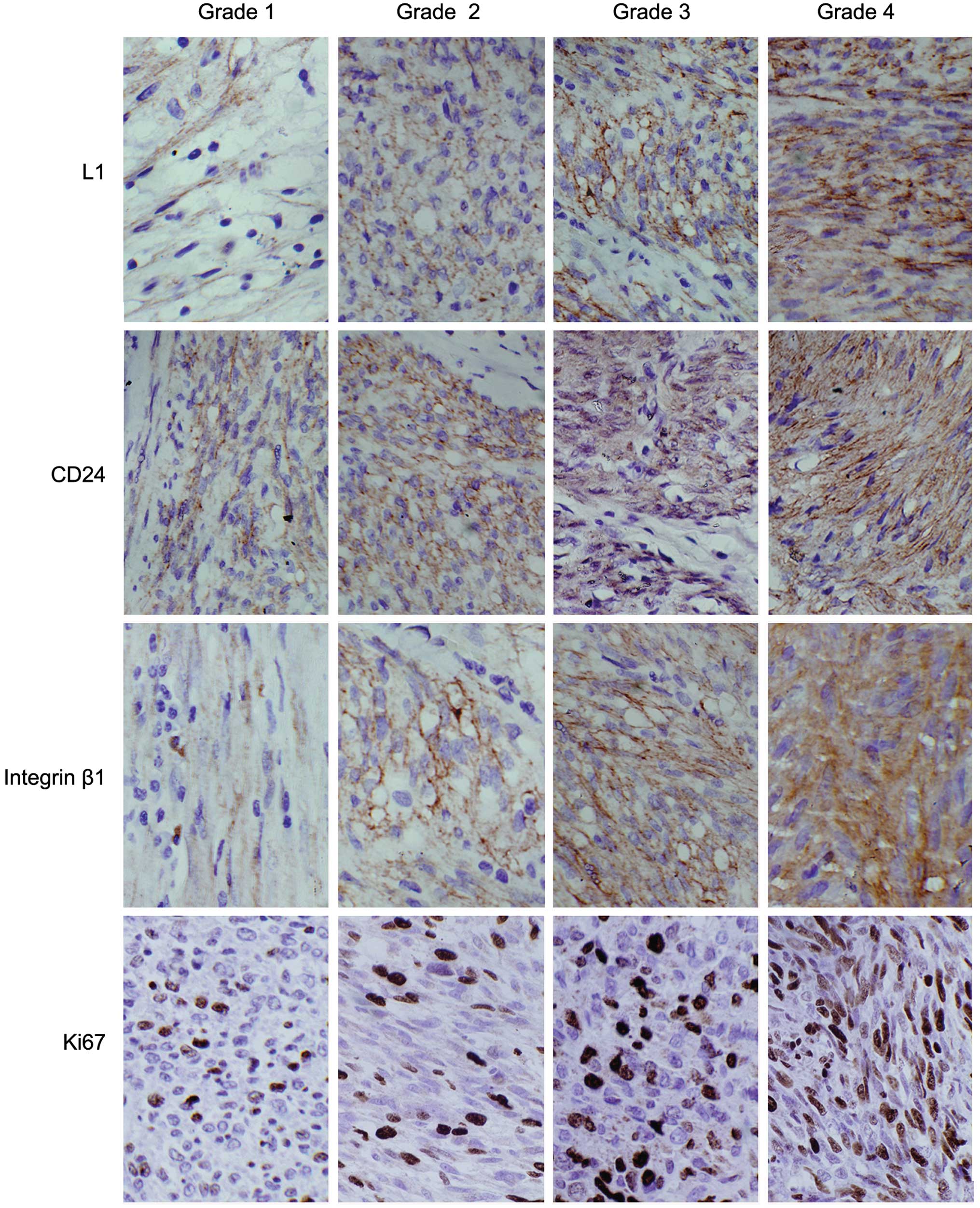
integrin β1 and Ki-67 in GISTs with different invasion risks
The expression rates for L1, CD24, integrin β1 and
Ki-67 in the GIST cases were 57.6% (38/66), 72.7% (48/66), 66.7%
(44/66) and 63.6% (42/66), respectively; all found elevated with an
increased risk of invasion. Statistical analyses revealed
significant differences in L1, integrin β1 and Ki-67 expression in
the different invasive GIST groups (P<0.05), while significant
difference in CD24 expression was found (P>0.05) (Table IV; Fig.
1).
| Table IV.Expression analysis of L1, CD24,
integrin β1 and Ki-67 in gastrointestinal stromal tumors with
different invasive risks. |
Table IV.
Expression analysis of L1, CD24,
integrin β1 and Ki-67 in gastrointestinal stromal tumors with
different invasive risks.
| | | IHC results, n | | | |
|---|
|
|---|
| Target | Grade | Total, n | - | + | ++ | +++ | Positive rates,
% | χ2 | P-value |
|---|
| L1 | I | 3 | 2 | 1 | 0 | 0 | 33.3 | 7.853 | <0.05 |
|
| II | 18 | 12 | 4 | 1 | 1 | 33.3 |
|
|
|
| III | 19 | 7 | 5 | 4 | 3 | 63.2 |
|
|
|
| IV | 26 | 7 | 6 | 7 | 6 | 73.1 |
|
|
| CD24 | I | 3 | 2 | 1 | 0 | 0 | 33.3 | 1.841 | >0.05 |
|
| II | 18 | 6 | 7 | 4 | 1 | 66.7 |
|
|
|
| III | 19 | 4 | 6 | 7 | 2 | 78.9 |
|
|
|
| IV | 26 | 6 | 7 | 7 | 6 | 76.9 |
|
|
| Integrin β1 | I | 3 | 2 | 1 | 0 | 0 | 33.3 | 13.088 | <0.05 |
|
| II | 18 | 10 | 5 | 2 | 1 | 44.4 |
|
|
|
| III | 19 | 6 | 5 | 5 | 3 | 68.4 |
|
|
|
| IV | 26 | 4 | 4 | 10 | 8 | 84.6 |
|
|
| Ki-67 | I | 3 | 3 | 0 | 0 | 0 |
0.0 | 17.682 | <0.05 |
|
| II | 18 | 12 | 4 | 2 | 0 | 33.3 |
|
|
|
| III | 19 | 5 | 6 | 5 | 3 | 73.7 |
|
|
|
| IV | 26 | 4 | 5 | 8 | 9 | 84.6 |
|
|
Correlation between L1, CD24, integrin
β1 and Ki-67 protein expression level in GISTs with increasing risk
of invasion
Spearman's rank correlation analysis was used to
determine the existence of any association between the expression
levels of L1, CD24, integrin β1 and Ki-67 in the GIST cases.
Expression levels of L1, CD24 and integrin β1 were correlated with
increasing invasiveness (r=0.312, P<0.05; r=0.323, P<0.05;
and r=0.298, P<0.05, respectively). L1 and integrin β1
expression levels were also correlated with the Ki-67 proliferation
index (r=0.291, P<0.05; and r=0.379, P<0.05,
respectively).
Expression levels of L1, CD24 and
integrin β1 in GIST, smooth muscle tumor and schwannoma
samples
While positive L1 expression ws identified in 38 out
of the 66 GIST cases and in 15 out of the 20 schwannoma cases, no
L1 expression was observed in the 20 cases of gastrointestinal
smooth muscle tumors. Statistically, there was a significant
difference in L1 expression between the GISTs and smooth muscle
tumors (P<0.05); however, no such significant difference was
identified when the GIST cases were compared with the schwannoma
cases (P>0.05). The expression levels of CD24 and integrin β1 in
the GIST, smooth muscle tumor and schwannoma cases were 72.7, 66.7
and 80.0%, and 70.0, 100.0 and 66.7%, respectively. No differences
in the expression levels of CD24 or integrin β1 were identified
among the three types of tumors (P>0.05) (Fig. 2).
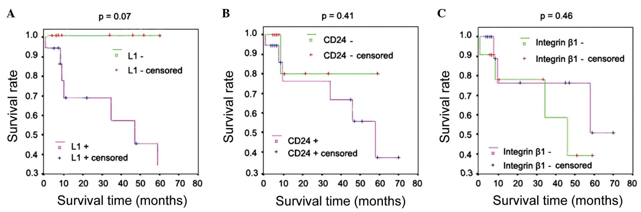
Follow-up results
Of the 66 patients with GISTs, 27 were followed up
over 1–71 months, with an average of 24 months. No significant
correlation was identified between the expression of L1, CD24 or
integrin β1 and patient survival (Table
V); Kaplan-Meier survival curve analysis was also performed
with an identical conclusion (Fig.
3).
| Table V.Correlation between the follow-up
results and the expression of L1, CD24 and integrin β1. |
Table V.
Correlation between the follow-up
results and the expression of L1, CD24 and integrin β1.
| Target | Type | Patients followed
up, n | Duration of
follow-up, months | Succumbed, n | Imatinib, n | Chemotherapy,
n | Radiotherapy,
n | χ2 | P-value |
|---|
| L1 | P | 16 | 1–71 | 7 | 1 | 0 | 1 | 3.26 | 0.07 |
|
| N | 11 | 4–59 | 0 | 4 | 3 | 0 |
|
|
| CD24 | P | 18 | 1–71 | 6 | 2 | 1 | 0 | 0.69 | 0.41 |
|
| N | 9 | 8–60 | 1 | 3 | 2 | 1 |
|
|
| Integrin β1 | P | 17 | 1–71 | 4 | 1 | 0 | 1 | 0.53 | 0.46 |
|
| N | 10 | 6–59 | 3 | 4 | 3 | 0 |
|
|
Discussion
In 1983, Mazur and Clark discovered that the gastric
stromal tumors once diagnosed as leiomyoma (smooth muscle tumors
and leiomyosarcoma) or schwannoma lacked immunohistochemical
staining for SMA or S-100 protein, and that no particular cell
features could be observed under the electron microscope (1). Therefore, the term ‘gastric stromal
tumor’ with a neutral tissue origin was suggested (1). Recently, a gain-of-function mutation of
c-kit has been found in the majority of GISTs, which results in the
ligand-independent activation of tyrosine kinase and the expression
of CD117 (KIT), providing the basis for the clinical diagnosis of
GISTs (2,20). However, a small proportion of GIST
cases with no CD117 expression remain, which introduces
difficulties for the diagnosis and differential diagnosis of GISTs.
The accurate evaluation of the biological behavior of GISTs is
associated with the treatment and outcome of patients. The dispute
has existed since the concept of GIST was proposed. Although it is
well-established that all the GISTs have malignant potential, its
malignant degree classification does not have a uniform standard.
The tumor size and mitotic activity are typically considered as the
most important criteria to differentiate between benign and
malignant tumors, which is largely consistent with the
classification standard proposed by Fletcher et al (12). However, studies have suggested that
certain tumors, with a size of <2 cm and without mitotic
activity, may also metastasize (21,22).
Therefore, a search for markers of the biological behavior of GIST
still has important research value for clinical application.
L1 is a transmembrane adhesion molecule originally
believed to be expressed only in the nervous system, where it is
involved in mediating adhesion between neurons, axonal spontaneous
contraction, synapse formation, axon growth and neuronal migration
(23). Expression of L1 has since
been identified in various tumors of the intestine, urogenital
tract, lung, rectum and kidney, and in rhabdomyosarcoma (24). High expression levels of L1 were first
identified in GISTs in 2006 using immunohistochemical staining,
with 73.6% (53/72) of sampled tumors expressing L1 at a high level
(25). However, L1 is not expressed
in fibromatosis or in gastrointestinal smooth muscle tumors.
CD24 is a highly glycosylated adhesion molecule of
low molecular weight, which has been found to be expressed at high
levels on the cell membrane within human hematopoietic system
tumors and solid tumors, including primary liver, gastric,
colorectal and breast cancers (26–28). As an
adhesion molecule receptor, integrin β1 plays a role in the
developmental processes of tumors, mainly by mediating the adhesion
between cells and between the cell and the ECM (29,30). The
present data found that L1, CD24 and integrin β1 were expressed at
a significantly higher level in GISTs than in normal control
tissues. Smooth muscle tumors had barely detectable L1 expression,
while there was no significant difference in L1 expression between
GISTs and schwannomas. No differences in CD24 and integrin β1
expression were identified across the three types of tumors. These
results suggested that L1, CD24 and integrin β1 could be useful
markers in the differential diagnosis of GISTs.
L1 is an adhesion molecule with a role in regulating
cell attachments and an important signaling molecule (31). CD24 is an adhesion molecule that
mainly localizes to the membrane surface (32), and integrin β1 is also an adhesion
molecule receptor (33). The
interaction of these three molecules has played a positive role in
the migration of tumor cells (34).
Fogel et al (35) found that
the overexpression of L1 in melanoma promoted tumor progression and
metastasis, and was closely associated with the prognosis of the
patients. Gavert et al (36)
determined that L1 could promote the progression and metastasis of
colon cancer with the use of cell culture and animal models, and
further elucidated the responsible mechanism. Firstly, L1 itself
has a weak adhesion capacity, but this can be enhanced through
interaction with homotypic or heterotypic molecules (37). The combination of L1 and CD24
molecules alters their conformation, facilitating the activation of
endothelial cells and platelets, and enhancing the attachment of L1
with ECM ligands (38). Furthermore,
the interaction of CD24 and L1 can increase the intracellular
calcium concentration and strengthen signal transduction, promoting
adhesion (39). Secondly, the
integrin family contains multiple L1 receptors, and L1 mainly
combines with integrin β1 in the presence of Mn2+. This
combination not only mediates the transfer of L1 into cells to
reduce attachments between the tumor cells, but also induces the
increased expression of adhesion molecule receptors in vascular
endothelial cells and the ECM, promoting the adhesion and migration
of tumor cells in these environments (40). Lastly, integrin β1 is the receptor for
CD24, and its interaction with L1 is regulated by CD24, likely
through a change in the conformation of an
arginine-glycine-aspartate sequence following the binding of CD24
and L1 (41). The present data
revealed that the expression levels of L1, CD24 and integrin β1
were significantly correlated in the GIST cases, and that they were
also associated with the expression of Ki-67. Moreover, L1 and
integrin β1 levels were significantly increased with increased
invasion risk in the GISTs. However, this was not the case for CD24
expression, indicating the synergistic effects of L1, CD24 and
integrin β1 in the progression of GISTs. Therefore, the detection
of L1 and integrin β1 in combination with Ki-67 favors an accurate
prediction of the biological behavior of GISTs, while elucidation
of the full function of CD24 in GIST requires further
investigation.
Acknowledgements
This study was supported by the China National
Natural Scientific Fund (grant no. 81000901), and the Tianjin
Binhai New Area Health Bureau Science and Technology Projects
(grant no. 2011BHKY009).
References
|
1
|
Mazur MT and Clark HB: Gastric stromal
tumors. Reappraisal of histogenesis. Am J Surg Pathol. 7:507–519.
1983. View Article : Google Scholar : PubMed/NCBI
|
|
2
|
Corless CL, Barnett CM and Heinrich MC:
Gastrointestinal stromal tumours: origin and molecular oncology.
Nat Rev Cancer. 11:865–878. 2011.PubMed/NCBI
|
|
3
|
Hamilton SR and Aaltonen LA: Mesenchymal
tumours of the stomachWorld Health Organization Classification of
Tumours. Pathology and Genetics of Tumours of the Digestive System.
IARC Press; Lyon: pp. 62–65. 2003
|
|
4
|
Kiśluk J, Gryko M, Guzińska-Ustymowicz K,
et al: Immunohistochemical diagnosis of gastrointestinal stromal
tumors-an analysis of 80 cases from 2004 to 2010. Adv Clin Exp Med.
22:33–39. 2013.PubMed/NCBI
|
|
5
|
Bülbül Doğusoy G: Turkish GIST Working
Group: Gastrointestinal stromal tumors: A multicenter study of 1160
Turkish cases. Turk J Gastroenterol. 23:203–211. 2012.PubMed/NCBI
|
|
6
|
Montgomery E, Torbenson MS, Kaushal M, et
al: Beta-catenin immunohistochemistry separates mesenteric
fibromatosis from gastrointestinal stromal tumor and sclerosing
mesenteritis. Am J Surg Pathol. 26:1296–1301. 2002. View Article : Google Scholar : PubMed/NCBI
|
|
7
|
Sugiyama T: Progress in new diagnosis and
therapeutic strategy for gastrointestinal malignancy: focus on new
molecular-targeted treatments. Digestion. 91:7–12. 2015.PubMed/NCBI
|
|
8
|
Miettinen M and Lasota J: Gastrointestinal
stromal tumors. Gastroenterol Clin North Am. 42:399–415. 2013.
View Article : Google Scholar : PubMed/NCBI
|
|
9
|
Patel S: Exploring novel therapeutic
targets in GIST: focus on the PI3K/Akt/mTOR pathway. Curr Oncol
Rep. 15:386–395. 2013. View Article : Google Scholar : PubMed/NCBI
|
|
10
|
Le Cesne A and Blay JY: Medical therapy of
GIST; from palliative to curative treatment. Bull Acad Natl Med.
196:861–874. 2012.[(In French)]. PubMed/NCBI
|
|
11
|
Emile JF: Histology and molecular biology
of GIST. Bull Acad Natl Med. 196:835–844. 2012.[(In French)].
PubMed/NCBI
|
|
12
|
Fletcher CD, Beman JJ, Corless C, et al:
Diagnosis of gastrointestinal stromal tumors: A consensus approach.
Hum Pathol. 33:459–465. 2002. View Article : Google Scholar : PubMed/NCBI
|
|
13
|
Bareck E, Ba-Ssalamah A, Brodowicz T, et
al: Austrian representatives of Medical and Surgical Oncology,
Pathology, Radiology, Nuclear Medicine, Gastroenterology, and
Laboratory Medicine: Gastrointestinal stromal tumors: diagnosis,
therapy and follow-up care in Austria. Wien Med Wochenschr.
163:137–152. 2013. View Article : Google Scholar : PubMed/NCBI
|
|
14
|
Rajendra R, Pollack SM and Jones RL:
Management of gastrointestinal stromal tumors. Future Oncol.
9:193–206. 2013. View Article : Google Scholar : PubMed/NCBI
|
|
15
|
Figge C, Loers G, Schachner M and Tilling
T: Neurite outgrowth triggered by the cell adhesion molecule L1
requires activation and inactivation of the cytoskeletal protein
cofilin. Mol Cell Neurosci. 49:196–204. 2012. View Article : Google Scholar : PubMed/NCBI
|
|
16
|
Siesser PF and Maness PF: L1 cell adhesion
molecules as regulators of tumor cell invasiveness. Cell Adh Migr.
3:275–277. 2009. View Article : Google Scholar : PubMed/NCBI
|
|
17
|
Tischler V, Pfeifer M, Hausladen S, et al:
L1CAM protein expression is associated with poor prognosis in
non-small cell lung cancer. Mol Cancer. 10:1272011. View Article : Google Scholar : PubMed/NCBI
|
|
18
|
Lieberoth A, Splittstoesser F,
Katagihallimath N, et al: Lewis(x) and alpha2,3-sialyl glycans and
their receptors TAG-1, Contactin, and L1 mediate CD24-dependent
neurite outgrowth. J Neurosci. 29:6677–6690. 2009. View Article : Google Scholar : PubMed/NCBI
|
|
19
|
Zhan P, Liu L, Liu B and Mao XG:
Expression of integrin β1 and its significance in squamous cell
carcinoma of the cervix. Mol Med Rep. 9:2473–2478. 2014.PubMed/NCBI
|
|
20
|
Hirota S, Isozaki K, Moriyama Y, et al:
Gain-of-function mutations of c-kit in human gastrointestinal
stromal tumors. Science. 279:577–580. 1998. View Article : Google Scholar : PubMed/NCBI
|
|
21
|
Miettinen M and Lasota J: Gastrointestinal
stromal tumors: pathology and prognosis at different sites. Semin
Diagn Pathol. 23:70–83. 2006. View Article : Google Scholar : PubMed/NCBI
|
|
22
|
Yi JH, Park BB, Kang JH, et al:
Retrospective analysis of extra-gastrointestinal stromal tumors.
World J Gastroenterol. 21:1845–1850. 2015. View Article : Google Scholar : PubMed/NCBI
|
|
23
|
Raveh S, Gavert N and Ben-Ze'ev A: L1 cell
adhesion molecule (L1CAM) in invasive tumors. Cancer Lett.
282:137–145. 2009. View Article : Google Scholar : PubMed/NCBI
|
|
24
|
Zhang C, Fan Y and Fu L: Research
development of L1-CAM (CD171) in human cancer. Zhonghua Bing Li Xue
Za Zhi. 42:574–576. 2013.[(In Chinese)]. PubMed/NCBI
|
|
25
|
Kaifi JT, Strelow A, Schurr PG, et al: L1
(CD171) is highly expressed in gastrointestinal stromal tumors. Mod
Pathol. 19:399–406. 2006. View Article : Google Scholar : PubMed/NCBI
|
|
26
|
Sagiv E and Arber N: The novel oncogene
CD24 and its arising role in the carcinogenesis of the GI tract:
from research to therapy. Expert Rev Gastroenterol Hepatol.
2:125–133. 2008. View Article : Google Scholar : PubMed/NCBI
|
|
27
|
Su MC, Hsu C, Kao HL and Jeng YM: CD24
expression is a prognostic factor in intrahepatic
cholangiocarcinoma. Cancer Lett. 235:34–39. 2006. View Article : Google Scholar : PubMed/NCBI
|
|
28
|
Jacob J, Bellach J, Grützmann R, et al:
Expression of CD24 in adenocarcinomas of the pancreas correlates
with higher tumor grades. Pancreatology. 4:454–460. 2004.
View Article : Google Scholar : PubMed/NCBI
|
|
29
|
Oh BY, Kim KH, Chung SS, et al: Role of
β1-integrin in colorectal cancer: case-control study. Ann
Coloproctol. 30:61–70. 2014. View Article : Google Scholar : PubMed/NCBI
|
|
30
|
Chen SY, Lin JS and Yang BC: Modulation of
tumor cell stiffness and migration by type IV collagen through
direct activation of integrin signaling pathway. Arch Biochem
Biophys. 555–556:1–8. 2014. View Article : Google Scholar
|
|
31
|
Hortsch M, Nagaraj K and Mualla R: The L1
family of cell adhesion molecules: a sickening number of mutations
and protein functions. Adv Neurobiol. 8:195–229. 2014. View Article : Google Scholar : PubMed/NCBI
|
|
32
|
Jaggupilli A and Elkord E: Significance of
CD44 and CD24 as cancer stem cell markers: an enduring ambiguity.
Clin Dev Immunol. 2012:7080362012. View Article : Google Scholar : PubMed/NCBI
|
|
33
|
Jahangiri A, Aghi MK and Carbonell WS: β1
integrin: Critical path to antiangiogenic therapy resistance and
beyond. Cancer Res. 74:3–7. 2014. View Article : Google Scholar : PubMed/NCBI
|
|
34
|
Liakhovich AV and Aksenov NL: Topography
of cell ahesion molecules CD9, CD24, L1 and N-CAM on the surface of
neuroblastoma cells studied using chemical cross linking.
Tsitologiia. 42:399–403. 2000.PubMed/NCBI
|
|
35
|
Fogel M, Mechtersheimer S, Huszar M, et
al: L1 adhesion molecule (CD 171) in development and progression of
human malignant melanoma. Cancer Lett. 189:237–247. 2003.
View Article : Google Scholar : PubMed/NCBI
|
|
36
|
Gavert N, Conacci-Sorrell M, Gast D, et
al: L1, a novel target of β-catenin signaling, transforms cells and
is expressed at the invasive front of colon cancers. J Cell Biol.
168:633–642. 2005. View Article : Google Scholar : PubMed/NCBI
|
|
37
|
Moulla A, Miliaras D, Sioga A, et al: The
immunohistochemical expression of CD24 and CD171 adhesion molecules
in borderline ovarian tumors. Pol J Pathol. 64:180–184. 2013.
View Article : Google Scholar : PubMed/NCBI
|
|
38
|
Baumann P, Cremers N, Kroese F, et al:
CD24 expression causes the acquisition of multiple cellular
properties associated with tumor growth and metastasis. Cancer Res.
65:10783–10793. 2005. View Article : Google Scholar : PubMed/NCBI
|
|
39
|
Kleene R, Yang H, Kutsche M and Schachner
M: The neural recognition molecule L1 is a sialic acid-binding
lectin for CD24, which induces promotion and inhibition of neurite
outgrowth. J Biol Chem. 276:21656–21663. 2001. View Article : Google Scholar : PubMed/NCBI
|
|
40
|
Sammar M, Aigner S and Altevogt P:
Heat-stable antigen (mouse CD24) in the brain: dual but distinct
interaction with P-selectin and L1. Biochim Biophys Acta.
1337:287–294. 1997. View Article : Google Scholar : PubMed/NCBI
|
|
41
|
Kadmon G, Imhof BA, Altevogt P and
Schachner M: Adhesive hierarchy involving the cell adhesion
molecules L1, CD24, and alpha 6 integrin in murine neuroblastoma
N2A cells. Biochem Biophys Res Commun. 214:94–101. 1995. View Article : Google Scholar : PubMed/NCBI
|