Introduction
The most common malignancy found in American males
is prostate cancer (PCa). The disseminated disease is responsible
for cancerous mortality and morbidity (1,2). With the
introduction and application of the screening for PCa using
prostate-specific antigen, the diagnosis of clinically localized
PCa has also been increased sharply (3). The use of biologically significant
biomarkers may therefore stratify the risk in this group of
patients.
Androgen receptor (AR) protein is expressed in
nearly all types of PCa, i.e., primary and metastatic (4). The study by Visakorpi et al was
the first to report the amplification of the AR gene together with
a gain of chromosome X, and suggested a possible mechanism for the
progression of PCa following hormone therapy (5). However, the precise mechanism for the
association between AR gene amplification and the development of
hormone-refractory PCa remains unknown. Androgens are significant
in the growth of normal and malignant prostate cells via AR; the AR
binds testosterone or dihydrotestosterone and then stimulates the
transcription of androgen-sensitive genes (6). Similar to other proteins in the steroid
receptor family, the AR contains separate regions responsible for
hormone binding, DNA binding and transcriptional activation
(transactivation). AR signaling has been widely studied in the
pathogenesis and progression of PCa, the underlying mechanisms of
which are believed to involve AR gene amplification, AR gene
mutations, elevated AR mRNA or ligand-independent AR activation by
growth factors/cytokines. Androgen deprivation therapy is the main
therapeutic option for the treatment of advanced cases (7–9). In other
words, the AR gene is an important factor in the growth and
progression of CaP and hence, the majority of PCa is
androgen-dependent (10,11). Additionally, in multiple studies, it
has been demonstrated that the amplification and overexpression of
the AR gene is evident in PCa patients whose tumors are refractory
to androgen ablation therapy (12).
The present study compared AR protein expression,
and clinical characteristics and survival in patients with PCa
whose tumors showed or did not show AR amplification and in
X-chromosome polysomy.
Materials and methods
Specimen source and tissue array
The present study was approved by the Ethics Review
Board of Wenzhou Central Hospital (Wenzhou, Zhejiang, China) and
all patients provided written informed consent. All samples were
collected from Wenzhou Central Hospital between July 2012 and
December 2013. A total of 37 male patients were selected by
analyzing their medical records. Specimens were collected following
detection of an increase in serum prostate-specific antigen (PSA)
level and urinary retention, treated by transurethral resection
following androgen deprivation therapy (Table I). The age range of the patients was
58–89 years (Table I).
 | Table I.Clinical characteristics and mean
optical density of AR expression. |
Table I.
Clinical characteristics and mean
optical density of AR expression.
| A, Non-amplified
AR |
|---|
|
|---|
|
| Clinical
information | AR | X | MOD |
|---|
|
|
|
|
|
|
|---|
| Spc | Age, years | Stage | PSA | GS | AD method | MOS B/w AD + TA | Sur A/f TA (MOS) | Sur A/f AD (MOS) | Copy | SR | % SR (>2) | % SR (>3) | Copy | % X Copy (>2) | % X Copy (>3) | AR exp |
|---|
| 1 | 62 | T3bN1M0 | 32.9 | 9 | FA | 17 | 9 | 26 | 1.10 | 1.00 | 3 | 0 | 1.13 | 23 | 0 | 0.2989 |
| 2 | 64 | T3bN1M0 | 31.8 | 9 | FA | 56 | 13 | 69 | 1.38 | 0.98 | 0 | 0 | 1.30 | 11 | 0 | 0.0923 |
| 3 | 58 | T4NxM1b | 6.2 | 9 | FA | 29 | 5 | 34 | 1.04 | 0.96 | 4 | 0 | 1.04 | 7 | 7 | 0.2333 |
| 4 | 62 | T2bNxM1b | 110 | 10 | PH | NI | 21 | 21 | 1.03 | 1.00 | 7 | 0 | 1.03 | 28 | 0 | 0.4345 |
| 5 | 64 | T3bN1M1b | 0.9 | 9 | Orchiectomy | 39 | 43 | 82 | 2.03 | 1.00 | 8 | 0 | 1.98 | 14 | 0 | 0.2134 |
| 6 | 79 | T3bN1M0 | 38 | 10 | LH-RH | 28 | 11 | 39 | 1.12 | 1.02 | 3 | 0 | 1.10 | 21 | 0 | 0.0896 |
| 7 | 78 | T1bNxM1b | 65.1 | 9 |
Diethylstilbesterol | 20 | 17 | 37 | 1.36 | 1.03 | 18 | 1 | 1.30 | 21 | 1 | 0.1844 |
| 8 | 83 | T3aNxM1b | 5.7 | 9 | Orchiectomy | 13 | 11 | 24 | 1.19 | 1.00 | 2 | 0 | 1.15 | 5 | 0 | 0.3912 |
| 9 | 87 | T3N2M1b | 51.2 | 9 | LH-RH+FA | 23 | 12 | 35 | 17.00 | 1.11 | 5 | 0 | 17.00 | 100 | 100 | 0.1832 |
| 10 | 73 | T3bNxM1b | 400 | 9 | FA | 16 | 4 | 20 | 1.60 | 0.91 | 6 | 0 | 1.50 | 13 | 0 | 0.2989 |
| 11 | 71 | T3N2M1b | 4 | 10 | LH-RH | 45 | 38 | 83 | 1.90 | 0.99 | 9 | 0 | 1.81 | 15 | 0 | 0.2344 |
| 12 | 66 | T2bNxM1b | 78.1 | 8 | Orchiectomy | 59 | 7 | 66 | 1.26 | 1.02 | 33 | 2 | 1.12 | 39 | 32 | 0.2283 |
| 13 | 79 | Tb3N1M0 | 45.3 | 9 | Orchiectomy | 45 | 37 | 82 | 1.50 | 0.99 | 0 | 0 | 1.49 | 1 | 0 | 0.1345 |
| 14 | 72 | T1bNxM1b | 23.6 | 9 |
Diethylstilbesterol | 27 | 13 | 40 | 2.09 | 1.00 | 0 | 0 | 2.01 | 9 | 0 | 0.2732 |
| 15 | 81 | T3bN1M1b | 8.9 | 9 | Orchiectomy | 33 | 29 | 62 | 1.40 | 1.21 | 2 | 0 | 1.34 | 21 | 0 | 0.2896 |
| 16 | 69 | T4NxM1b | 6 | 9 | LH-RH+FA | 29 | 8 | 37 | 2.10 | 1.00 | 56 | 4 | 2.11 | 61 | 4 | 0.1891 |
| 17 | 65 | T2bNxMx | 145 | 8 | Orchiectomy | 9 | 15 | 24 | 1.90 | 1.24 | 5 | 0 | 1.89 | 9 | 0 | 0.1892 |
| 18 | 76 | T3bN1M0 | 0.7 | 8 | FA | 11 | 14 | 25 | 1.98 | 1.02 | 7 | 0 | 1.91 | 22 | 0 | 0.2985 |
| 19 | 80 | T1bNxM1b | 67.3 | 10 | LH-RH+FA | 19 | 18 | 37 | 1.78 | 1.01 | 8 | 0 | 1.78 | 34 | 0 | 0.1189 |
| 20 | 86 | T3aNxM1b | 65.0 | 10 | Orchiectomy+FA | 67 | 56 | 123 | 17.00 | 1.00 | 9 | 0 | 17.00 | 100 | 100 | 0.2177 |
| 21 | 73 | T2bNxM1b | 45.5 | 8 |
Diethylstilbesterol | 78 | 15 | 93 | 1.34 | 1.11 | 5 | 0 | 1.31 | 61 | 0 | 0.1365 |
| 22 | 79 | T3bN1M0 | 5.8 | 9 | LH-RH | 81 | 17 | 98 | 1.42 | 1.00 | 13 | 1 | 1.40 | 43 | 1 | 0.2111 |
| 23 | 70 | T2bNxMx | 5.2 | 9 | Orchiectomy | 69 | 6 | 75 | 1.78 | 0.98 | 7 | 0 | 1.71 | 9 | 0 | 0.2832 |
| 24 | 84 | T3aNxM1b | 34.1 | 9 | Orchiectomy | 16 | 19 | 35 | 16.00 | 1.00 | 8 | 0 | 16.00 | 81 | 80 | 0.4512 |
|
| B, Amplified
AR |
|
|
| Clinical
information | AR | X | MOD |
|
|
|
|
|
|
| Spc | Age, years | Stage | PSA | GS | AD method | MOS B/w AD +
TA | Sur A/f TA
(MOS) | Sur A/f AD
(MOS) | Copy | SR | % SR (>2) | % SR (>3) | Copy | % X Copy
(>2) | % X Copy
(>3) | AR exp |
|
| 25 | 65 | TXNM1b | 14.9 | 8 | Orchiectomy+FA | 41 | 9 | 50 | 3.24 | 2.12 | 48 | 39 | 1.40 | 5 | 0 | 0.3176 |
| 26 | 69 | T4NxM0 | 28.3 | 8 | LH-RH | 42 | 11 | 53 | 4.12 | 3.42 | 42 | 30 | 1.13 | 9 | 0 | 0.3809 |
| 27 | 80 | TxN1Mx | 9.9 | 9 |
Diethylstilbesterol | 39 | 19 | 58 | 4.32 | 3.43 | 39 | 29 | 1.39 | 36 | 4 | 0.3302 |
| 28 | 89 | T4NxM1b | 23.6 | 9 | Orchiectomy | 56 | 6 | 62 | 3.12 | 2.98 | 36 | 32 | 1.02 | 17 | 0 | 0.382 |
| 29 | 72 | TXNM1b | 190 | 9 | Orchiectomy+FA | 28 | 17 | 45 | 2.60 | 1.96 | 37 | 29 | 1.22 | 47 | 9 | 0.329 |
| 30 | 79 | T3bN0M0 | 15.2 | 10 | LH-RH+FA | 13 | 10 | 23 | 2.78 | 1.98 | 89 | 58 | 1.56 | 5 | 0 | 0.48 |
| 31 | 73 | T4NxM1b | 8.8 | 8 | Orchiectomy | 40 | 23 | 63 | 13.10 | 11.6 | 66 | 44 | 1.27 | 8 | 0 | 0.4591 |
| 32 | 68 | T4NxM0 | 11.2 | 9 | LH-RH | 37 | 31 | 68 | 14.6 | 13.8 | 86 | 71 | 2.56 | 49 | 7 | 0.2901 |
| 33 | 70 | T3bN0M0 | 17 | 8 | LH-RH+FA | 13 | 12 | 25 | 12.2 | 11.6 | 56 | 46 | 2.58 | 5 | 0 | 0.23 |
| 34 | 65 | TXNM1b | 21.2 | 9 | Orchiectomy+FA | 23 | 19 | 42 | 11.3 | 10.9 | 87 | 67 | 1.18 | 18 | 0 | 0.3567 |
| 35 | 76 | T4NxM1b | 16.6 | 10 | Orchiectomy | 28 | 25 | 53 | 4.33 | 4.67 | 61 | 49 | 1.91 | 60 | 45 | 0.378 |
| 36 | 66 | TxN1Mx | 26.7 | 9 |
Diethylstilbesterol | 33 | 29 | 62 | 2.67 | 1.97 | 34 | 29 | 1.87 | 4 | 0 | 0.3871 |
| 37 | 78 | TXNM1b | 8.2 | 9 | Orchiectomy+FA | 37 | 28 | 65 | 2.98 | 1.89 | 41 | 38 | 1.05 | 14 | 3 | 0.3409 |
The tissue array was constructed from 37 duplicate
samples of recurrent PCa and 9 samples of adenofibromyomatous
hyperplasia. Tissue preparation artifacts were marked for sampling
and 2-mm diameter punch biopsies were obtained. The tissue areas
were free of necrosis and cauterization injury. Sections (5-µm
thick) were cut from each paraffin block containing a recurrent PCa
or adenofibromyomatous hyperplasia sample, and mounted on slides,
followed by staining with hematoxylin and eosin, and
immunohistochemical analysis.
Immunohistochemical detection
The tissue array sections (BioMax; Guangzhou, China)
were used for immunohistochemical detection according to the
manufacturer's instructions. Briefly, the sections underwent
antigen retrieval in Citra buffer (Biogenex, Shanghai, China) and
were cooled to room temperature for pre-incubation with 2% normal
horse serum for 15 min at 37°C followed by incubation with
monoclonal anti-human AR antibody (F39.4.1; 1:200; Biogenex) for 1
h. The sections were then treated with biotinylated anti-mouse
immunoglobulin G (1:200) for 15 min at 37°C, followed by
avidin-biotin complex amplification (Vector, Beijing, China). The
signals were visualized using diaminobenzidine (Vector). The mean
optical density (MOD) of immunostaining was measured using a Zeiss
Axioskop microscope (Zeiss, Beijing, China), a 3-chip CCD camera
C5810, (Hamamatsu, Beijing, China) and a camera control unit
(Hamamatsu). Immunopositivity and immunonegativity were determined
using a linear discriminant analysis method.
Fluorescence in situ hybridization
(FISH) protocol
The 5-µm PCa tissue slides were deparaffinized,
treated with 0.2 N HCl, incubated in 1 M sodium thiocyanate and
immersed in protease solution (Vysis Inc., Downers Grove, IL, USA)
for 10 min at 37°C. The tissues were then fixed with 10% formalin
for 10 min, denatured for 5 min at 72°C and sequentially incubated
in 75, 90 and 100% ethanol. The tissues were then treated with
proteinase K for 6 min at 37°C, followed by their dehydration and
hybridization.
Next, two-colored FISH was performed, as described
previously (13), using spectrum
orange-labeled AR and spectrum green-labeled X-chromosome
centromere region DNA probes (Vysis). Briefly, tissue array
sections were hybridized with 3 µl of each probe and Cot1-DNA (1
µg/µl; Vysis) overnight at 37°C in a humidified atmosphere. The
slides were then was hed for counterstaining with 0.2 µM
4′,6′-diamidino-2-phenylindole hydrochloride. FISH signals were
scored with a fluorescence microscope (Zeiss) equipped with two
double-band pass filters using 40X and 100X objective lenses. The
number of AR gene signals and X centromere signals was evaluated by
visual analysis of 800 to 1,200 nuclei per specimen. AR
amplification was present if the AR to X ratio exceeded 1.5
(14), and X polysomy was present if
the number of X centromere signals exceeded an average of 2 signals
per cell (15).
Statistical data analysis
Medians and interquartile ranges are used to
describe the data. Optical density is presented as the mean ±
standard deviation. The data were not evenly distributed, so a
Wilcoxin analysis was used for comparison of the groups.
Kaplan-Meier plot was used for comparison of the survival rates via
log-rank analysis.
Results
Immunohistochemistry image
analysis
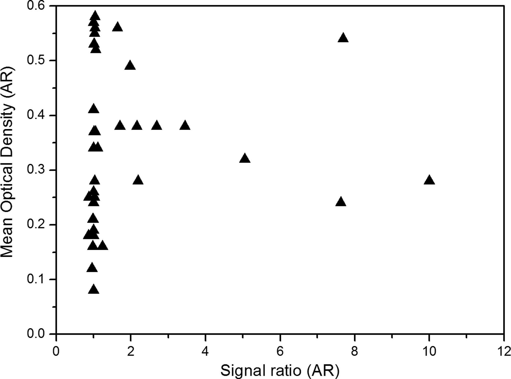
The clinical characteristics and mean optical
density of AR expression for the 37 patients are summarized in
Table I. Out of 37 subjects with PCa
recurrence, 13 (35%) exhibited amplified AR expression, while 24
did not (Table I). Amplified AR
(optical density, 0.45±0.03) was more intensely immunostained than
non-amplified AR (optical density, 0.21±0.06) in the chosen tumor
specimens. The intensity of AR immunostaining and the degree of AR
amplification did not exhibit any association or trend. There was
additionally no association between the protein expression of AR
and the X-chromosome copy number.
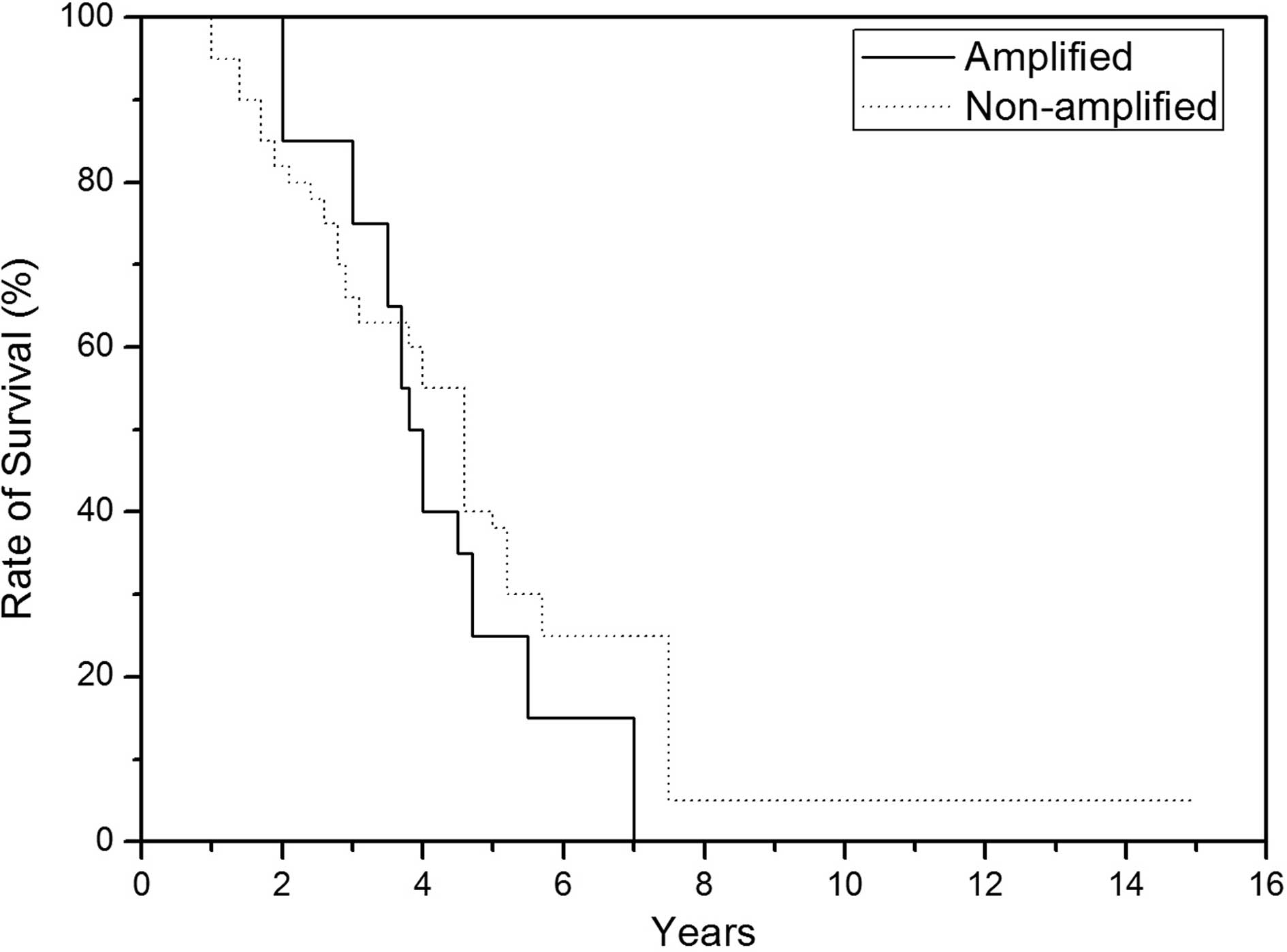
No statistically significant differences were
observed between amplified AR and non-amplified AR tumors specimens
with regard to serum PSA levels, clinical tumor-node-metastasis
stage, Gleason sum, time from androgen deprivation therapy to
recurrence and survival following androgen deprivation (Table II and Fig.
1).
| Table II.Clinical characteristics and survival
of specimens with amplified and non-amplified AR. |
Table II.
Clinical characteristics and survival
of specimens with amplified and non-amplified AR.
| Entry | Non-amplified
AR | Amplified AR |
|---|
| Number of
specimens | 24.0 | 13.00 |
| Mean age of Spc ±
SD | 73.40±8.40 | 73.10±7.10 |
| Mean GS ± SD | 8.90±0.70 | 9.00±0.60 |
| Mean PSA ± SD | 53.20±88.80 | 30.10±46.60 |
| Mean MOS B/w AD +
TA ± SD | 36.00±21.80 | 33.10±11.60 |
| Mean survival A/f
AD (MOS) ± SD | 52.80±28.50 | 51.50±13.90 |
| Mean AR expression
(MOD) ± SD | 0.24±0.10 | 0.36±0.07 |
Additionally, no association with X polysomy was
observed for these clinical parameters. Nevertheless, 6 of the
recurrent PCa specimens (16% of the chosen patients) exhibited
X-chromosome 2.5 (copy number) and greater, while no differences
were found when clinical characteristics between these groups were
compared.
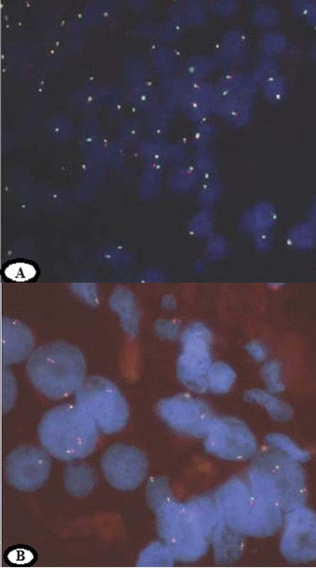
FISH analysis
AR FISH analysis results for tumors with normal AR
and AR amplification are shown in Fig. 2A
and B, respectively. The majority of the nuclei in the normal
cells showed one green and one red signal, indicating that each
nucleus had one X chromosome with one AR gene (Fig. 2A). The presence of two red signals in
the FISH section in the majority of the PCa cells indicated the
presence of AR gene amplification (Fig.
3).
Discussion
Since an effective therapy for the treatment of PCa
recurrence has yet to be identified, an improved understanding of
the mechanism behind the transition from androgen-dependent PCa to
PCa recurrence may provide novel treatment targets (16). The only possible explanation for PCa
recurrence is that the increased expression of AR protein through
AR amplification may allow the expression of androgen-regulated
genes despite castration levels of serum androgens. The present
study found that the AR gene was amplified in ~35% of 37 PCa
recurrence specimens; these results are far indicate an improved
outcome compared with previous studies showing AR amplification in
23% of 47 (13), 25% of 16 (17), 28% of 54 (18), 30% of 23 (5) and 31% of 13 (19) patients. The results compared AR
expression levels between tumors with AR amplification and with a
single AR signal. Reverse transcriptase polymerase chain reaction
was previously used in 13 patients to demonstrate that AR mRNA
expression occurred in the recurrence of 4 tumors and demonstrated
that AR amplification was higher than AR mRNA in the recurrence of
9 tumors that were not amplified (19). When immunohistochemistry was used to
compare AR protein expression between amplified and non-amplified
tumors, the study by Visakorpi et al (5) found that ~80% of primary tumor cells (as
well as recurrence) expressed nuclear AR protein, but that there
was no significant difference in the level of protein expression in
primary tumors compared with other tumors (exhibiting recurrence or
recurrence with AR amplification). In another study, the
hybridization intensity for AR mRNA (ISH for 5 PCa reccurence
specimens) was higher than that of the original PCa non-amplified
specimens (18). The results showed
different results for the length of survival in patients with
advanced PCa treated with androgen deprivation based on the
amplified or non-amplified AR in recurrent tumors. However, the
present study found that amplified AR had no association with the
duration of survival following androgen deprivation (Fig. 1), and no association was found between
survival times following androgen deprivation and X polysomy.
Koivisto et al reported that AR amplification occurred more
often in males who exhibited a complete response to or longer
interval between androgen deprivation and recurrence (18). However, the present study found no
difference in the interval between androgen deprivation and
recurrence. Recently, FISH was used to appraise the attainability
of characterizing gene copy number alterations of circulating tumor
cells isolated using the Cell Search system in specimens with PCa
(particularly progressive castration-resistant metastatic PCa)
(20); this study reported high-level
chromosomal amplification of AR in 38% of the analyzed samples and
relative gain of MYC in 56% of the samples, which also supports the
present results.
Immunohistochemical detection was also used to
optimize the production of the linear association between AR
protein and its DAPI immunostaining (21) using automated digital video image
analysis for precise results. It was observed that those patients
whose tumors demonstrated AR amplification exhibited a 5-month
faster recurrence than those whose had non-amplified tumors. The
amplified AR PCa recurrent tumors exhibited greater levels of AR
protein expression, but this was not associated with survival. It
was also demonstrated that AR protein expression was 60% higher in
tumors with an AR copy number >2.1. Additionally, the study
found that X-chromosome copy number was increased in up to 13.8% of
cancer specimens, which corresponds with the fact that AR copy
number can be increased by X-chromosome polysomy, but will not
impact on AR protein expression (5,15,21).
In conclusion, this study is the first of its type,
quantitatively comparing AR protein expression and AR amplification
in PCa recurrence. This study demonstrated that AR influences tumor
growth and progression even where androgen is deprived.
Furthermore, the results indicated a potential contribution of AR
amplification to AR activation in the relative absence of
androgen.
Acknowledgements
The authors would like to thank the staff of the
Wenzhou Central Hospital for their assistance in subject
recruitment throughout the study period.
References
|
1
|
Lassi K and Dawson NA: Update on
castrate-resistant prostate cancer: 2010. Curr Opin Oncol.
22:263–267. 2010. View Article : Google Scholar : PubMed/NCBI
|
|
2
|
Jemal A, Siegel R, Xu J and Ward E: Cancer
statistics, 2010. CA Cancer J Clin. 60:277–300. 2010. View Article : Google Scholar : PubMed/NCBI
|
|
3
|
Shahrokh F, Shariat, Axel S, et al: Tumor
markers in prostate cancer I: Blood-based markers. Acta Oncol.
50:61–75. 2011. View Article : Google Scholar
|
|
4
|
Culig Z, Hobisch A, Bartsch G and Klocker
H: Androgen receptor - an update of mechanisms of action in
prostate cancer. Urol Res. 28:211–219. 2000. View Article : Google Scholar : PubMed/NCBI
|
|
5
|
Visakorpi T, Hyytinen E, Koivisto P, et
al: In vivo amplification of the androgen receptor gene and
progression of human prostate cancer. Nat Genet. 9:401–406. 1995.
View Article : Google Scholar : PubMed/NCBI
|
|
6
|
Cude KJ, Montgomery JS, Price DK, et al:
The role of an androgen receptor polymorphism in the clinical
outcome of patients with metastatic prostate cancer. Urol Int.
68:16–23. 2002. View Article : Google Scholar : PubMed/NCBI
|
|
7
|
Heinlein CA and Chang C: Androgen receptor
in prostate cancer. Endocr Rev. 25:276–308. 2004. View Article : Google Scholar : PubMed/NCBI
|
|
8
|
Chen CD, Welsbie DS, Tran C, et al:
Molecular determinants of resistance to antiandrogen therapy. Nat
Med. 10:33–39. 2004. View
Article : Google Scholar : PubMed/NCBI
|
|
9
|
Gelmann EP: Molecular biology of the
androgen receptor. J Clin Oncol. 20:3001–3015. 2002. View Article : Google Scholar : PubMed/NCBI
|
|
10
|
Shaffer DR and Scher HI: Prostate cancer:
a dynamic illness with shifting targets. Lancet Oncol. 4:407–414.
2003. View Article : Google Scholar : PubMed/NCBI
|
|
11
|
Linja MJ and Visakorpi T: Alterations of
androgen receptor in prostate cancer. J Steroid Biochem Mol Biol.
92:255–264. 2004. View Article : Google Scholar : PubMed/NCBI
|
|
12
|
Kiessling A, Hogrefe C, Erb S, et al:
Expression, regulation and function of the ISGylation system in
prostate cancer. Oncogene. 28:2606–2620. 2009. View Article : Google Scholar : PubMed/NCBI
|
|
13
|
Bubendorf L, Kononen J, Koivisto P, et al:
Survey of gene amplifications during prostate cancer progression by
high-throughout fluorescence in situ hybridization on tissue
microarrays. Cancer Res. 59:803–806. 1999.PubMed/NCBI
|
|
14
|
Edwards J, Krishna NS, Mukherjee R,
Watters AD, Underwood MA and Bartlett JM: Amplification of the
androgen receptor may not explain the development of
androgen-independent prostate cancer. BJU Int. 88:633–637. 2001.
View Article : Google Scholar : PubMed/NCBI
|
|
15
|
Koivisto P, Hyytinen E, Palmberg C, et al:
Analysis of genetic changes underlying local recurrence of prostate
carcinoma during androgen deprivation therapy. Am J Pathol.
147:1608–1614. 1995.PubMed/NCBI
|
|
16
|
Feldman BJ and Feldman D: The development
of androgen-independent prostate cancer. Nat Rev Cancer. 1:34–45.
2001. View
Article : Google Scholar : PubMed/NCBI
|
|
17
|
Hyytinen ER, Haapala K, Thompson J, et al:
Pattern of somatic androgen receptor gene mutations in patients
with hormone-bractory prostate cancer. Lab Invest. 82:1591–1598.
2002. View Article : Google Scholar : PubMed/NCBI
|
|
18
|
Koivisto P, Kononen J, Palmberg C, et al:
Androgen receptor gene amplification: A possible molecular
mechanism for androgen deprivation therapy failure in prostate
cancer. Cancer Res. 57:314–319. 1997.PubMed/NCBI
|
|
19
|
Linja MJ, Savinainen KJ, Saramaki OR,
Tammela TL, Vessella RL and Visakorpi T: Amplification and
overexpression of androgen receptor gene in hormone-bractory
prostate cancer. Cancer Res. 61:3550–3555. 2001.PubMed/NCBI
|
|
20
|
Leversha MA, Han J, Asgari Z, et al:
Fluorescence in situ hybridization analysis of circulating tumor
cells in metastatic prostate cancer. Clin Cancer Res. 15:2091–2097.
2009. View Article : Google Scholar : PubMed/NCBI
|
|
21
|
Brown RS, Edwards J, Dogan A, et al:
Amplification of the androgen receptor gene in bone metastases from
hormone-bractory prostate cancer. J Pathol. 198:237–244. 2002.
View Article : Google Scholar : PubMed/NCBI
|