Introduction
Worldwide, glioma is one of the most frequent types
of brain tumor in adults. The current standard therapeutic regime
includes maximal safe surgical resection combined with radiotherapy
and temozolomide chemotherapy (1).
Due to the development of such treatment strategies, the percentage
of patients alive two years after diagnosis has increased to 26%
(2). However, the prognosis of
patients with glioma remains unsatisfactory. For example, the
median survival time of patients with glioblastoma multiforme is
only 14.6 months subsequent to standard therapy (3). Therefore, the identification of more
effective treatment strategies for patients with glioma is
required.
All-trans retinoic acid (ATRA) is a
derivative of vitamin A that exerts its effects through retinoic
acid receptors (RARs) and retinoic X receptors. Various studies
have demonstrated that ATRA can induce differentiation and
apoptosis (4,5), as well as induce cell growth arrest, in
glioma cells (6). Furthermore, a
previous study revealed that ATRA may enhance the bystander effect
of suicide-gene therapy against medulloblastoma (7), with a number of studies demonstrating
that treatment with ATRA in combination with specific
chemoimmunotherapeutic agents may significantly enhance its
antitumor effect on glioma (8–12). These
results indicate the therapeutic potential of ATRA for patients
with glioma.
The invasion of glioma is crucial to tumor
progression. Malignant glioma, such as glioblastoma, can rapidly
invade into neighboring brain structures and spread through
infiltration. This invasion is largely dependent on matrix
metalloproteinases (MMPs). MMPs, particularly MMP-2 and MMP-9, are
known to be major glioma invasion-mediating factors that degrade
the extracellular matrix to create space for invading glioma cells
(13). However, the effect of ATRA on
the migration and invasion of glioma cells remains poorly
understood. In addition, although it is universally accepted that
ATRA can induce the apoptosis and inhibit the proliferation of
glioma cells, the association between the concentration and effects
of ATRA remain unclear. Therefore, the present study aimed to
investigate the effects of ATRA treatment on the migration,
invasion, apoptosis and proliferation of glioma cells, with the
intention of partially revealing the anti-glioma mechanisms of
ATRA.
Materials and methods
Materials
U-87MG and SHG44 human glioma cell lines were
purchased from the Cell Resource Center of the Chinese Academy of
Sciences (Shanghai, China).
Cell culture
U87 cells were cultured in DMEM (Hyclone
Laboratories, Inc., Beijing, China) supplemented with 10% fetal
bovine serum (FBS; Hyclone Laboratories, Inc.) in an atmosphere of
5% CO2 at 37°C. The SHG44 cells were cultured in RPMI
1640 (Hyclone Laboratories, Inc.) supplemented with 10% FBS in a 5%
CO2 atmosphere at 37°C.
Scratch wound healing assay
The U87MG and SHG44 cells were seeded at a density
of 1×10⁵ cells/well in 24-well plates (Corning Life
Sciences, Lowell, MA, USA). After 24 h, the cell monolayer was
scraped in a straight line using a 20-µl pipette tip and the cells
were was hed three times with phosphate-buffered saline. ATRA
(Sigma-Aldrich, St. Louis, MO, USA) was dissolved in dimethyl
sulfoxide (DMSO; Sigma-Aldrich) and stored in light-protected vials
at −20°C as stock solution. The stock solution was diluted to the
desired concentrations immediately prior to use and all experiments
were performed under low-light conditions to minimize ATRA
photoisomerization. The cells were incubated in low-serum media
containing 0.5% FBS and various concentrations of ATRA (5, 10, 20
and 40 µmol/l) and the control group was treated with an equal
volume of solvent (DMSO) in culture medium. All the plates were
placed in a culture incubator at 37°C and at 0, 6, 12 and 24 h
after incubation, the plates were removed from the incubator for
images to be captured under a phase-contrast microscope (CKX41;
Olympus Corporation, Tokyo, Japan). Image-Pro Plus software (Media
Cybernetics, Inc., Rockville, MD, USA) was used to measure the
relative migration distances.
Matrigel invasion assay
The U87MG and SHG44 cells were plated at a density
of 5×10⁵ cells/well on Transwell chambers (Corning Life
Sciences) precoated with 50 µl Matrigel (BD Biosciences, Bedford,
MA, USA) diluted with culture medium. Serum-free culture medium
containing 5, 10, 20 or 40 µmol/l ATRA was used to incubate the
cells of each group and medium containing 20% FBS in the lower
chamber served as the chemoattractant. Non-invading cells were
removed with cotton swabs after 24 h, while invading cells were
dyed with 0.1% crystal violet (Amresco, Inc., Solon, OH, USA) and
counted using Image-Pro Plus software under a phase-contrast
microscope (magnification, x200). The average cell number in five
random visual fields was considered to be the number of invading
cells of each chamber.
Flow cytometry
The U87MG and SHG44 cells were seeded at a density
of 3×10⁵ cells/well in six-well plates (Corning Life
Sciences) with 5, 10, 20 or 40 µmol/l ATRA for 24 h. An Annexin
V-fluorescein isothiocyanate cell apoptosis detection kit (Beyotime
Institute of Biotechnology, Haimen, Jiangsu, China) was used to
detect cell apoptosis and a cell cycle analysis kit (Beyotime
Institute of Biotechnology) was used to detect the cell cycle
position of each group, according to the manufacturers'
instructions. Furthermore, the proliferation index (PI) was used to
indicate the proliferation level of each group, according to the
following equation: PI = (S + G2/M) / (G0/G1 + S + G2/M) ×
100%.
Quantitative polymerase chain reaction
(qPCR)
The U87MG and SHG44 cells were seeded at a density
of 3×10⁵ cells/well in six-well plates with various
concentrations of ATRA for 24 h, as aforementioned. The cells were
then lysed and the total RNA was isolated using RNA Fast 200 kit
(Shanghai Fastagen Biotechnology Co., Ltd., Shanghai, China),
according to the manufacturer's instructions. RNA was
reverse-transcribed using a PrimeScript RT Master Mix kit (Takara
Biotechnology Co., Ltd., Dalian, China). qPCR was performed using
SYBR Premix Ex Taq II (Takara Biotechnology Co., Ltd.) on an iQ5
thermal cycler and analyzed using iQ5 software, version 2.0
(Bio-Rad, Hercules, CA, USA). Gene expression was compared using
the cycle threshold (Ct). Ct was defined using the following
equation: ∆Ct = CtTarget – Ctβ-actin, where
β-actin expression was used as the endogenous reference gene.
Change in gene expression was evaluated using the 2−∆∆Ct
method (14). All primers were
designed and synthesized by Takara Biotechnology Co., Ltd. (Table
I).
Western blotting
The U87MG and SHG44 cells were cultured in 25 ml
cell culture flasks (Corning Life Sciences) with various
concentration of ATRA for 24 h. The cells were then harvested for
use in assays. The cells were was hed twice with PBS and then
scraped in 300 µl radioimmunoprecipitation assay lysis buffer
(Beyotime Institute of Biotechnology) with 1 mmol/l
phenylmethanesulfonyl fluoride on ice. The lysates were cleared of
insoluble material by centrifugation, the protein concentration was
determined using a Bradford protein assay kit (Beyotime Institute
of Biotechnology) and samples were boiled in 1X SDS-PAGE sample
loading buffer, resolved by SDS-PAGE, and then transferred to
nitrocellulose membranes (Bio-Rad Laboratories). The membranes were
probed with rabbit anti-human MMP-2 (dilution, 1:500; cat. no.
BS1236), rabbit anti-human MMP-9 (dilution, 1:500; cat. no. BS1241)
and rabbit anti-human GAPDH (dilution, 1:5000; cat. no. ap0063)
antibodies, all purchased from Bioworld Technology, Inc. (St. Louis
Park, MN, USA). Subsequent to was hing in Tris-buffered saline
containing 0.02% Tween 20 (Sigma-Aldrich), the membranes were
incubated with a secondary polyclonal anti-rabbit IgG antibody
conjugated to horseradish peroxidase (dilution, 1:2000; Thermo
Fisher Scientific, Pittsburgh, PA, USA). Membranes were developed
in Supersignal West Pico chemiluminescent reagent (Pierce
Biotechnology, Inc., Rockford, IL, USA).
Statistical analysis
The data are presented as the mean ± standard
deviation and were analyzed using SPSS software, version 17.0
(SPSS, Inc., Chicago, IL, USA). One-way analysis of variance was
used to compare groups and Fisher's least significant difference
tests were performed for subsequent comparisons between groups.
P<0.05 was considered to indicate a statistically significant
difference.
Results
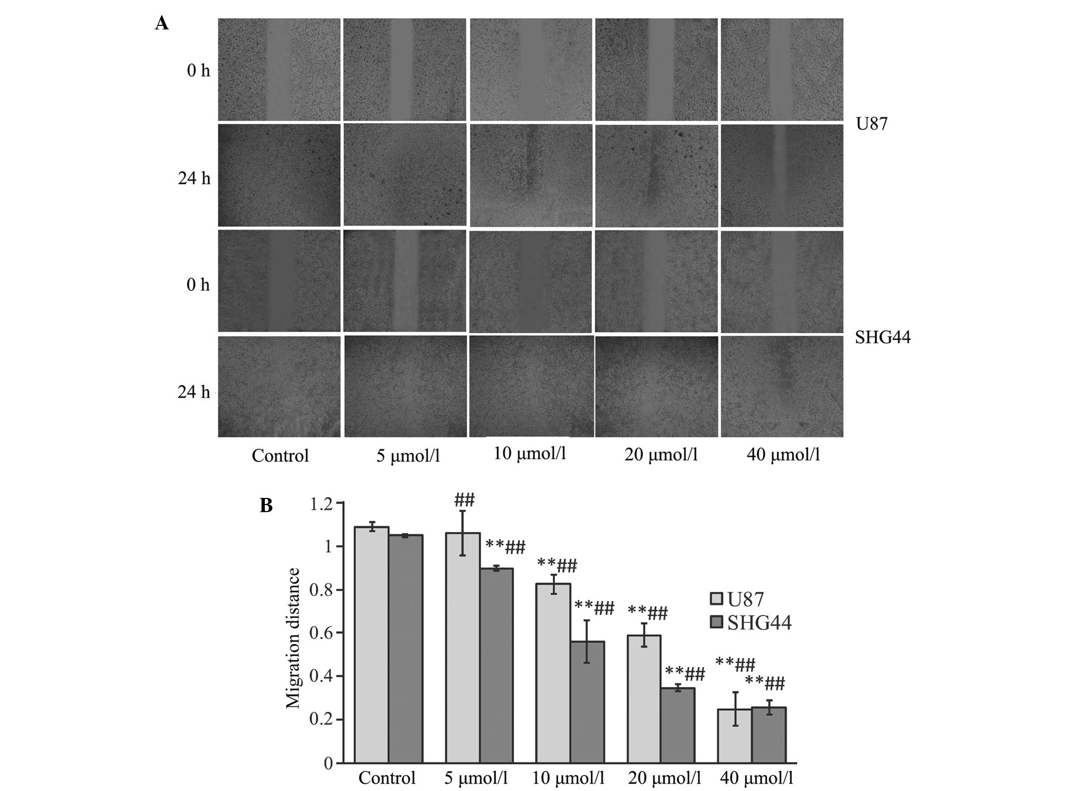
ATRA significantly inhibits the
migration of glioma cells
Subsequent to treatment with various concentrations
of ATRA for 24 h, the migration distance of glioma cells was
significantly reduced compared with the control group (P<0.01),
excluding U87 glioma cells treated with 5 µmol/l ATRA (P>0.05).
Furthermore, the migration distance significantly decreased with
each increase in ATRA concentration (P<0.01; Fig. 1).
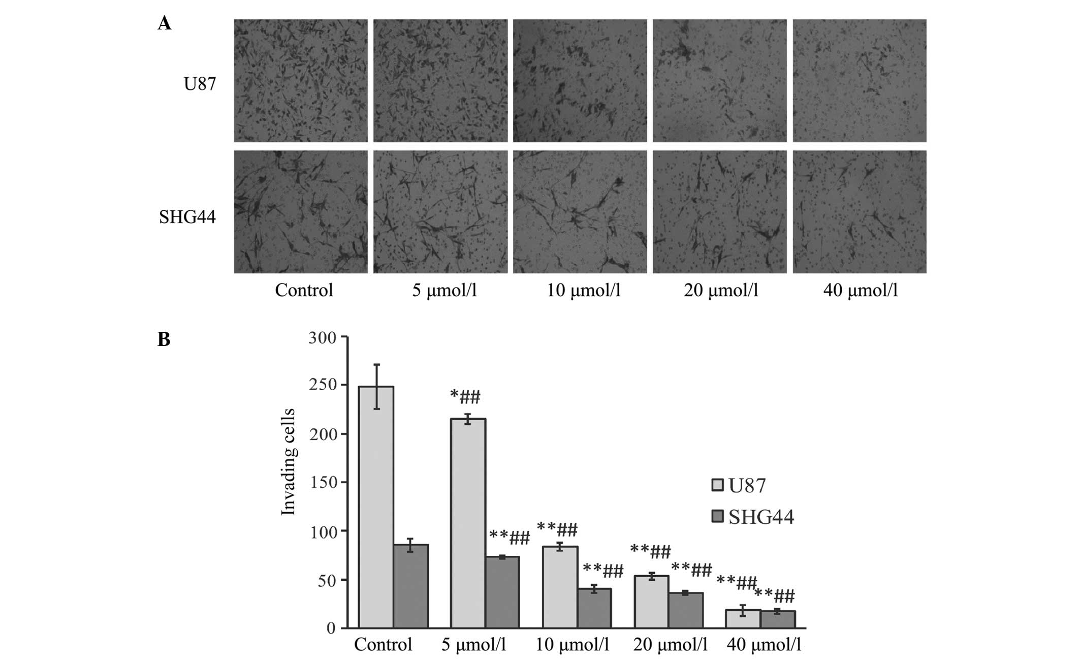
ATRA significantly inhibits the
invasion of glioma cells
Following treatment with different concentrations of
ATRA for 24 h, the number of invading cells was significantly
reduced compared with that of the control group, particularly after
treatment with high concentrations of ATRA, consisting of 20 and 40
µmol/l ATRA. A statistically significant difference was identified
between all groups (P<0.01; Fig.
2).
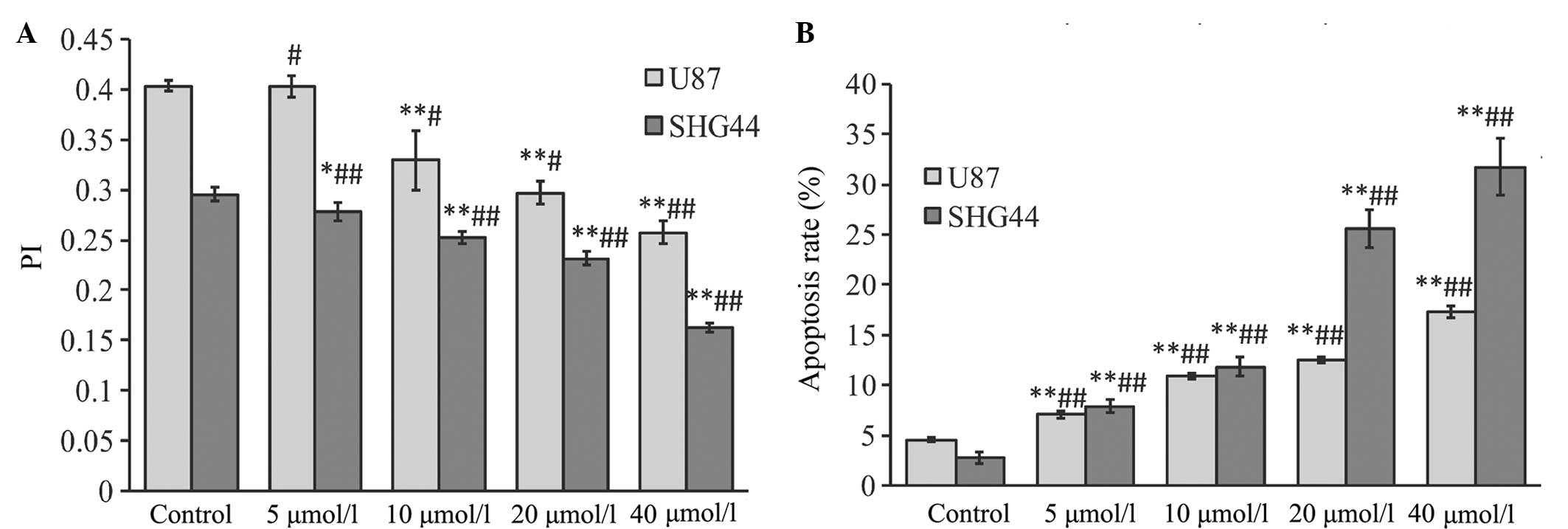
ATRA significantly inhibits
proliferation and promotes apoptosis in glioma cells
Following treatment with various concentrations of
ATRA for 24 h, the PI of glioma cells in each treatment group was
significantly decreased (P<0.01), particularly subsequent to
treatment with 20 and 40 µmol/l ATRA (Fig. 3A).
Following treatment with various concentrations of
ATRA for 24 h, the apoptosis rate of glioma cells in each treatment
group was significantly increased compared with the control group
(P<0.01). Furthermore, a statistically significant difference
was identified between each treatment group and all other treatment
groups (P<0.01; Fig. 3B).
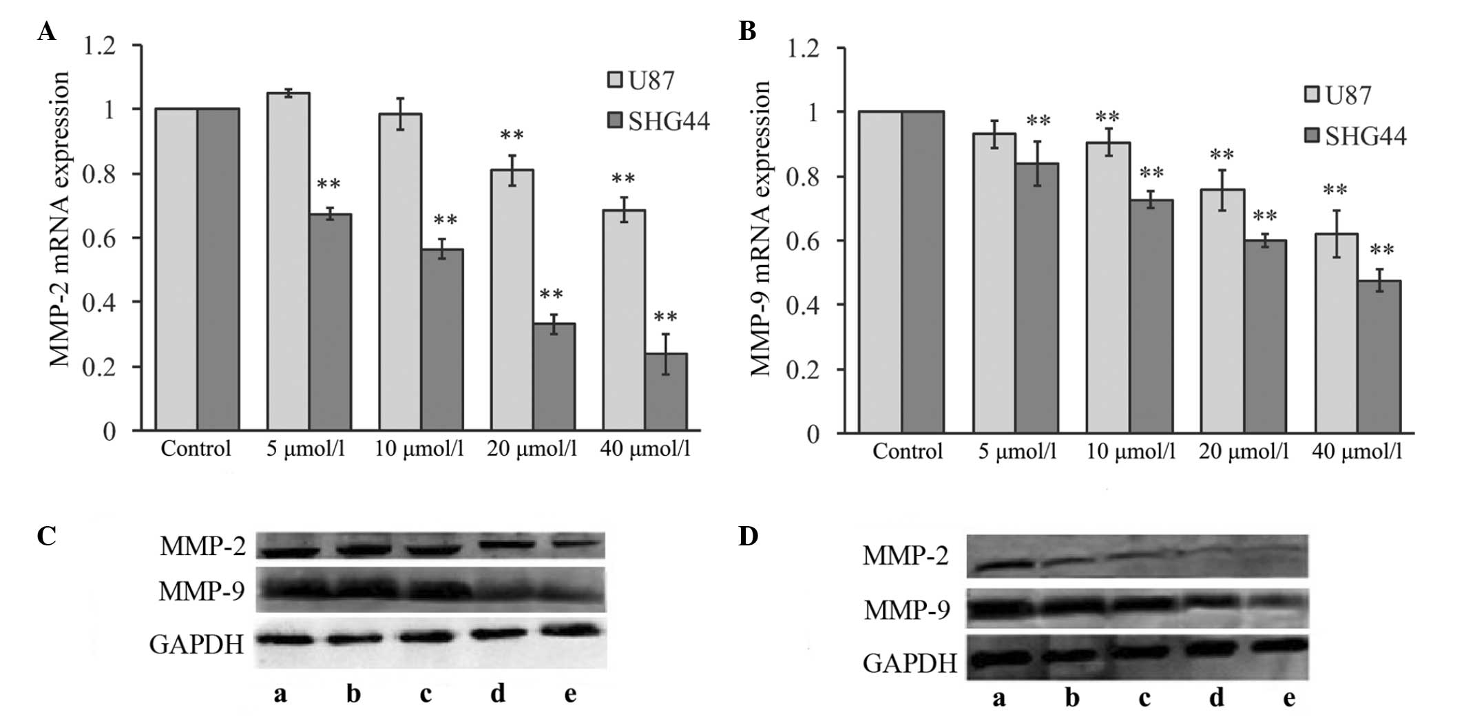
ATRA significantly inhibits the
expression of MMP-2 and MMP-9
Following a 24-h incubation with the aforementioned
concentrations of ATRA, various effects occurred on the MMP-2 mRNA
expression level in glioma cells, as indicated in Fig. 4A. Lower concentrations of ATRA (5 and
10 µmol/l) did not influence the expression of MMP-2 mRNA in U87
glioma cells (P>0.05 vs. control group). However, following
treatment with 20 or 40 µmol/l ATRA, the MMP-2 mRNA expression
levels were significantly downregulated (P<0.01 vs. control
group). By contrast, treating SHG44 glioma cells with various
concentrations of ATRA for 24 h resulted in a significant
downregulation of MMP-2 mRNA expression levels at all ATRA
concentrations (P<0.01 vs. control group; Fig. 4A).
Following treatment with various concentrations of
ATRA, the variations in MMP-9 mRNA expression in the glioma cell
lines were similar to those of MMP-2. The MMP-9 mRNA expression was
significantly downregulated in glioma cells following treatment
with different concentrations of ATRA, compared with the control
group (P<0.01; Fig. 4B).
MMP protein in each group was examined by western
blotting, as indicated in Fig. 4C and
D. Changes in MMP-2 and MMP-9 protein expression levels
exhibited a similar trend to the changes in MMP-2 and MMP-9 mRNA
expression levels. Thus, high concentrations of ATRA may
significantly downregulate the protein expression levels of MMP-2
and MMP-9 in each glioma cell line.
Discussion
Numerous studies have reported that ATRA can inhibit
the migration and invasion of various types of tumor cell lines,
such as human breast cancer cells (15), gastric cancer cells (16), thyroid cancer cells (17) and colon cancer cells (18). However, the effects of ATRA on the
migration and invasiveness of glioma remain poorly understood. In
the present study, it was identified that ATRA could significantly
inhibit the migration and invasiveness of glioma cell lines in a
dose-dependent manner. As gelatinases are important in the
migration and invasiveness, the effects of ATRA treatment on the
expression of MMP-2 and MMP-9 gelatinases were examined in two
glioma cell lines. Following treatment with ATRA, MMP-2 expression
was inconsistent between the various glioma cell lines. ATRA
appeared to significantly inhibit MMP-2 expression in the SHG44
glioma cell line in a dose-dependent manner. By contrast, only high
concentrations of ATRA inhibited MMP-2 expression in the U87 glioma
cell line. Furthermore, MMP-9 expression was significantly
decreased following treatment with ATRA in the two glioma cell
lines. However, there was no statistically significant difference
between the 5 and 10 µmol/l group of U87 glioma cells. The
mechanisms that resulted in the various gelatinase expression
levels induced by ATRA in the two glioma cell lines remains
unclear. According to existing evidence, this may be partially
associated with variations in the distribution of RAR isoforms
between cell lines (19).
As ATRA inhibits the expression of gelatinases in
glioma cells, it may be proposed that the inhibitory effects of
ATRA on the migration and invasion of glioma cells may partially
depend on the inhibitory effects of ATRA on the expression of MMP-2
and MMP-9. However, the present study identified that the
expression of MMP-2 and MMP-9 in the U87 glioma cell line was not
completely compatible with the variation in invasion and migration
ability following treatment with various concentrations of ATRA.
These results indicate that ATRA may also regulate the migration
and invasion of glioma cells using alternate mechanisms. Previous
studies have reported that ATRA can inhibit the activity of MMP-2
(20) and MMP-9 (15) via tissue inhibitor of
metalloproteinase, a protein that may be involved in the regulation
of migration and invasion of ATRA-treated glioma cells.
Although it is universally accepted that ATRA can
induce apoptosis and inhibit proliferation in glioma cells
(4,20,21), the
association between the concentration of ATRA and its effects
remain unclear. In the present study, ATRA treatment significantly
inhibited the proliferation and increased the apoptosis of glioma
cells in a dose-dependent manner. Previous studies reported similar
results in melanoma cells treated with various concentrations of
ATRA (22). The mechanisms by which
ATRA regulates the apoptosis of glioma cells may be associated with
the caspase-3/poly(ADP-ribose) polymerase-1 signaling pathway
(23–25). In addition, the downregulation of B
cell lymphoma-2 (Bcl-2) and the upregulation of Bcl-2-associated X
protein may be involved in this process (4,20,24). Furthermore, it has been reported that
ATRA can inhibit the expression of cyclin D1 and c-myc, proteins
that are important in the regulation of the cell cycle and can
inhibit cellular proliferation (6).
In conclusion, the present study identified that
ATRA treatment may inhibit the migration and invasion of glioma
cells in a dose-dependent manner, and these effects may be
partially associated with the effect of ATRA on the expression of
gelatinases. Furthermore, ATRA may inhibit proliferation and
increase apoptosis in glioma cells. Thus, the results of the
present study reveal additional information regarding the
anti-glioma mechanisms of ATRA.
References
|
1
|
Nishikawa R: Standard therapy for
glioblastoma - a review of where we are. Neurol Med Chir (Tokyo).
50:713–719. 2010. View Article : Google Scholar : PubMed/NCBI
|
|
2
|
Clarke J, Butowski N and Chang S: Recent
advances in therapy for glioblastoma. Arch Neurol. 67:279–283.
2010. View Article : Google Scholar : PubMed/NCBI
|
|
3
|
Stupp R, Mason WP, van den Bent MJ, et al:
European Organisation for Research and Treatment of Cancer Brain
Tumor and Radiotherapy Groups; National Cancer Institute of Canada
Clinical Trials Group: Radiotherapy plus concomitant and adjuvant
temozolomide for glioblastoma. N Engl J Med. 352:987–996. 2005.
View Article : Google Scholar : PubMed/NCBI
|
|
4
|
Zang C, Wachter M, Liu H, et al: Ligands
for PPARgamma and RAR cause induction of growth inhibition and
apoptosis in human glioblastomas. J Neuroncol. 65:107–118. 2003.
View Article : Google Scholar
|
|
5
|
Tang K, Cao L, Fan SQ, et al: Effect of
all-trans-retinoic acid on C6 glioma cell proliferation and
differentiation. Zhong Nan Da Xue Xue Bao Yi Xue Ban. 33:892–897.
2008.PubMed/NCBI
|
|
6
|
Chang Q, Chen Z, You J, et al:
All-trans-retinoic acid induces cell growth arrest in a human
medulloblastoma cell line. J Neurooncol. 84:263–267. 2007.
View Article : Google Scholar : PubMed/NCBI
|
|
7
|
Li S, Gao Y, Pu K, Ma L, Song X and Liu Y:
All-trans retinoic acid enhances bystander effect of suicide-gene
therapy against medulloblastomas. Neurosci Lett. 503:115–119. 2011.
View Article : Google Scholar : PubMed/NCBI
|
|
8
|
Das A, Banik NL and Ray SK: Molecular
mechanisms of the combination of retinoid and interferon-gamma for
inducing differentiation and increasing apoptosis in human
glioblastoma T98G and U87MG cells. Neurochem Res. 34:87–101. 2009.
View Article : Google Scholar : PubMed/NCBI
|
|
9
|
Haque A, Banik NL and Ray SK: Emerging
role of combination of all-trans retinoic acid and interferon-gamma
as chemoimmunotherapy in the management of human glioblastoma.
Neurochem Res. 32:2203–2209. 2007. View Article : Google Scholar : PubMed/NCBI
|
|
10
|
Zhang R, Banik NL and Ray SK: Combination
of all-trans retinoic acid and interferon-gamma upregulated
p27(kip1) and down regulated CDK2 to cause cell cycle arrest
leading to differentiation and apoptosis in human glioblastoma LN18
(PTEN-proficient) and U87MG (PTEN-deficient) cells. Cancer
Chemother Pharmacol. 62:407–416. 2008. View Article : Google Scholar : PubMed/NCBI
|
|
11
|
Karmakar S, Banik NL, Patel SJ and Ray SK:
Combination of all-trans retinoic acid and taxol regressed
glioblastoma T98G xenografts in nude mice. Apoptosis. 12:2077–2087.
2007. View Article : Google Scholar : PubMed/NCBI
|
|
12
|
Karmakar S, Banik NL and Ray SK:
Combination of all-trans retinoic acid and paclitaxel-induced
differentiation and apoptosis in human glioblastoma U87MG
xenografts in nude mice. Cancer. 112:596–607. 2008. View Article : Google Scholar : PubMed/NCBI
|
|
13
|
Onishi M, Ichikawa T, Kurozumi K and Date
I: Angiogenesis and invasion in glioma. Brain Tumor Pathol.
28:13–24. 2011. View Article : Google Scholar : PubMed/NCBI
|
|
14
|
Livak KJ and Schmittgen TD: Analysis of
relative gene expression data using real-time quantitative PCR and
the 2(-Delta Delta C(T)) method. Methods. 25:402–408. 2001.
View Article : Google Scholar : PubMed/NCBI
|
|
15
|
Dutta A, Sen T and Chatterjee A: All-trans
retinoic acid (ATRA) downregulates MMP-9 by modulating its
regulatory molecules. Cell Adhes Migr. 4:409–418. 2010. View Article : Google Scholar
|
|
16
|
Yang P, Liu Z, Wang H, et al: Enhanced
activity of very low density lipoprotein receptor II promotes
SGC7901 cell proliferation and migration. Life Sci. 84:402–408.
2009. View Article : Google Scholar : PubMed/NCBI
|
|
17
|
Lan L, Cui D, Luo Y, Shi BY, Deng LL,
Zhang GY and Wang H: Inhibitory effects of retinoic acid on
invasiveness of human thyroid carcinoma cell lines in vitro. J
Endocrinol Invest. 32:731–738. 2009. View Article : Google Scholar : PubMed/NCBI
|
|
18
|
Adachi Y, Itoh F, Yamamoto H, Iku S,
Matsuno K, Arimura Y and Imai K: Retinoic acids reduce matrilysin
(matrix metalloproteinase 7) and inhibit tumor cell invasion in
human colon cancer. Tumour Biol. 22:247–253. 2001. View Article : Google Scholar : PubMed/NCBI
|
|
19
|
Zhou TB and Qin YH: The potential
mechanism for the different expressions of gelatinases induced by
all-trans retinoic acid in different cells. J Recept Signal
Transduct Res. 32:129–133. 2012. View Article : Google Scholar : PubMed/NCBI
|
|
20
|
Papi A, Bartolini G, Ammar K, et al:
Inhibitory effects of retinoic acid and IIF on growth, migration
and invasiveness in the U87MG human glioblastoma cell line. Oncol
Rep. 18:1015–1021. 2007.PubMed/NCBI
|
|
21
|
Haque A, Das A, Hajiaghamohseni LM,
Younger A, Banik NL and Ray SK: Induction of apoptosis and immune
response by all-trans retinoic acid plus interferon-gamma in human
malignant glioblastoma T98 G and U87MG cells. Cancer Immunol
Immunother. 56:615–625. 2007. View Article : Google Scholar : PubMed/NCBI
|
|
22
|
Zhang H, Satyamoorthy K, Herlyn M and
Rosdahl I: All-trans retinoic acid (atRA) differentially induces
apoptosis in matched primary and metastatic melanoma cells - a
speculation on damage effect of atRA via mitochondrial dysfunction
and cell cycle redistribution. Carcinogenesis. 24:185–191. 2003.
View Article : Google Scholar : PubMed/NCBI
|
|
23
|
Gumireddy K, Sutton LN, Phillips PC and
Reddy CD: All-trans-retinoic acid-induced apoptosis in human
medulloblastoma: activation of caspase-3/poly (ADP-ribose)
polymerase 1 pathway. Clin Cancer Res. 9:4052–4059. 2003.PubMed/NCBI
|
|
24
|
Ran L, Tan W, Tan S, Zhang R, Wang W and
Zeng W: Effects of ATRA, acitretin and tazarotene on growth and
apoptosis of Tca8113 cells. J Huazhong Univ Sci Technolog Med Sci.
25:393–396. 2005. View Article : Google Scholar : PubMed/NCBI
|
|
25
|
Yu Z, Han J, Lin J, Xiao Y, Zhang X and Li
Y: Apoptosis induced by atRA in MEPM cells is mediated through
activation of caspase and RAR. Toxicol Sci. 89:504–509. 2006.
View Article : Google Scholar : PubMed/NCBI
|