Introduction
Gastric cancer has one of the highest incidence
rates of any cancer type worldwide. In 2008, 989,600 new cases of
gastric cancer were reported globally, accounting for 8% of all
cancer cases and resulting in 738,000 mortalities (10% of all
cancer-related mortalities) (1). The
current treatment strategy for gastric cancer is surgical
resection. Surgical resection is associated with a ≤90%
postoperative 5-year survival rate for patients diagnosed with
early-stage gastric cancer. However, the majority of patients with
gastric cancer are diagnosed at an advanced stage, and, thus, are
no longer suitable to undergo radical surgery, resulting in a
decreased 5-year survival rate of 11–40%. Therefore, improvement in
the early-stage diagnosis of gastric cancer, the development of
novel molecular-targeted therapies, and the identification of early
indicators of cancer metastasis and recurrence are important
research topics. These issues may be resolved by improved
investigation into the pathogenesis of gastric cancer as well as
the finding of specific molecular targets for gastric cancer
therapy.
microRNAs (miRNAs/miRs) are small, non-coding,
single-stranded RNAs (length, 19–25 nt) that are expressed by
plants and animals. miRNAs inhibit the transcription and
translation of specific target genes by incomplete base pairing
with their target mRNA molecules (2).
Over 2,000 types of miRNA have been found thus far, and numerous
studies are currently being conducted to identify more (http://www.mirbase.org/). Previous studies have
demonstrated that miRNAs regulate the expression of approximately
two-thirds of all proteins, and almost every vital cell activity,
including development, apoptosis, cell proliferation and division
(3).
miRNAs are extensively correlated with specific
pathological processes, such as tumor development, metastasis and
tolerance, and ~50% of miRNAs are located within tumor-associated
fragile sites, oncogenes or tumor suppressor breakpoints (4). Furthermore, various studies (3,4) have
indicated that miRNAs have been well-conserved during evolution, as
demonstrated by their strict spatial and temporal specificity. The
majority of aberrantly expressed miRNAs in tumors can be detected
in the peripheral blood, and these serum miRNAs can tolerate
multi-gelation, variations in pH and treatment with DNAases. In
addition, miRNA appears to be more stable following treatment with
RNAases compared with mRNA (5,6). In
consideration of the aforementioned studies, miRNAs appear to
exhibit properties that potentially enable their exploitation as
biological targets in tumor diagnostics, prognostic evaluation and
treatment.
Previous studies have screened for and identified a
subset of miRNAs that are associated with gastric cancer,
indicating their extensive participation in the development and
progression of gastric cancer (7,8). However,
the biological function and mechanisms of many of these miRNAs
remains unknown. Therefore, the present study aimed to screen for
additional miRNAs that may be associated with gastric cancer using
gene chromatin immunoprecipitation (ChIP) technology. In addition,
the present study aimed to identify the association between these
miRNAs, and the clinicopathological characteristics of gastric
cancer to demonstrate the specific functions and mechanisms of
these miRNAs during tumor development. To the best of our
knowledge, the present study provides novel hypotheses regarding
the mechanism of gastric cancer as well as proposing possible
future targeted treatment strategies. Subsequent to their
verification, these specific markers of gastric cancer may serve as
criteria in the diagnosis and prognostic evaluation of gastric
cancer.
Materials and methods
Patients and tissue sample
collection
The present study was approved by the Ethics
Committee of the First Hospital of Lanzhou University (Lanzhou,
China). Tissues were collected from 56 gastric cancer patients who
received surgical treatment at the Wuwei Tumor Hospital of Gansu
(Wuwei, China) between October 2009 and April 2010. Matched gastric
cancer and adjacent healthy (n=56) tissue samples were obtained for
use in the present study subsequent to written informed consent
being provided by all the patients, and samples were stored at
−80°C immediately after surgical resection. The sixth edition of
the International Union Against Cancer and the American Joint
Committee on Cancer pathological staging systems were used to
determine the tumor node metastasis (TNM) stage of the tissues
samples. In addition, clinical data were recorded for all patients,
including gender, age, tumor diameter, tumor location, degree of
differentiation, TNM stage and lymph-node metastasis. The samples
were obtained from 32 male patients and 24 female patients;
therefore, the male:female ratio was 1.3:1. Furthermore, the
patients were aged between 35 and 76 years, and the median age was
56 years. No patients received radiotherapy or chemotherapy prior
to undergoing surgery.
miRNA microarray
RNAiso Plus (Takara Bio, Inc., Otsu, Japan) was used
to treat the cancer and adjacent healthy tissue samples from two
representative patients, according to the manufacturer's
instructions. The microarray experiments were performed by the
Shanghai Bohao Biotechnological Co., Ltd (Shanghai, China) using an
8*60 K human miRNA microarray chip (version 18.0), a microarray
scanner, Feature Extraction software (version 10.7) and GeneSpring
GX software (version 11.0; Agilent Technologies, Inc., Santa Clara,
CA, USA), according to the manufacturer's instructions.
Reverse transcription-quantitative
polymerase chain reaction (RT-qPCR)
A PrimeScript® First Strand cDNA Synthesis kit and
miRNA qPCR primer mix (Takara Bio, Inc.) were used to perform the
qRT-PCR assay, according to the manufacturer's instructions.
Statistical analysis
SPSS software (version 15.0; SPSS, Inc., Chicago,
IL, USA) was used to perform the data analysis. T-tests were used
to evaluate the differences between the mean values. Values were
presented as the mean ± standard error of the mean. P<0.05 was
considered to indicate a statistically significant difference.
Results
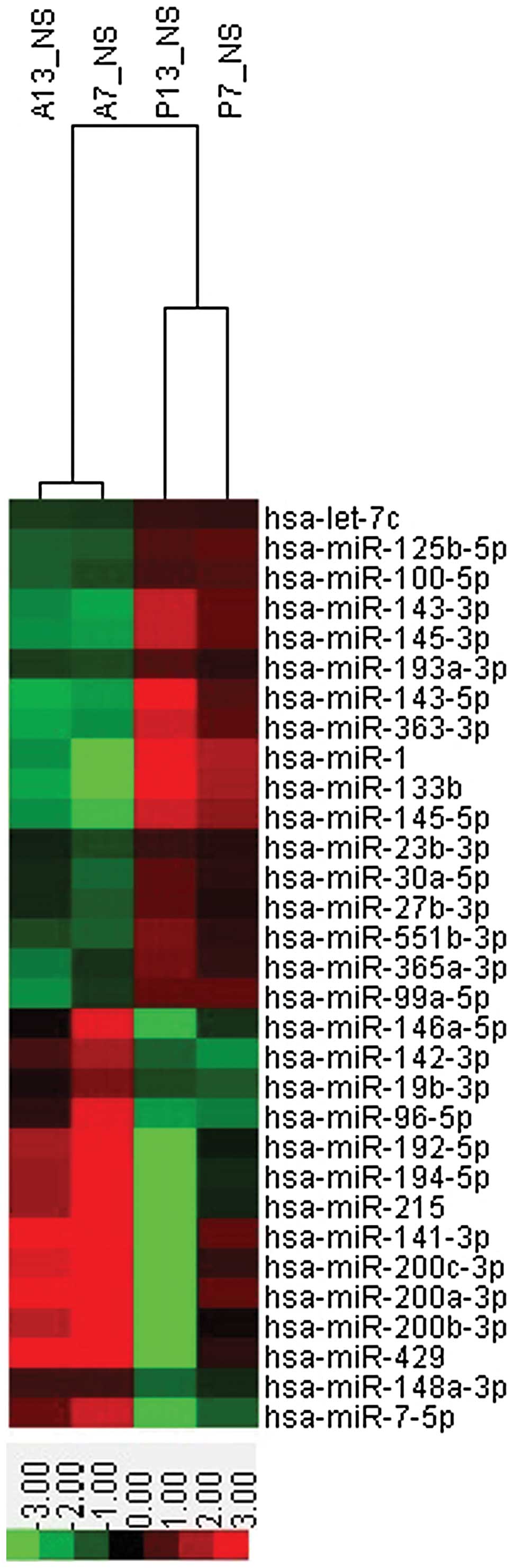
Screening for gastric
cancer-associated miRNAs
The 8*60 K human miRNA microarray chip (version
18.0; Agilent Technologies, Inc), which contains 1,907 human miRNA
genes, was used to screen for miRNAs that are differentially
expressed between gastric cancer and healthy adjacent tissues.
The screening criteria used to compare the gastric
cancer tissue samples with the adjacent healthy mucosa samples
were: i) In the two paired samples, the expression of a miRNA was
upregulated or downregulated >2-fold; and ii) in every four
samples, there were a minimum of two samples with a signal value of
>6. It was identified that the expression levels of 31 miRNAs
were abnormal in the gastric cancer tissues. Of these, 14 miRNAs
were overexpressed and 17 were underexpressed (Table I). Clustering analysis was
subsequently performed for all 31 abnormally expressed miRNAs
(Fig. 1).
 | Table I.Differential expression of
preliminarily screened miRNAs in gastric cancer. |
Table I.
Differential expression of
preliminarily screened miRNAs in gastric cancer.
| No. | Gene name | Expression in gastric
cancer tissue |
|---|
| 1 | Hsa-miR-192-5p | Upregulated |
| 2 | Hsa-miR-200b-3p | Upregulated |
| 3 | Hsa-miR-200c-3p | Upregulated |
| 4 | Hsa-miR-19b-3p | Upregulated |
| 5 | Hsa-miR-141-3p | Upregulated |
| 6 | Hsa-miR-215 | Upregulated |
| 7 | Hsa-miR-194-5p | Upregulated |
| 8 | Hsa-miR-200a-3p | Upregulated |
| 9 |
Hsa-miR-148a-3p | Upregulated |
| 10 | Hsa-miR-429 | Upregulated |
| 11 | Hsa-miR-142-3p | Upregulated |
| 12 | Hsa-miR-7-5p | Upregulated |
| 13 |
Hsa-miR-146a-5p | Upregulated |
| 14 | Hsa-miR-96-5p | Upregulated |
| 15 | Hsa-miR-145-5p | Downregulated |
| 16 | Hsa-miR-27b-3p | Downregulated |
| 17 | Hsa-miR-143-3p | Downregulated |
| 18 | Hsa-miR-23b-3p | Downregulated |
| 19 |
Hsa-miR-125b-5p | Downregulated |
| 20 | Hsa-let-7c | Downregulated |
| 21 | Hsa-miR-1 | Downregulated |
| 22 | Hsa-miR-133b | Downregulated |
| 23 | Hsa-miR-100-5p | Downregulated |
| 24 | Hsa-miR-99a-5p | Downregulated |
| 25 | Hsa-miR-30a-5p | Downregulated |
| 26 | Hsa-miR-145-3p | Downregulated |
| 27 | Hsa-miR-143-5p | Downregulated |
| 28 | Hsa-miR-363-3p | Downregulated |
| 29 |
Hsa-miR-365a-3p | Downregulated |
| 30 |
Hsa-miR-193a-3p | Downregulated |
| 31 |
Hsa-miR-551b-3p | Downregulated |
Verification of the expression of the
selected miRNAs in gastric cancer tissue
RT-qPCR was performed to select 31 miRNA targets
from the ChIP, and to examine the miRNA expression of the 56
gastric cancer tissues and healthy adjacent mucosa.
The results demonstrated that 23/31 miRNAs were
differentially expressed between the gastric cancer and healthy
tissue samples. Ten miRNAs were overexpressed and 13 miRNAs
demonstrated a lower expression in the gastric cancer samples
compared with the healthy tissue samples. No significant difference
was identified for the remaining 8 miRNAs. Thus, the sensitivity of
the results from the two experiments was 74.2% (23/31 miRNAs;
Table II).
| Table II.Expression of screened miRNAs in
gastric cancer versus adjacent healthy tissue samples. |
Table II.
Expression of screened miRNAs in
gastric cancer versus adjacent healthy tissue samples.
|
|
| miRNA expression,
ΔCt (mean ± SE) |
|
|
|---|
|
|
|
|
|
|
|---|
| No. | Gene name | Tumor tissue | Adjacent healthy
tissue | Change in
expression in gastric cancer | P-value |
|---|
| 1 | Hsa-miR-192-5p |
−1.967±0.812 |
1.648±0.611 | ▲ | 0.001* |
| 2 |
Hsa-miR-200b-3p |
−1.644±0.369 |
0.134±0.676 | ▲ | 0.024* |
| 3 |
Hsa-miR-200c-3p |
−0.476±0.347 |
1.242±0.688 | ▲ | 0.029* |
| 4 | Hsa-miR-19b-3p |
3.153±0.312 |
3.149±0.351 |
| 0.994 |
| 5 | Hsa-miR-141-3p |
3.572±0.619 |
3.533±0.503 |
| 0.962 |
| 6 | Hsa-miR-215 |
3.859±0.407 |
6.590±0.622 | ▲ | 0.000* |
| 7 | Hsa-miR-194-5p |
−1.178±0.299 |
0.897±0.592 | ▲ | 0.002* |
| 8 |
Hsa-miR-200a-3p |
1.726±0.320 |
3.511±0.673 | ▲ | 0.018* |
| 9 |
Hsa-miR-148a-3p |
0.709±0.330 |
0.707±0.451 |
| 0.997 |
| 10 | Hsa-miR-429 |
2.699±0.362 |
4.585±0.477 | ▲ | 0.012* |
| 11 | Hsa-miR-142-3p |
0.903±0.296 |
1.784±0.290 | ▲ | 0.036* |
| 12 | Hsa-miR-7-5p |
3.220±0.333 |
4.833±0.491 | ▲ | 0.008* |
| 13 |
Hsa-miR-146a-5p |
2.219±0.269 |
3.437±0.476 | ▲ | 0.028* |
| 14 | Hsa-miR-96-5p |
4.640±0.317 |
5.048±0.487 |
| 0.484 |
| 15 | Hsa-miR-145-5p |
−0.384±0.329 |
−2.605±0.275 | ▼ | 0.000* |
| 16 | Hsa-miR-27b-3p |
4.938±0.215 |
4.671±0.291 |
| 0.462 |
| 17 | Hsa-miR-143-3p |
−1.205±0.337 |
−3.392±0.199 | ▼ | 0.000* |
| 18 | Hsa-miR-23b-3p |
−1.295±0.691 |
−2.159±0.792 |
| 0.413 |
| 19 |
Hsa-miR-125b-5p |
−0.605±0.498 |
−1.327±0.379 |
| 0.251 |
| 20 | Hsa-let-7c |
−0.098±0.225 |
−0.669±0.252 |
| 0.094 |
| 21 | Hsa-miR-1 |
18.273±0.447 |
11.937±0.516 | ▼ | 0.000* |
| 22 | Hsa-miR-133b |
8.942±0.356 |
3.620±0.939 | ▼ | 0.000* |
| 23 | Hsa-miR-100-5p |
2.324±0.332 |
1.152±0.204 | ▼ | 0.003* |
| 24 | Hsa-miR-99a-5p |
2.102±0.336 |
0.873±0.211 | ▼ | 0.003* |
| 25 | Hsa-miR-30a-5p |
0.992±0.289 |
0.160±0.272 | ▼ | 0.039* |
| 26 | Hsa-miR-145-3p |
2.221±0.313 |
0.444±0.224 | ▼ | 0.000* |
| 27 | Hsa-miR-143-5p |
4.236±0.317 |
2.384±0.271 | ▼ | 0.000* |
| 28 | Hsa-miR-363-3p |
6.890±0.309 |
2.703±0.892 | ▼ | 0.000* |
| 29 |
Hsa-miR-365a-3p |
3.936±0.261 |
2.535±0.349 | ▼ | 0.002* |
| 30 |
Hsa-miR-193a-3p |
6.188±0.279 |
5.236±0.319 | ▼ | 0.027* |
| 31 |
Hsa-miR-551b-3p |
8.733±0.309 |
6.449±0.356 | ▼ | 0.000* |
Association between the identified
miRNAs and the clinicopathological characteristics of gastric
cancer
The correlation between the expression of the 23
selected miRNAs in the gastric cancer samples and associated
clinicopathological characteristics were analyzed. The results
identified that four miRNAs, miR-142-3p, miR-146a-5p, miR-145-5p
and miR-1, were not associated with the following
clinicopathological characteri stics: Gender, age, tumor size,
differentiation/grade, clinical stage and lymph-node metastasis.
However, the remaining 19 miRNAs were significantly associated with
lymph-node metastasis, and miR-551b-3p, miR-133b, miR-100-5p,
miR-363-3p and miR-215 were significantly correlated with tumor
differentiation/grade and TNM stage (P<0.05; Tables III–VII). In addition, it was identified that
the abnormal expression of miR-200a-3p and miR-429 was
significantly associated with the gender and age of the gastric
cancer patients, respectively (P<0.05; Tables VIII–IX).
| Table III.Association between the expression of
miR-551b-3p and clinicopathological characteristics of gastric
cancer. |
Table III.
Association between the expression of
miR-551b-3p and clinicopathological characteristics of gastric
cancer.
| Variable | Cases, n (%) | Expression of
miR-551b-3p, median ± range | P-value |
|---|
| Gender |
|
| 0.857 |
|
Male | 32 (57) |
8.684±0.417 |
|
|
Female | 24 (43) |
8.798±0.470 |
|
| Age, years |
|
| 0.785 |
|
≤50 | 18 (32) |
8.857±0.362 |
|
|
>50 | 38 (68) |
8.674±0.425 |
|
| Tumor size, cm |
|
| 0.360 |
| ≤5 | 31 (55) |
8.475±0.445 |
|
|
>5 | 25 (45) |
9.052±0.420 |
|
| Degree of
differentiation |
|
| 0.015* |
| Well
and moderately | 17 (30) |
7.148±0.601 |
|
|
Poorly | 39 (70) |
9.988±0.356 |
|
| TNM stage |
|
| 0. 039* |
|
I/II | 27 (48) |
5.902±0.436 |
|
|
III/IV | 29 (52) |
8.575±0.442 |
|
| Lymph-node
status |
|
| 0.013* |
| No
metastasis | 6 (11) |
8.470±0.310 |
|
|
Metastasis | 50 (89) |
10.924±0.937 |
|
| Table VII.Association between the expression of
miR-215 and clinicopathological parameters of gastric cancer. |
Table VII.
Association between the expression of
miR-215 and clinicopathological parameters of gastric cancer.
| Variable | Cases, n (%) | Expression of
miR-215, median ± range | P-value |
|---|
| Gender |
|
| 0.134 |
|
Male | 32 (57) |
4.750±0.840 |
|
|
Female | 24 (43) |
3.437±0.441 |
|
| Age, years |
|
| 0.285 |
|
≤50 | 18 (32) |
3.479±0.553 |
|
|
>50 | 38 (68) |
4.366±0.596 |
|
| Tumor size, cm |
|
| 0.171 |
| ≤5 | 31 (55) |
4.448±0.561 |
|
|
>5 | 25 (45) |
3.355±0.571 |
|
| Degree of
differentiation |
|
| 0. 039* |
| Well
and moderately | 17 (30) |
5.408±0.704 |
|
|
Poorly | 39 (70) |
3.056±0.500 |
|
| TNM stage |
|
| 0.047* |
|
I/II | 27 (48) |
4.985±0.674 |
|
|
III/IV | 29 (52) |
3.042±0.484 |
|
| Lymph-node
status |
|
| 0.001* |
| No
metastasis | 6 (11) |
7.583±0.386 |
|
|
Metastasis | 50 (89) |
3.413±0.383 |
|
| Table VIII.Association between the expression of
miR-200a-3p and clinicopathological parameters of gastric
cancer. |
Table VIII.
Association between the expression of
miR-200a-3p and clinicopathological parameters of gastric
cancer.
| Variable | Cases, n (%) | Expression of
miR-200a-3p, median ± range | P-value |
|---|
| Gender |
|
| 0.048* |
|
Male | 32 (57) |
1.178±0.377 |
|
|
Female | 24 (43) |
2.456±0.527 |
|
| Age, years |
|
| 0.376 |
|
≤50 | 18 (32) |
2.143±0.674 |
|
|
>50 | 38 (68) |
1.528±0.351 |
|
| Tumor size, cm |
|
| 0.909 |
| ≤5 | 31 (55) |
1.759±0.470 |
|
|
>5 | 25 (45) |
1.684±0.430 |
|
| Degree of
differentiation |
|
| 0.458 |
| Well
and moderately | 17 (30) |
1.885±0.394 |
|
|
Poorly | 39 (70) |
1.361±0.552 |
|
| TNM stage |
|
| 0.377 |
|
I/II | 27 (48) |
2.023±0.568 |
|
|
III/IV | 29 (52) |
1.449±0.325 |
|
| Lymph-node
status |
|
| 0.003* |
| No
metastasis | 6 (11) |
4.379±0.597 |
|
|
Metastasis | 50 (89) |
1.407±0.280 |
|
| Table IX.Association between the expression of
miR-429 and clinicopathological parameters of gastric cancer. |
Table IX.
Association between the expression of
miR-429 and clinicopathological parameters of gastric cancer.
| Variable | Cases, n (%) | Expression of
miR-429, median ± range | P-value |
|---|
| Gender |
|
| 0.208 |
|
Male | 32 (57) |
2.272±0.334 |
|
|
Female | 24 (43) |
3.130±0.637 |
|
| Age, years |
|
| 0.047* |
|
≤50 | 18 (32) |
3.602±0.763 |
|
|
>50 | 38 (68) |
2.184±0.317 |
|
| Tumor size, cm |
|
| 0.746 |
| ≤5 | 31 (55) |
2.739±0.531 |
|
|
>5 | 25 (45) |
2.518±0.368 |
|
| Degree of
differentiation |
|
| 0.705 |
| Well
and moderately | 17 (30) |
2.445±0.521 |
|
|
Poorly | 39 (70) |
2.725±0.426 |
|
| TNM stage |
|
| 0.363 |
|
I/II | 27 (48) |
2.959±0.584 |
|
|
III/IV | 29 (52) |
2.343±0.351 |
|
| Lymph-node
status |
|
| 0.074 |
| No
metastasis | 6 (11) |
3.169±0.756 |
|
|
Metastasis | 50 (89) |
2.216±0.262 |
|
Discussion
miRNAs regulate endogenous gene expression in
humans, affecting numerous cell functions (for example, growth,
differentiation, apoptosis and stress responses) by downregulating
the expression of target genes (9).
miRNAs were found to be important in the process of tumor
development, and multiple miRNAs may be directly associated with
the development of human tumors (for example, pancreatic,
colorectal, gastric, liver, breast and lung cancer, and lymphoma)
(10–12). The role of miRNAs in tumor
pathogenesis is complex, as they can regulate or suppress
tumor-associated genes to promote or inhibit cancer, respectively.
In addition, miRNAs may serve as oncogenes or tumor suppressor
genes and, thus, participate in the development of various types of
cancer. Within a particular tissue type, one miRNA targets multiple
genes, as each target mRNA can potentially interact with multiple
miRNAs. Therefore, miRNAs compose a complex regulatory network that
may contribute to the development of tumors (13).
Significant progress has been made in the field of
miRNA. For example, previous studies have screened for and
identified a variety of functional miRNAs in a number of different
tumor types. The identification of these miRNAs may facilitate the
elucidation of the mechanisms of tumorigenesis, and may provide
novel targets for early diagnosis and treatment (14,15). It is
possible that numerous miRNAs have yet to be identified; therefore,
the correlation between the expression of specific miRNAs and
tumors is unknown. However, significant progress has been made in
this field.
In the current study, miRNA ChIP technology was used
to evaluate the expression levels of miRNAs in carcinoma and
adjacent healthy tissue from two patients who were diagnosed with
gastric cancer to identify the miRNAs that are associated with this
cancer type. A total of 31 miRNAs were identified that exhibited
differential expression levels between the tumor and healthy tissue
samples. RT-qPCR was subsequently performed to verify the
differential expression of these miRNAs in samples from 56 patients
diagnosed with gastric cancer. The results demonstrated that only
23 miRNAs exhibited significantly differential expression while the
remaining 8 miRNAs demonstrated no significant difference in
expression between the gastric cancer and healthy tissue samples.
Therefore, the results from the microarray and RT-qPCR experiments
were 74.2% in agreement, indicating error in the miRNA array. The
two primary reasons for this experimental error may be: i) The
limited number of test samples analyzed; and ii) the shortcomings
of the gene ChIP technique, including its relatively poor stability
and reproducibility, as well as the fact that false positive
detection cannot be avoided (16).
Correlation analysis between the expression levels
of the 23 differentially expressed miRNAs and the associated
clinicopathological characteristics was performed. It was
identified that four miRNAs, miR-142-3p, miR-146a-5p, miR-145-5p
and miR-1, were not associated with the following analyzed
clinicopathological parameters: Gender, age, tumor size,
differentiation/grade, clinical stage and lymph-node metastasis.
The remaining 19 miRNAs were associated with lymph-node metastasis,
of which miR-551b-3p, miR-133b, miR-100-5p, miR-363-3p and miR-215
were significantly associated with tumor differentiation/grade and
TNM stage (P<0.05), indicating that these miRNAs are possibly
involved in the invasion and metastasis of gastric cancer. Notably,
the abnormal expression levels of miR-200a-3p and miR-429 were
significantly associated with the gender and age of the gastric
cancer patients in the present study, respectively (P<0.05).
According to epidemiological statistics, the incidence of gastric
cancer significantly increases with age, with the peak age of
gastric cancer occurrence ranging between 50 and 80 years.
Furthermore, male individuals are 1.5- to 2.5-fold more likely to
develop gastric cancer compared with females (1). Thus, identification of the significant
association between abnormal miR-200a-3p and miR-429 expression in
gastric cancer tissue, and respective differences in age and
gender, may be useful during diagnosis.
A recent study demonstrated that the expression of
miR-551a is downregulated in gastric cancer tissues and cells, and
that miR-551a may inhibit cell invasion and migration by
downregulating the expression of phosphatase of regenerating
liver-3 (17). This finding indicates
that miR-551a is important in gastric cancer invasion and
migration. Similarly, the present study identified that miR-551b-3p
exhibits different expression levels in gastric cancer tissues
compared with healthy tissues. Furthermore, the expression of
miR-551b-3p appears to be significantly associated with the tumor
differentiation/grade, TNM stage and lymph-node metastasis. These
results indicate that miR-551b-3p is an important factor in the
development of gastric cancer, as well as in the invasion and
metastasis of this cancer type.
The miR-133 family consists of miR-133a-1,
miR-133a-2 and miR-133b. Originally, a number of studies determined
that miR-133 is critical in regulating heart and skeletal muscle
development (18,19). However, a more recent study
demonstrated that miR-133 is aberrantly expressed in tumors, and
that miR-133a/b can inhibit the proliferation, and metastasis of
bladder and prostate cancer by regulating the expression of the
epidermal growth factor receptor (20,21). In
addition, previous studies have observed a significant
downregulation of miR-133a/b in head and neck neoplasms, lung
cancer, esophageal cancer, colon cancer, renal carcinoma, prostate
cancer, and other malignancies (22–25). The
present study indicated that miR-133b may act as a tumor suppressor
while regulating the invasion and metastasis of cancer cells.
Previous studies have indicated that the expression
level of miR-100 varies between tumor types and, therefore, this
miRNA may have different functions in different cancer types. For
example, miR-100 expression is downregulated in lung, cervical,
ovarian and bladder cancer, and acts as a tumor suppressor
(26–28). However, in various types of cancer,
such as acute myeloid leukemia, miR-100 is upregulated and
functions as an oncogene (29). The
present study indicated that miR-100-5p is downregulated in gastric
cancer, and may be involved in the infiltration and metastasis of
gastric cancer.
Studies regarding the function of miR-363-3p in the
development of cancer are limited. Sun et al (30) demonstrated that miR-363-3p expression
is downregulated in squamous cell carcinoma of the head and neck
(SCCHN), and is involved in the development and metastasis of SCCHN
via the regulation of podoplanin. Furthermore, Georgieva et
al (31) used high-throughput
sequencing to detect the abnormal expression of miR-363 in uterine
fibroids. However, to the best of our knowledge, no studies have
thus far investigated the function of miR-363-3p in gastric cancer.
The present study demonstrated that miR-363-3p is significantly
downregulated in gastric cancer, and that its expression level is
significantly associated with the degree of tumor differentiation,
TNM stage and lymph node metastasis (P<0.05. Therefore, the
current results indicate that miR-364-3p is a novel molecule that
may be involved in the infiltration and metastasis of gastric
cancer.
miR-215 functions differently in different tumor
types. It was initially identified to be a P53-induced miRNA that
functions as a tumor suppressor by regulating the expression of
various proteins involved in cell cycle checkpoints (32). Alternative studies have demonstrated
that miR-215 downregulates thymidylate synthetase, dihydrofolate
reductase, thymidylate synthetase and denticleless E3 ubiquitin
protein ligase homolog, and functions in the development of
chemotherapy resistance (33,34). Furthermore, the upregulation of
miR-215 was observed in esophageal, kidney and colon cancer
(35–37). In contrast to its role in other solid
tumors, miR-215 functions as an oncogene in gastric cancer by
promoting cell proliferation and cancer cell metastasis (38–40);
however, the underlying mechanism has yet to be defined. In
agreement with previous reports, the current study demonstrated
that the relatively high expression of miR-215 in gastric cancer is
significantly associated with the degree of tumor differentiation,
TNM stage and lymph node metastasis P<0.05). Therefore, miR-215
could potentially be used as a biomarker in the diagnosis of
gastric cancer due to its significantly upregulated expression in
this cancer type (P<0.05).
In conclusion, the present study successfully
identified a number of miRNAs that are involved in the development
of gastric cancer. However, additional studies are required to
delineate the underlying mechanisms of the association, and to
explore their potential as valid biomarkers in the diagnosis,
classification and prognosis of gastric cancer.
Acknowledgements
The present study was supported by the National
Natural Science Foundation of China (grant no. 81372145); the
Research Fund of Personnel training plan of West Light (grant no.
201218), Chinese Academy of Sciences; and the Gansu Provincial
Natural Science Foundation (grant no. 1308RJZA223).
References
|
1
|
Jemal A, Bray F, Center MM, et al: Global
cancer statistics. CA Cancer J Clin. 61:69–90. 2011. View Article : Google Scholar : PubMed/NCBI
|
|
2
|
Treiber T, Treiber N and Meister G:
Regulation of microRNA biogenesis and function. Thromb Haemost.
107:605–610. 2012. View Article : Google Scholar : PubMed/NCBI
|
|
3
|
Anglicheau D, Muthukumar T and
Suthanthiran M: MicroRNAs: small RNAs with big effects.
Transplantation. 90:105–112. 2010. View Article : Google Scholar : PubMed/NCBI
|
|
4
|
Farazi TA, Hoell JI, Morozov P and Tuschl
T: MicroRNAs in human cancer. Adv Exp Med Biol. 774:1–20.
2013.PubMed/NCBI
|
|
5
|
Mitchell PS, Parkin RK, Kroh EM, et al:
Circulating microRNAs as stable blood-based markers for cancer
detection. In: Proc Natl Acad Sci USA. 105. pp. 10513–10518. 2008;
View Article : Google Scholar : PubMed/NCBI
|
|
6
|
Ng EK, Chong WW, Jin H, et al:
Differential expression of microRNAs in plasma of patients with
colorectal cancer: a potential marker for colorectal cancer
screening. Gut. 58:1375–1381. 2009. View Article : Google Scholar : PubMed/NCBI
|
|
7
|
Wang HJ, Ruan HJ, He XJ, et al:
MicroRNA-101 is down-regulated in gastric cancer and involved in
cell migration and invasion. Eur J Cancer. 46:2295–2303. 2010.
View Article : Google Scholar : PubMed/NCBI
|
|
8
|
Yao Y, Suo AL, Li ZF, et al: MicroRNA
profiling of human gastric cancer. Mol Med Rep. 2:963–970.
2009.PubMed/NCBI
|
|
9
|
Ambros V: MicroRNA pathways in flies and
worms: growth, death, fat, stress and timing. Cell. 113:673–676.
2003. View Article : Google Scholar : PubMed/NCBI
|
|
10
|
Rachagani S, Kumar S and Batra SK:
MicroRNA in pancreatic cancer: pathological, diagnostic and
therapeutic implications. Cancer Lett. 292:8–16. 2010. View Article : Google Scholar : PubMed/NCBI
|
|
11
|
Liu R, Zhang C, Hu Z, et al: A
five-microRNA signature identified from genome-wide serum microRNA
expression profiling serves as a fingerprint for gastric cancer
diagnosis. Eur J Cancer. 47:784–791. 2011. View Article : Google Scholar : PubMed/NCBI
|
|
12
|
Xiong Y, Fang JH, Yun JP, et al: Effects
of MicroRNA-29 on apoptosis, tumorigenicity, and prognosis of
hepatocellular carcinoma. Hepatology. 51:836–845. 2010.PubMed/NCBI
|
|
13
|
Synnergren J, Améen C, Lindahl A, et al:
Expression of microRNAs and their target mRNAs in human stem
cell-derived cardiomyocyte clusters and in heart tissue. Physiol
Genomics. 43:581–594. 2011. View Article : Google Scholar : PubMed/NCBI
|
|
14
|
Calin GA and Croce CM: MicroRNA signatures
in human cancers. Nat Rev Cancer. 6:857–866. 2006. View Article : Google Scholar : PubMed/NCBI
|
|
15
|
Lovat F, Valeri N and Croce CM: MicroRNAs
in the pathogenesis of cancer. Semin Oncol. 38:724–733. 2011.
View Article : Google Scholar : PubMed/NCBI
|
|
16
|
Simon R: Challenges of microarray data and
the evaluation of gene expression profile signatures. Cancer
Invest. 26:327–332. 2008. View Article : Google Scholar : PubMed/NCBI
|
|
17
|
Li Z, Cao Y, Jie Z, et al: miR-495 and
miR-551a inhibit the migration and invasion of human gastric cancer
cells by directly interacting with PRL-3. Cancer Lett. 323:41–47.
2012. View Article : Google Scholar : PubMed/NCBI
|
|
18
|
Yin VP, Lepilina A, Smith A and Poss KD:
Regulation of zebrafish heart regeneration by miR-133. Dev Biol.
365:319–327. 2012. View Article : Google Scholar : PubMed/NCBI
|
|
19
|
Chen JF, Mandel EM, Thomson JM, et al: The
role of microRNA-1 and microRNA-133 in skeletal muscle
proliferation and differentiation. Nat Genet. 38:228–233. 2006.
View Article : Google Scholar : PubMed/NCBI
|
|
20
|
Zhou Y, Wu D, Tao J, et al: MicroRNA-133
inhibits cell proliferation, migration and invasion by targeting
epidermal growth factor receptor and its downstream effector
proteins in bladder cancer. Scand J Urol. 47:423–432. 2013.
View Article : Google Scholar : PubMed/NCBI
|
|
21
|
Tao J, Wu D, Xu B, et al: MicroRNA-133
inhibits cell proliferation, migration and invasion in prostate
cancer cells by targeting the epidermal growth factor receptor.
Oncol Rep. 27:1967–1975. 2012.PubMed/NCBI
|
|
22
|
Kinoshita T, Nohata N, Watanabe-Takano H,
et al: Actin-related protein 2/3 complex subunit 5 (ARPC5)
contributes to cell migration and invasion and is directly
regulated by tumor-suppressive microRNA-133a in head and neck
squamous cell carcinoma. Int J Oncol. 40:1770–1778. 2012.PubMed/NCBI
|
|
23
|
Moriya Y, Nohata N, Kinoshita T, et al:
Tumor suppressive microRNA-133a regulates novel molecular networks
in lung squamous cell carcinoma. J Hum Genet. 57:38–45. 2012.
View Article : Google Scholar : PubMed/NCBI
|
|
24
|
Kawakami K, Enokida H, Chiyomaru T, et al:
The functional significance of miR-1 and miR-133a in renal cell
carcinoma. Eur J Cancer. 48:827–836. 2012. View Article : Google Scholar : PubMed/NCBI
|
|
25
|
Wu ZS, Wang CQ, Xiang R, et al: Loss of
miR-133a expression associated with poor survival of breast cancer
and restoration of miR-133a expression inhibited breast cancer cell
growth and invasion. BMC Cancer. 12:512012. View Article : Google Scholar : PubMed/NCBI
|
|
26
|
Liu J, Lu KH, Liu ZL, et al: MicroRNA-100
is a potential molecular marker of non-small cell lung cancer and
functions as a tumor suppressor by targeting polo-like kinase 1.
BMC Cancer. 12:5192012. View Article : Google Scholar : PubMed/NCBI
|
|
27
|
Li BH, Zhou JS, Ye F, et al: Reduced
miR-100 expression in cervical cancer and precursors and its
carcinogenic effect through targeting PLK1 protein. Eur J Cancer.
47:2166–2174. 2011. View Article : Google Scholar : PubMed/NCBI
|
|
28
|
Peng DX, Luo M, Qiu LW, et al: Prognostic
implications of microRNA-100 and its functional roles in human
epithelial ovarian cancer. Oncol Rep. 27:1238–1244. 2012.PubMed/NCBI
|
|
29
|
Zheng YS, Zhang H, Zhang XJ, et al:
MiR-100 regulates cell differentiation and survival by targeting
RBSP3, a phosphatase-like tumor suppressor in acute myeloid
leukemia. Oncogene. 31:80–92. 2012. View Article : Google Scholar : PubMed/NCBI
|
|
30
|
Sun Q, Zhang J, Cao W, et al: Dysregulated
miR-363 affects head and neck cancer invasion and metastasis by
targeting podoplanin. Int J Biochem Cell Biol. 45:513–520. 2013.
View Article : Google Scholar : PubMed/NCBI
|
|
31
|
Georgieva B, Milev I, Minkov I, et al:
Characterization of the uterine leiomyoma microRNAome by deep
sequencing. Genomics. 99:275–281. 2012. View Article : Google Scholar : PubMed/NCBI
|
|
32
|
Georges SA, Biery MC, Kim SY, et al:
Coordinated regulation of cell cycle transcripts by p53-Inducible
microRNAs, miR-192 and miR-215. Cancer Res. 68:10105–10112. 2008.
View Article : Google Scholar : PubMed/NCBI
|
|
33
|
Song B, Wang Y, Titmus MA, et al:
Molecular mechanism of chemoresistance by miR-215 in osteosarcoma
and colon cancer cells. Mol Cancer. 9:962010. View Article : Google Scholar : PubMed/NCBI
|
|
34
|
Boni V, Bitarte N, Cristobal I, et al:
miR-192/miR-215 influence 5-fluorouracil resistance through cell
cycle-mediated mechanisms complementary to its post-transcriptional
thymidilate synthase regulation. Mol Cancer Ther. 9:2265–2275.
2010. View Article : Google Scholar : PubMed/NCBI
|
|
35
|
Wijnhoven BP, Hussey DJ, Watson DI, et al:
South Australian Oesophageal Research Group: MicroRNA profiling of
Barrett's oesophagus and oesophageal adenocarcinoma. Br J Surg.
97:853–861. 2010. View
Article : Google Scholar : PubMed/NCBI
|
|
36
|
White NM, Khella HW, Grigull J, et al:
miRNA profiling in metastatic renal cell carcinoma reveals a
tumour-suppressor effect for miR-215. Br J Cancer. 105:1741–1749.
2011. View Article : Google Scholar : PubMed/NCBI
|
|
37
|
Faltejskova P, Svoboda M, Srutova K, et
al: Identification and functional screening of microRNAs highly
deregulated in colorectal cancer. J Cell Mol Med. 16:2655–2666.
2012. View Article : Google Scholar : PubMed/NCBI
|
|
38
|
Jin Z, Selaru FM, Cheng Y, et al:
MicroRNA-192 and −215 are upregulated in human gastric cancer in
vivo and suppress ALCAM expression in vitro. Oncogene.
30:1577–1585. 2011. View Article : Google Scholar : PubMed/NCBI
|
|
39
|
Ueda T, Volinia S, Okumura H, et al:
Relation between microRNA expression and progression and prognosis
of gastric cancer: a microRNA expression analysis. Lancet Oncol.
11:136–146. 2010. View Article : Google Scholar : PubMed/NCBI
|
|
40
|
Tsukamoto Y, Nakada C, Noguchi T, Tanigawa
M, et al: MicroRNA-375 is downregulated in gastric carcinomas and
regulates cell survival by targeting PDK1 and 14-3-3zeta. Cancer
Res. 70:2339–2349. 2010. View Article : Google Scholar : PubMed/NCBI
|