Introduction
Gastric cancer is the fourth most common malignancy
and the second leading cause of cancer-associated mortality
worldwide, with an overall survival (OS) rate of 20–25% reported in
the majority of countries (1,2). Despite continued improvements in medical
technologies and treatment strategies, major obstacles to the
treatment of gastric cancer remain, including delayed diagnosis,
recurrence, metastasis and a poor prognosis (3,4).
Therefore, the elucidation of novel molecular mechanisms and the
identification of biomarkers for the development and progression of
gastric cancer should be investigated.
In the human genome, protein-coding genes account
for ~2% of the total genome sequence. However, ≥90% of gene
sequences are primarily transcribed into non-coding RNAs (ncRNAs)
(5). Over the past several years,
short ncRNAs, such as microRNAs (miRNAs), have been comprehensively
studied. Long ncRNAs (lncRNAs) are generally considered to be
mRNA-like transcripts longer than ~200 nucleotides (nt), but they
do not function as templates for protein synthesis (6). An increasing number of studies have
focused on lncRNAs, which have been demonstrated to contribute to
the onset and progression of numerous diseases (7). Functional analyses have indicated that
lncRNAs are involved in the regulation of a large number of
biological processes, including cancer development and progression
(8,9).
Expression of the lncRNA, HOTAIR, has been correlated with tumor
metastasis and poor prognosis in hepatocellular (10), pancreatic (11), breast (12) and colorectal cancer (13), and gastrointestinal stromal tumors
(14). Furthermore, aberrant
expression of certain lncRNAs, including ANRIL, MALAT1, H19, GAS5
and MEG3, have been reported in a number of different types of
cancer (15–19). However, the roles of lncRNAs in
gastric cancer and their association with clinicopathological
characteristics in this disease remain largely unknown and
therefore require further study.
The RP11-119F7.4 gene is located on chromosome 10
(70451421-70452698), and codes for an antisense lncRNA with a
length of 349 nts. However, whether there is an association between
this specific lncRNA and cancer is unclear. Therefore, the primary
aim of the current study was to investigate the expression pattern
of lncRNA RP11-119F7.4 in gastric cancer tissues and matched
non-tumor adjacent tissues (NATs) as a possible biomarker; and to
evaluate the correlation between its expression level and
clinicopathological characteristics of patients with gastric
cancer.
Materials and methods
Tissue samples
Human gastric cancer tissue samples and pair-matched
NATs were obtained from 96 patients who underwent radical resection
of gastric cancer between November 2007 and May 2009 at the First
Hospital of China Medical University (Shenyang, Liaoning, China).
Informed consent was obtained from all patients and the study
protocol was approved by the Research Ethics Committee of China
Medical University. All resected tissue samples were immediately
frozen in liquid nitrogen and stored at −80°C until used for
analysis. Matching NAT mucosal specimens from at least 5 cm from
the tumor edge were also collected. Hematoxylin-eosin staining of
each sample was performed for histopathological evaluation. The
histological cancer grade was assessed following guidelines
established by the World Health Organization. The depth of wall
invasion (pT classification) and the extent of regional lymph node
metastasis (pN classification) were staged according to the 7th
edition of the tumor-node-metastasis (TNM) staging system (20,21).
Total RNA extraction and reverse
transcription (RT)
Total RNA was extracted from the gastric cancer
tissues and corresponding NATs using TRIzol reagent (Invitrogen
Life Technologies, Carlsbad, CA, USA) following the manufacturer's
instructions. The concentration and purity of the total RNA were
detected by UV spectrophotometry (A260/A280 >1.9) using a
NanoPhotometer UV/Vis spectrophotometer (Implen GmbH, Munich,
Germany). The total RNA was dissolved in diethyl
pyrocarbonate-treated water and reverse transcription (RT) was
performed using the Super Script III First-Stand Synthesis System
(Invitrogen Life Technologies) for use in polymerase chain reaction
(PCR) according to the manufacturer's instructions. In brief, a
10-µl RT reaction mixture containing 1 µg total RNA, 1 µl oligo(dT)
primer, 1 µl 10 mM deoxyribonucleotide triphosphate mix and
diethylpyrocarbonate-treated water was incubated at 65°C for 5 min.
Next, a 10-µl mixture of 2 µl 10X RT buffer, 4 µl 25 mM
MgCl2, 2 µl 0.1 M dopachrome tautomeras, 1 µl RNaseOUT
(40 U/µl) and 1 µl SuperScript III RT (200 U/µl) was added.
Following the addition of 1 µl RNase H, the total mixture was
incubated in a GeneAmp PCR 9700 Thermocycler (Applied Biosystems
Life Technologies, Foster City, CA, USA) for 50 min at 50°C, 5 min
at 85°C and 20 min at 37°C. A no-template control was run with the
RT reaction.
Quantitative PCR (qPCR)
Quantitative PCR was performed using the SYBR Premix
Ex Taq kit (Takara Biotenchnology, Co., Ltd., Shiga, Japan) on a
Light Cycler 480 II Real-Time PCR system (Roche Diagnostics, Basel,
Switzerland). Each 25-µl PCR reaction mixture contained 2 µl of
reverse-transcribed product, 12.5 µl SYBR Green mix, 9.5 µl
RNase-free water, and 0.5 µl forward and 0.5 µl reverse primers.
The reaction was incubated in a 96-well optical plate and amplified
in 45 cycles of 95°C for 5 sec, 58°C for 30 sec and 72°C for 30
sec. The primers were designed by Primer Premier software, version
5.0 (Premier Biosoft International, Palo Alto, CA, USA). The primer
sequences used were as follows: RP11-119F7.4 forward,
5′-CAGACTTGAAATGGGTGAGATGA-3′ and reverse,
5′-TGCCTTTGCTAGTGCAGTGTAT-3′; and glyceraldehyde 3-phosphate
dehydrogenase (GAPDH) forward, 5′-CGGATTTGGTCGTATTGGG-3′ and
reverse, 5′-CTGGAAGATGGTGATGGGATT-3′. GAPDH was used as an
endogenous reference to normalize lncRNA expression levels using
the 2−∆∆Ct method, where ∆Ct was the difference in
threshold cycle (Ct) values. The relative lncRNA expression is
presented as the fold-change normalized to the endogenous reference
(GAPDH) and relative to the non-tumorous control. Each sample was
analyzed in triplicate and repeated three times.
Statistical analysis
RNA expression levels in gastric cancer tissues
relative to the control were calculated by the 2−ΔΔCt
formula, where ΔΔCt is the difference in the ΔCt values between the
treatment tumor cells and the control (ΔΔCt = ΔCttumor
lncRNA - ΔCtNAT lncRNA), and ΔCt is the difference
in the Ct value between the target and endogenous reference (GAPDH;
ΔCt = CtlncRNA - CtGAPDH). If the expression
levels of the treatment and control cells were equal, ΔΔCt was
equal to zero and 2−ΔΔCT was equal to one. The relative
expression levels of lncRNA RP11-119F7.4 in the gastric cancer
tissues were determined using the ΔCttumor lncRNA and
ΔCtNAT lncRNA values. Statistical differences in lncRNA
expression levels in cancer tissues relative to the non-tumor
control were analyzed using Student's t-test, and correlations
between lncRNA expression levels and clinicopathological parameters
were analyzed using non-parametric tests: The Mann-Whitney U test
between two groups and the Krushal-Wallis test for three or more
groups. Survival curves were estimated by the Kaplan-Meier method.
The log-rank test was used to estimate the statistical differences
between survival curves. A receiver operating characteristic (ROC)
curve was established to evaluate the diagnostic value for
differentiating between gastric cancer and benign diseases.
Statistical analysis was performed using SPSS software, version 8.0
(SPSS, Inc., Chicago, IL, USA) and GraphPad Prism, version 5.0
(GraphPad Software, La Jolla, CA, USA). P<0.05 was considered to
indicate a statistically significant difference.
Results
General characteristics of the
subjects
Of the 96 patients (median age, 61 years; range,
26–84 years) with gastric cancer included for analysis, the
majority (n=72, 75%) were male, 73 presented with lymph node
metastasis (pN stage, N1-N3)and 92 patients had completed follow-up
examinations (Table I). OS time was
defined as the time between surgery and mortality, whereas
disease-free survival (DFS) time was defined as the time between
surgery and cancer-specific mortality.
 | Table I.Association between the expression of
long non-coding RNA RP11-119F7.4 and the clinicopathological
features in 96 patients with gastric cancer. |
Table I.
Association between the expression of
long non-coding RNA RP11-119F7.4 and the clinicopathological
features in 96 patients with gastric cancer.
| Characteristics | No of patients | Expression
levela | P-value |
|---|
| Gender |
|
| 0.148 |
| Male | 72 | 0.55 (0.16–1.03) |
|
|
Female | 24 | 1.11 (0.27–2.31) |
|
| Age, years |
|
| 0.138 |
| ≤60 | 46 | 0.79
(0.26–1.69) |
|
|
>60 | 50 | 0.55
(0.13–1.00) |
|
| Tumor size, cm |
|
| 0.204 |
|
<6 | 40 | 0.72
(0.37–1.59) |
|
| ≥6 | 56 | 0.43
(0.14–1.21) |
|
| Tumor location |
|
| 0.865 |
| Upper
stomach | 5 | 0.62
(0.29–1.21) |
|
| Middle
stomach | 35 | 0.78
(0.15–1.70) |
|
| Lower
stomach | 55 | 0.48
(0.16–1.11) |
|
| Entire
stomach | 1 | 0.68 |
|
| Macroscopic
type |
|
| 0.041b |
|
Borrmann I-III | 85 | 0.54
(0.15–1.20) |
|
|
Borrmann IV | 11 | 0.90
(0.50–4.11) |
|
| Histological
grade |
|
| 0.197 |
|
Good | 21 | 0.54
(0.04–0.86) |
|
|
Poor | 75 | 0.72
(0.22–1.36) |
|
| Lauren grade |
|
| 0.020b |
|
Intestinal type | 32 | 0.41
(0.04–0.95) |
|
| Diffuse
type | 64 | 0.76
(0.27–1.64) |
|
| pT stage |
|
| 0.882 |
|
T1+T2 | 17 | 0.62
(0.21–1.18) |
|
|
T3+T4 | 79 | 0.60
(0.16–1.23) |
|
| pN stage |
|
| 0.414 |
| N0 | 23 | 0.52
(0.16–1.11) |
|
| N1 | 13 | 0.37
(0.07–1.27) |
|
| N2 | 19 | 0.80
(0.42–1.70) |
|
| N3 | 41 | 0.68
(0.20–1.52) |
|
| pTNM stage |
|
| 0.678 |
| I | 9 | 0.52
(0.13–1.01) |
|
| II | 25 | 0.42
(0.11–1.17) |
|
|
III | 62 | 0.72
(0.21–1.69) |
|
| Invasion into
lymphatic vessels |
|
| 0.062 |
|
Negative | 16 | 0.28
(0.10–0.84) |
|
|
Positive | 80 | 0.72
(0.22–1.47) |
|
RP11-119F7.4 expression is
downregulated in gastric cancer tissues
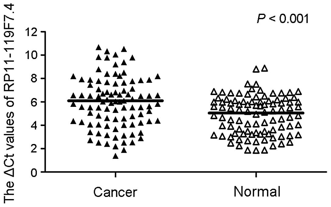
As presented in Fig.
1, statistically significant differences were observed in the
RP11-119F7.4 expression levels between the 96 paired tumor tissues
and matched NATs, as determined by qPCR (P<0.001; Student's
t-test). RP11-119F7.4 expression levels in the gastric cancer
tissues were reduced compared with those in the corresponding
normal tissues. Furthermore, the RP11-119F7.4 expression levels in
64/96 (66.7%) tumor tissues were downregulated compared with the
corresponding NATs.
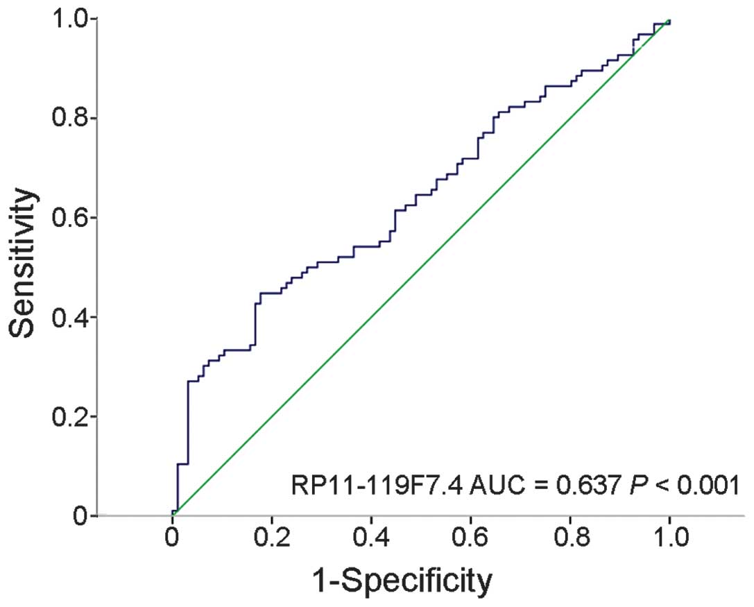
Diagnostic value of lncRNA
RP11-119F7.4 as a biomarker of gastric cancer
The suitability of lncRNA RP11-119F7.4 as a
potential biomarker of gastric cancer was evaluated using
corresponding NATs as controls to produce an ROC curve. The cut off
value was 6.445. The area under the ROC curve was 0.637 (95%
confidence interval, 0.558–0.715; P<0.001). The sensitivity and
specificity were 0.448 and 0.823, respectively. The Youden index
was 0.271 (Fig. 2). The ROC curve
indicated that RP11-119F7.4 has potential diagnostic value in
gastric cancer.
Correlations between RP11-119F7.4
levels in cancer tissues and clinicopathological characteristics of
patients with gastric cancer
The present study also investigated whether the
lncRNA RP11-119F7.4 expression levels were correlated with the
clinicopathological characteristics of gender, age, tumor size,
macroscopic type and TNM stage in gastric cancer. As shown in
Table I, the RP11-119F7.4 levels were
associated with macroscopic type (P=0.041), and the patients with
Borrmann type IV gastric cancer tended to have increased
RP11-119F7.4 expression levels compared with patients with Borrmann
type I-III gastric cancer. Furthermore, it was also observed that
the RP11-119F7.4 expression levels of diffuse-type gastric cancer
were increased compared with those of intestinal-type gastric
cancer (P=0.020). There were no significant correlations between
RP11-119F7.4 expression levels and the clinicopathological
characteristics of gender (P=0.148), age (P=0.138), tumor size
(P=0.204), tumor location (P=0.865), histological grade (P=0.197),
pT stage (P=0.882), pN stage (P=0.414), pTNM stage (P=0.678) or
invasion into the lymphatic vessels (P=0.062).
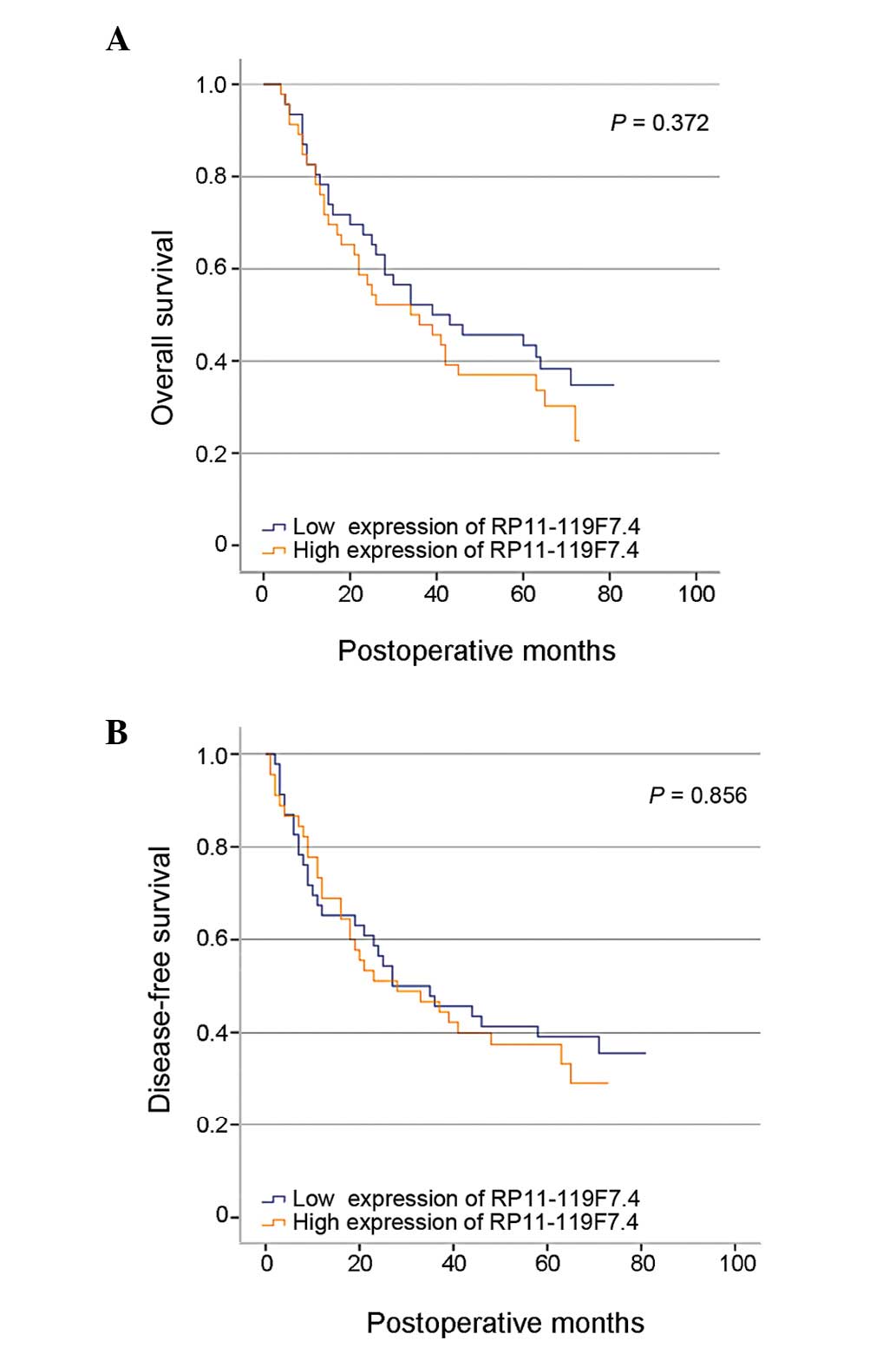
Association between RP11-119F7.4
expression and patient survival
Univariate analysis was performed to investigate
whether RP11-119F7.4 expression impacted OS and DFS. The
RP11-119F7.4 expression levels were determined by qPCR, as
aforementioned. Of the 96 included patients, 48 were allocated to
the high RP11-119F7.4 expression group, and the remaining 48 to the
low RP11-119F7.4 expression group. The median relative expression
levels of RP11-119F7.4 were used to determine the cut-off values of
the high and low RP11-119F7.4 expression groups in the present
study. However, from the results presented in Fig. 3, no significant differences were
observed in OS (P=0.372) and DFS (P=0.658) between the patients in
the high and low RP11-119F7.4 expression groups.
Discussion
Tumors associated with gastric cancer are among the
most malignant and tend to be asymptomatic in the early stages of
disease; thus, they are often not detected late until the disease
has progressed to an advanced stage (4). Japan and South Korea provide a
government-sponsored screening program for gastric cancer due to
the high incidence of this disease in these two countries. However,
in these countries, gastric cancer is associated with a low
mortality-to-incidence ratio, which may be partly due to the
aforementioned population-based screening in regions with a high
prevalence of disease (3). Therefore,
biomarkers to indicate morbidity associated with gastric cancer are
required, particularly for use among populations with a high
prevalence of disease. A previous study demonstrated that lncRNAs
may function as a novel class of diagnostic and treatment
indicators of several types of cancer (8). However, the role of lncRNA expression in
gastric cancer remains largely unstudied. In fact, to the best of
our knowledge, the present study is the first to investigate the
association between RP11-119F7.4 expression and the
clinicopathological characteristics and prognosis of gastric cancer
based on qPCR data. The results of the present study indicated that
lncRNA RP11-119F7.4 was more frequently downregulated in the
gastric cancer tissues compared with their matched NATs.
Furthermore, the results demonstrated that the RP11-119F7.4
expression levels were associated with macroscopic type and Lauren
grade.
Increasing evidence has indicated that lncRNAs
participate in a wide variety of physiological and pathological
processes by modulating gene expression at the transcriptional,
post-transcriptional and epigenetic levels (22–24). At
the transcriptional level, a number of lncRNAs regulate gene
expression depending on the sequence and relative position, for
example, by binding to the promoters of target genes and forming
stable DNA-RNA triplex complexes to affect transcription
initiation; this process may also inhibit the transcription process
of targeted genes and subsequently silence expression (25). At the post-transcriptional level,
lncRNAs may competitively bind to target mRNA and block the binding
sites of transcription factors, leading to dysregulation of mRNA
translation, splicing and degradation (26). lncRNAs regulate gene expression at the
epigenetic level through numerous processes, including DNA
methylation, histone modification and chromatin remodeling
(27).
A number of previous studies have demonstrated that
lncRNA dysfunction is closely associated with transcriptional
regulation (12,28,29). In
addition, other previous studies have reported that ectopic lncRNA
expression is associated with tumorigenesis, tumor proliferation,
invasion and metastasis (8,30,31). In
the present study, differences in the expression levels of lncRNA
RP11-119F7.4 were observed between the gastric cancer tissues and
matched NATs. For patients with gastric cancer, diagnosis and
treatment in the early stages of disease may improve their survival
time. At present, the most common methods to detect gastric cancer
are electronic gastroscopy and histopathological analysis (3). It is therefore necessary to identify
novel diagnostic biomarkers of gastric cancer. To this end, ROC
curves were used to evaluate the potential diagnostic suitability
of RP11-119F7.4 in the present study. The results demonstrated that
RP11-119F7.4 had potential diagnostic value in gastric cancer and
further indicated that increased expression levels tended to be
associated with Borrmann type IV and diffuse-type gastric cancer. A
number of previous studies have indicated that Borrmann type IV and
diffuse Lauren-type gastric cancers were associated with a worse
prognosis compared with other types (32,33).
According to the results of the present study, RP11-119F7.4
expression levels may be associated with tumorigenesis and the
level of malignancy in gastric cancer. Although statistically
significant differences were not observed in the survival analysis,
a trend towards a worse prognosis was observed in the patients with
relatively increased RP11-119F7.4 expression levels. These findings
were supported by the correlation between the clinicopathological
characteristics and the RP11-119F7.4 expression levels.
Numerous factors have been observed to downregulate
lncRNA expression. Yang et al (34) demonstrated that lncRNA-LET was
downregulated in a number of different tumor types and indicated
that the reduced expression of lncRNA-LET may be a result of
dysregulated histone acetylation. Another previous study by Liu
et al (35) indicated that p53
directly interacts with the p53 response element in the upstream
region of lncRNA loc285194 and regulates lncRNA expression in
colorectal cancer. Competing endogenous RNAs (ceRNAs) are
hypothesized to form large-scale regulatory networks and may be
important in pathological processes, including cancer development
and progression (36). miRNAs are key
components of the ceRNA network and may affect the expression of
lncRNA in cancer. For example, members of the miR-200 family have
been reported to regulate lncRNA-ATB expression in hepatocellular
carcinoma (37). As aforementioned,
lncRNAs are likely to be regulated by numerous factors, therefore
further investigations are required to identify mechanisms that
participate in lncRNA RP11-119F7.4 dysregulation in gastric
cancer.
In conclusion, the results of the present study
demonstrated that lncRNA RP11-119F7.4 expression was downregulated
in gastric cancer, which was associated with the macroscopic type
and Lauren grade. RP11-119F7.4 is therefore a potential novel
biomarker and diagnostic target in gastric cancer. However, the
molecular mechanisms that regulate RP11-119F7.4 expression in
gastric cancer require further study.
Acknowledgements
The authors would like to thank the Department of
Surgical Oncology of the First Hospital of China Medical University
for providing human gastric tissue samples, and the College of
China Medical University for providing technical assistance in the
experiments. The present study was supported by the National
Science Foundation of China (grant nos. 81201888, 81372549 and
81172370), the Specialized Research Fund for the Doctoral Program
of Higher Education (grant no. 20122104110009) and the Natural
Science Foundation of Liaoning Province (grant no. 2014029201).
Abbreviations:
|
lncRNA
|
long non-coding RNA
|
|
NATs
|
non-tumor adjacent tissues
|
|
OS
|
overall survival
|
|
ROC
|
receiver operating characteristic
|
|
miRNA
|
microRNA
|
|
TNM
|
tumor-node-metastasis
|
|
GAPDH
|
glyceraldehyde 3-phosphate
dehydrogenase
|
|
DFS
|
disease-free survival
|
|
ceRNA
|
competing endogenous RNA
|
References
|
1
|
Hartgrink HH, Jansen EP, van Grieken NC
and van de Velde CJ: Gastric cancer. Lancet. 374:477–490. 2009.
View Article : Google Scholar : PubMed/NCBI
|
|
2
|
Ferlay J, Shin HR, Bray F, Forman D,
Mathers C and Parkin DM: Estimates of worldwide burden of cancer in
2008: GLOBOCAN 2008. Int J Cancer. 127:2893–2917. 2010. View Article : Google Scholar : PubMed/NCBI
|
|
3
|
Shen L, Shan YS, Hu HM, et al: Management
of gastric cancer in Asia: resource-stratified guidelines. Lancet
Oncol. 14:e535–e547. 2013. View Article : Google Scholar : PubMed/NCBI
|
|
4
|
Thiel A and Ristimaki A: Gastric cancer:
basic aspects. Helicobacter. 17(Suppl 1): 26–29. 2012.PubMed/NCBI
|
|
5
|
Yan B and Wang Z: Long noncoding RNA: its
physiological and pathological roles. DNA Cell Biol. 31(Suppl 1):
34–41. 2012.
|
|
6
|
Qi P and Du X: The long non-coding RNAs, a
new cancer diagnostic and therapeutic gold mine. Mod Pathol.
26:155–165. 2013. View Article : Google Scholar : PubMed/NCBI
|
|
7
|
Wapinski O and Chang HY: Long noncoding
RNAs and human disease. Trends Cell Biol. 21:354–361. 2011.
View Article : Google Scholar : PubMed/NCBI
|
|
8
|
Qiu MT, Hu JW, Yin R and Xu L: Long
noncoding RNA: An emerging paradigm of cancer research. Tumour
Biol. 34:613–620. 2013. View Article : Google Scholar : PubMed/NCBI
|
|
9
|
Maruyama R and Suzuki H: Long noncoding
RNA involvement in cancer. BMB Rep. 45:604–611. 2012. View Article : Google Scholar : PubMed/NCBI
|
|
10
|
Yang Z, Zhou L, Wu LM, et al:
Overexpression of long non-coding RNA HOTAIR predicts tumor
recurrence in hepatocellular carcinoma patients following liver
transplantation. Ann Surg Oncol. 18:1243–1250. 2011. View Article : Google Scholar : PubMed/NCBI
|
|
11
|
Kim K, Jutooru I, Chadalapaka G, et al:
HOTAIR is a negative prognostic factor and exhibits pro-oncogenic
activity in pancreatic cancer. Oncogene. 32:1616–1625. 2013.
View Article : Google Scholar : PubMed/NCBI
|
|
12
|
Gupta RA, Shah N, Wang KC, et al: Long
non-coding RNA HOTAIR reprograms chromatin state to promote cancer
metastasis. Nature. 464:1071–1076. 2010. View Article : Google Scholar : PubMed/NCBI
|
|
13
|
Kogo R, Shimamura T, Mimori K, Kawahara K,
Imoto S, Sudo T, et al: Long noncoding RNA HOTAIR regulates
polycomb-dependent chromatin modification and is associated with
poor prognosis in colorectal cancers. Cancer Res. 71:6320–6326.
2011. View Article : Google Scholar : PubMed/NCBI
|
|
14
|
Niinuma T, Suzuki H, Nojima M, et al:
Upregulation of miR-196a and HOTAIR drive malignant character in
gastrointestinal stromal tumors. Cancer Res. 72:1126–1136. 2012.
View Article : Google Scholar : PubMed/NCBI
|
|
15
|
Tano K, Mizuno R, Okada T, et al: MALAT-1
enhances cell motility of lung adenocarcinoma cells by influencing
the expression of motility-related genes. FEBS Lett. 584:4575–4580.
2010. View Article : Google Scholar : PubMed/NCBI
|
|
16
|
Zhang EB, Kong R, Yin DD, et al: Long
noncoding RNA ANRIL indicates a poor prognosis of gastric cancer
and promotes tumor growth by epigenetically silencing of
miR-99a/miR-449a. Oncotarget. 5:2276–2292. 2014.PubMed/NCBI
|
|
17
|
Bi HS, Yang XY, Yuan JH, et al: H19
inhibits RNA polymerase II-mediated transcription by disrupting the
hnRNP U-actin complex. Biochim Biophys Acta. 1830:4899–4906. 2013.
View Article : Google Scholar : PubMed/NCBI
|
|
18
|
Mourtada-Maarabouni M, Pickard MR, Hedge
VL, Farzaneh F and Williams GT: GAS5, a non-protein-coding RNA,
controls apoptosis and is downregulated in breast cancer. Oncogene.
28:195–208. 2009. View Article : Google Scholar : PubMed/NCBI
|
|
19
|
Jia LF, Wei SB, Gan YH, et al: Expression,
regulation and roles of miR-26a and MEG3 in tongue squamous cell
carcinoma. Int J Cancer. 135:2282–2293. 2014. View Article : Google Scholar : PubMed/NCBI
|
|
20
|
Hamilton SR and Aaltonen LA: World Health
Organization Classification of TumoursPathology and genetics of
Tumours of the Digestive System. IARC Press; Lyon, Paris: 2000
|
|
21
|
Sobin L, Gospodarowicz M and Wittekind C:
International Union Against Cancer (UICC)TNM classification of
malignant tumours. 7th. Wiley-Liss; NewYork, NY, USA: pp. 117–126.
2010
|
|
22
|
Tsai MC, Manor O, Wan Y, et al: Long
noncoding RNA as modular scaffold of histone modification
complexes. Science. 329:689–693. 2010. View Article : Google Scholar : PubMed/NCBI
|
|
23
|
Gupta RA, Shah N, Wang KC, et al: Long
non-coding RNA HOTAIR reprograms chromatin state to promote cancer
metastasis. Nature. 464:1071–1076. 2010. View Article : Google Scholar : PubMed/NCBI
|
|
24
|
Cesana M, Cacchiarelli D, Legnini I, et
al: A long noncoding RNA controls muscle differentiation by
functioning as a competing endogenous RNA. Cell. 147:358–369. 2011.
View Article : Google Scholar : PubMed/NCBI
|
|
25
|
Martianov I, Ramadass A, Serra Barros A,
Chow N and Akoulitchev A: Repression of the human dihydrofolate
reductase gene by a non-coding interfering transcript. Nature.
445:666–670. 2007. View Article : Google Scholar : PubMed/NCBI
|
|
26
|
Gong Z, Zhang S, Zhang W, et al: Long
non-coding RNAs in cancer. Sci China Life Sci. 55:1120–1124. 2012.
View Article : Google Scholar : PubMed/NCBI
|
|
27
|
Yap KL, Li S, Munoz-Cabello AM, et al:
Molecular interplay of the noncoding RNA ANRIL and methylated
histone H3 lysine 27 by polycomb CBX7 in transcriptional silencing
of INK4a. Mol Cell. 38:662–674. 2010. View Article : Google Scholar : PubMed/NCBI
|
|
28
|
Hung T and Chang HY: Long noncoding RNA in
genome regulation: prospects and mechanisms. RNA Biol. 7:582–585.
2010. View Article : Google Scholar : PubMed/NCBI
|
|
29
|
Yang L, Lin C, Jin C, et al:
lncRNA-dependent mechanisms of androgen-receptor-regulated gene
activation programs. Nature. 500:598–602. 2013. View Article : Google Scholar : PubMed/NCBI
|
|
30
|
Esteller M: Non-coding RNAs in human
disease. Nat Rev Genet. 12:861–874. 2011. View Article : Google Scholar : PubMed/NCBI
|
|
31
|
Gibb EA, Vucic EA, Enfield KS, et al:
Human cancer long non-coding RNA transcriptomes. PLoS One.
6:e259152011. View Article : Google Scholar : PubMed/NCBI
|
|
32
|
An JY, Kang TH, Choi MG, Noh JH, Sohn TS
and Kim S: Borrmann type IV: an independent prognostic factor for
survival in gastric cancer. J Gastrointest Surg. 12:1364–1369.
2008. View Article : Google Scholar : PubMed/NCBI
|
|
33
|
Bravo Neto GP, dos Santos EG, Victer FC
and Carvalho CE: Lymph node metastasis in early gastric cancer. Rev
Col Bras Cir. 41:11–17. 2014. View Article : Google Scholar : PubMed/NCBI
|
|
34
|
Yang F, Huo XS, Yuan SX, et al: Repression
of the long noncoding RNA-LET by histone deacetylase 3 contributes
to hypoxia-mediated metastasis. Mol Cell. 49:1083–1096. 2013.
View Article : Google Scholar : PubMed/NCBI
|
|
35
|
Liu Q, Huang J, Zhou N, et al: LncRNA
loc285194 is a p53-regulated tumor suppressor. Nucleic Acids Res.
41:4976–4987. 2013. View Article : Google Scholar : PubMed/NCBI
|
|
36
|
Salmena L, Poliseno L, Tay Y, Kats L and
Pandolfi PP: A ceRNA hypothesis: The Rosetta Stone of a hidden RNA
language? Cell. 146:353–358. 2011. View Article : Google Scholar : PubMed/NCBI
|
|
37
|
Yuan JH, Yang F, Wang F, et al: A long
noncoding RNA activated by TGF-beta promotes the
invasion-metastasis cascade in hepatocellular carcinoma. Cancer
Cell. 25:666–681. 2014. View Article : Google Scholar : PubMed/NCBI
|