Introduction
Esophageal cancer is one of the most common
malignancies, its incidence rate ranks eighth among cancers
worldwide (1). The incidence rates
vary widely between countries, with approximately half of all the
diagnosed cases occurring in China (2). In addition, esophageal cancer is ~3
times more common in men than in women (3). The majority of patients are diagnosed
with advanced stage disease, since the early symptoms are rarely
detected. Systemic metastasis is the major cause of mortality in
patients with esophageal cancer (4,5).
Therefore, developing novel methods for the early diagnosis of the
disease is pivotal for the treatment of esophageal carcinoma
patients; however, there is a lack of molecular targets for the
diagnosis of esophageal cancer in clinical practice.
Activin A, which is a member of the transforming
growth factor β superfamily, was initially isolated from the
porcine pituitary follicle and has the ability to induce
differentiation of embryonic stem cells (6). There are three types of receptors for
activin A: type I, II and III. Activin A binds to the type II
receptor with the aid of the type Ⅲ receptor, while it can cause
the phosphorylation of type I receptor and then form a ternary
complex with the phosphorylated type I receptor (7). Smad2 and Smad3 in the cytosol can be
phosphorylated by this complex and subsequently transferred into
the nucleus, regulating the expression of certain genes (8–11). Activin
A plays an important role in cell proliferation, differentiation,
apoptosis and adhesion (12–14). Recent studies have demonstrated that
activin A expression is significantly increased in various types of
cancer and is correlated with cancer progression and metastasis
(15–17). However, to the best of our knowledge
no studies have previously investigated the expression of activin A
in esophageal cancer tissues. In particular, the association of
activin A with esophageal carcinoma differentiation and metastasis,
as well as the survival and recurrence rates in esophageal
carcinoma patients, remain to be elucidated.
In present study, the expression of activin A in
esophageal cancer tissues was detected and its association with
tumor differentiation, metastasis, postoperative survival and
recurrence was analyzed, which may provide evidence for the
development of novel clinical diagnosis methods for esophageal
cancer.
Patients and methods
Patients
A total of 57 esophageal cancer tissue specimens
were included in the study, which had been resected from patients
receiving surgery at Xinxiang Central Hospital (Xinxiang, China),
between June 2010 and June 2013. All the samples were
pathologically diagnosed as esophageal carcinomas and termed the
observation group. The 57 patients included 36 males and 21
females, with an age range of 44–67 years and mean age of 50.4±10.4
years. In total, 17 tumor tissue specimens were
well-differentiated, 25 were moderately-differentiated and 15 were
poorly-differentiated. Lymph node metastasis was observed in 37
patients, while the other 20 patients presented no metastasis. All
these patients were followed-up for 3 years after surgery. In
addition, 36 esophageal tissue samples from individuals who
underwent physical examination but did not present any pathological
changes were selected as the control group. The 36 control patients
included 25 males and 11 females, with an age range of 42–69 years
and mean ages of 51.7±11.1 years. The two groups demonstrated no
statistically significant differences in their age, gender and
other indicators (P>0.05). This study was conducted in
accordance with the Declaration of Helsinki (The Seventh Revision,
2013) and with approval from the Ethics Committee of Xinxiang
Central Hospital (Xinxiang, China). Written informed consent was
obtained from all participants.
Reverse transcription-quantitative
polymerase chain reaction (RT-qPCR)
The esophageal tumor tissues or normal esophageal
tissues were homogenized in 1 ml TRIzol (Invitrogen Life
Technologies, Carlsbad, CA, USA), and then 200 µl chloroform was
added and mixed. The mixture was naturally stratified on ice and
then centrifuge at 15,000 × g for 15 min, and 500 µl of the
supernatant was transferred and mixed with an equal volume of
isopropanol. Next, the mixture was placed on ice for 15 min,
followed by collection of total RNA by centrifugation at 15,000 × g
for 10 min. The mixture was then washed twice with 75% cooling
ethanol, and the precipitation was redissolved in
diethylpyrocarbonate-treated sterilized water. Total RNA was
quantified and reverse-transcribed into cDNA using an M-MLV Reverse
Transcription kit. (Takara Biotechnology Co., Ltd., Dalian, China),
and then used for qPCR.
Primers for qPCR were designed according to the mRNA
sequence from GenBank (accession no. AY578797.1) using Primer
Premier software, version 5.0 (Premier Biosoft, Palo Alto, CA,
USA). The primers used were as follows: activin A forward,
5′-TTCTCGCTGTACTGCTGCAGA-3′, and reverse,
5′-CTTCCTGCATGTCTTCAAGAGATG-3′; β-actin (internal control) forward,
5′-GCGGGAAATCGTGCGTGAC-3′, and reverse, 5′-CGTCATACTCCTGCTTGCTG-3′.
The reaction mixture was prepared with SYBR Green Master Mix
(Takara Biotechnology Co., Ltd.), 10 µmmol/l of each primer and
20–50 µg cDNA, with a final volume of 20 µl. qPCR was performed
under the following cycling parameters: 30 sec at 95°C, followed by
40 cycles of 3 sec at 95°C and 30 sec at 60°C. The specificity of
the product was determined by melting curve analysis. Data acquired
were analyzed using the 2−ΔΔCt method (18).
Immunohistochemical assays
The tissue samples were embedded in paraffin and cut
into 5-µm sections, then mounted on polylysine-treated glass slides
and dried for 1 h at 50°C. Next, the slides were deparaffinized in
xylene, rehydrated by graded ethanol, washed three times with
distilled water and then heated for 8 min in sodium citrate
solution. Subsequent to washing three times in phosphate-buffered
saline (PBS), the tissues were quenched in 3%
H2O2 in methanol and then washed three times
in PBS for 5 min each time. The tissues were blocked with PBS-Tween
20 containing 10% goat serum for 30 min at 37°C and then incubated
with mouse anti-human activin A antibody (1:100; sc-35644; Santa
Cruz Biotechnology, Inc., Santa Cruz, CA, USA) at 4°C overnight.
After washing three times in PBS to remove excess antibodies, the
slides were incubated with diluted goat anti-mouse antibody (1:100;
sc-38462; Santa Cruz Biotechnology, Inc.) for 30 min at 37°C.
Subsequent to washing for a further three times, 50 µl of
3,3′-diaminobenzidine was used as the chromogen. Finally, the
slides were rinsed with water, dehydrated in ethanol gradient and
xylene, and then mounted using neutral resin (Dingguo Inc.,
Beijing, China).
Observation indexes and evaluation
criteria
The mRNA and protein expression levels of activin A
in the two groups (esophageal carcinoma or normal tissues) were
observed. The expression levels of activin A in esophageal
carcinoma patients with different degrees of differentiation and
metastasis status were analyzed, as well as their association with
the survival and tumor recurrence of patients. Tissue samples with
>50% activin A-positive cells in the immunohistochemical assay
were defined as activin A-positive patients, while samples with
<50% positive cells were defined as activin A-negative
patients.
Statistical analysis
The data are expressed as the mean ± standard
deviation and analyzed using the SPSS version 13.0 software (SPSS,
Inc., Chicago, IL, USA). Measurement data was performed using
Student's t-test. Comparisons of cumulative recurrence rate or
cumulative survival rate were performed using the Kaplan-Meier
method and log-rank test, respectively. P<0.05 was considered to
indicate a statistically significant difference.
Results
mRNA expression levels of activin A in
esophageal carcinoma tissues
The mRNA level of activin A was detected using the
RT-qPCR method. As shown in Fig. 1,
the primers used for activin A were specific to the activin A gene
and the melting curve was a simplex. In esophageal carcinoma
tissues, the mRNA expression of activin A was significantly
increased when compared with that in the normal esophageal tissues,
and the difference was statistically significant (P<0.05).
Protein expression of activin A in
esophageal carcinoma tissues
As shown in Fig. 2,
activin A was expressed mainly in the cytoplasm, which appeared as
brown staining in the immunohistochemical assay. A low expression
of activin A was observed in the normal esophageal tissues, while
the expression was evidently increased in esophageal carcinoma
tissues. Quantitative analysis revealed that activin A expression
in esophageal cancer was significantly higher compared with that in
normal esophageal tissues, and the difference was statistically
significant (P<0.05).
Expression levels of activin A in
esophageal carcinoma tissues with various differentiation
degrees
As shown in Fig. 3,
the expression of activin A was relatively low in
well-differentiated esophageal carcinoma tissues, higher in
moderately-differentiated esophageal carcinoma tissues and the
highest in poorly-differentiated tumors. Quantitative analysis
demonstrated that the expression of activin A was significantly
higher in poorly- and moderately-differentiated tumor tissues
compared with that in well-differentiated tumors and normal
esophageal tissues (P<0.05). By contrast, activin A expression
in poorly-differentiated carcinoma tissues was significantly higher
compared with that in moderately-differentiated tumor tissues
(P<0.05); in addition, the expression in well-differentiated
tumors was significantly higher compared with the control normal
tissues (P<0.05).
Correlation between activin A and
metastasis in esophageal cancer
Among the 57 patients included in the current study,
37 subjects presented lymph node metastasis and the remaining 20
patients presented no metastasis. Patients were divided into two
groups according to the presence or absence of lymph node
metastasis. The two groups demonstrated no statistically
significant differences in the gender and age index, and were
considered to be comparable (P>0.05). The results revealed that
the expression of activin A was markedly higher in metastatic
esophageal carcinomas compared with tumors without metastasis, and
the difference was statistically significant (P<0.05; Fig. 4).
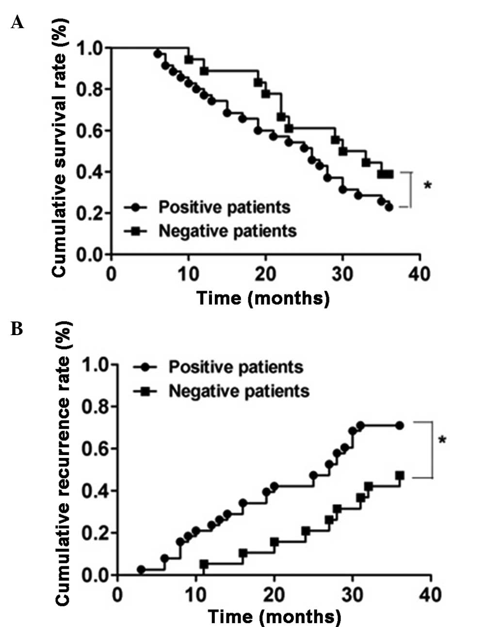
Correlation of activin A with survival
and recurrence rates
The 57 patients were divided into two groups
according to the evaluation standard for activin A expression in
the immunohistochemical assay, which included 38 activin A-positive
subjects or 19 activin A-negative subjects. Subsequently, the
recurrence and survival rates of each group were analyzed. As shown
in Fig. 5, the cumulative survival
rates at 1, 2 and 3 years postoperatively in the positive group
were 71.05, 50.00 and 21.05%, respectively, while in the negative
group the rates were 84.21, 63.16 and 36.84%, respectively.
Patients with a positive expression of activin A had a
significantly shorter survival time compared with that of patients
with a negative expression of activin A (P<0.05). Furthermore,
the cumulative recurrence rates at 1, 2 and 3 years postoperatively
in the positive group were found to be 23.68, 42.11 and 71.00%,
respectively, while in the negative group the rates were 5.26,
21.05 and 47.37%, respectively. The positive group presented an
evidently higher cumulative recurrence rate compared with the
negative group (P<0.05).
Discussion
Esophageal carcinoma is one of the most common
cancer types. A previous study has reported that abnormal
expression of activin A was detected in the serum of patients with
esophageal carcinoma (19).
Therefore, investigating the correlation of the activin A
expression with the tumorigenicity and metastasis of human
esophageal cancer is essential. However, only a limited number of
studies have been conducted on the expression of activin A in
esophageal tumor tissues. The present study detected the mRNA and
protein expression levels of activin A in esophageal tumor and
normal esophageal tissues. The results revealed that the mRNA and
protein expression levels of activin A were significantly higher in
esophageal cancer compared with those in normal esophageal tissues,
which is consistent with the findings of previous studies (20,21).
In addition, the expression of activin A in
esophageal tumor tissues with various degrees of differentiation
was investigated. The immunohistochemical assay demonstrated that
the protein expression of activin A was the highest in
poorly-differentiated tumor tissues and the lowest in
well-differentiated esophageal tumor tissues, which revealed an
association between activin A and the differentiation degree of
esophageal carcinoma. Thus, a high expression of activin A can be
considered to represent a high degree of malignancy, while a low
expression of activin A may indicate a benign tumor. Furthermore,
analysis of the lymph node metastasis demonstrated that the
expression of activin A was higher in esophageal tumors with
presence of lymph node metastasis compared with absence of
metastasis. This result indicates that activin A may be involved in
the metastasis of esophageal cancer, which is consistent with the
aforementioned association with the differentiation degree of
esophageal carcinoma. Malignant tumors are generally more likely to
metastasize, while benign tumors demonstrate few metastases. The
results of the present study revealed that activin A plays an
important role in tumor metastasis and grade malignancy, which has
also been observed in numerous other types of cancer (22–24).
The primary measure for the treatment of esophageal
carcinoma is surgery; however, recurrence is observed
postoperatively in certain patients and affects the survival of
these patients (25). The present
study confirmed that patients with high expression of activin A
preserved a lower cumulative survival rate and a higher cumulative
recurrence rate compared with patients with low expression of
activin A. These results suggest that activin A is closely
associated with the recurrence and survival of esophageal cancer
patients, which may be due to its correlation with the metastasis
and malignancy of esophageal tumors.
In conclusion, the present study identified that
activin A was abnormally expressed in esophageal carcinoma tissues
and correlated with the tumor differentiation, metastasis,
recurrence and survival of esophageal carcinoma patients.
Therefore, activin A may be a potential molecular marker for the
diagnosis and postoperative prognosis of esophageal cancer.
References
|
1
|
Siegel R, Naishadham D and Jemal A: Cancer
statistics, 2012. CA Cancer J Clin. 62:10–29. 2012. View Article : Google Scholar : PubMed/NCBI
|
|
2
|
Jiang L, Zhao X, Meng X and Yu J: Involved
field irradiation for the treatment of esophageal cancer: Is it
better than elective nodal irradiation. Cancer Lett. 357:69–74.
2015. View Article : Google Scholar : PubMed/NCBI
|
|
3
|
McK Manson J and Beasley W: A personal
perspective on controversies in the surgical management of
oesophageal cancer. Ann R Coll Surg Engl. 96:575–578. 2014.
View Article : Google Scholar : PubMed/NCBI
|
|
4
|
Baba Y, Watanabe M, Yoshida N and Baba H:
Neoadjuvant treatment for esophageal squamous cell carcinoma. World
J Gastrointest Oncol. 6:121–128. 2014. View Article : Google Scholar : PubMed/NCBI
|
|
5
|
Napier KJ, Scheerer M and Misra S:
Esophageal cancer: A Review of epidemiology, pathogenesis, staging
workup and treatment modalities. World J Gastrointest Oncol.
6:112–120. 2014. View Article : Google Scholar : PubMed/NCBI
|
|
6
|
Ling N, Ying SY, Ueno N, Shimasaki S, Esch
F, Hotta M and Guillemin R: A homodimer of the beta-subunits of
inhibin A stimulates the secretion of pituitary follicle
stimulating hormone. Biochem Biophys Res Commun. 138:1129–1137.
1986. View Article : Google Scholar : PubMed/NCBI
|
|
7
|
Lin HS, Gong JN, Su R, et al: miR-199a-5p
inhibits monocyte/macrophage differentiation by targeting the
activin A type 1B receptor gene and finally reducing C/EBPα
expression. J Leukoc Biol. 96:1023–1035. 2014. View Article : Google Scholar : PubMed/NCBI
|
|
8
|
Kariyawasam HH, Semitekolou M, Robinson DS
and Xanthou G: Activin-A: a novel critical regulator of allergic
asthma. Clin Exp Allergy. 41:1505–1514. 2011. View Article : Google Scholar : PubMed/NCBI
|
|
9
|
Maeshima A, Miya M, Mishima K, Yamashita
S, Kojima I and Nojima Y: Activin A: autocrine regulator of kidney
development and repair. Endocr J. 55:1–9. 2008. View Article : Google Scholar : PubMed/NCBI
|
|
10
|
Xu J, Oakley J and McGee EA:
Stage-specific expression of Smad2 and Smad3 during
folliculogenesis. Biol Reprod. 66:1571–1578. 2002. View Article : Google Scholar : PubMed/NCBI
|
|
11
|
Zalzman M, Anker-Kitai L and Efrat S:
Differentiation of human liver-derived, insulin-producing cells
toward the beta-cell phenotype. Diabetes. 54:2568–2575. 2005.
View Article : Google Scholar : PubMed/NCBI
|
|
12
|
Ferreira MC, Witz CA, Hammes LS, Kirma N,
Petraglia F, Schenken RS and Reis FM: Activin A increases
invasiveness of endometrial cells in an in vitro model of
human peritoneum. Mol Hum Reprod. 14:301–307. 2008. View Article : Google Scholar : PubMed/NCBI
|
|
13
|
Schwall RH, Robbins K, Jardieu P, Chang L,
Lai C and Terrell TG: Activin induces cell death in hepatocytes
in vivo and in vitro. Hepatology. 18:347–356. 1993.
View Article : Google Scholar : PubMed/NCBI
|
|
14
|
Thissen JP and Loumaye A: Role of activin
A and myostatin in cancer cachexia. Ann Endocrinol (Paris).
74:79–81. 2013.(In French). View Article : Google Scholar : PubMed/NCBI
|
|
15
|
Hoda MA, Münzker J, Ghanim B, Schelch K,
Klikovits T, Laszlo V, Sahin E, Bedeir A, Lackner A, Dome B, et al:
Suppression of activin A signals inhibits growth of malignant
pleural mesothelioma cells. Br J Cancer. 107:1978–1986. 2012.
View Article : Google Scholar : PubMed/NCBI
|
|
16
|
Hofland J, van Weerden WM, Steenbergen J,
Dits NF, Jenster G and de Jong FH: Activin A stimulates AKR1C3
expression and growth in human prostate cancer. Endocrinology.
153:5726–5734. 2012. View Article : Google Scholar : PubMed/NCBI
|
|
17
|
Kim YI, Kim BH, Khang I, Cho BN and Lee
HK: Cell growth regulation through apoptosis by activin in human
gastric cancer SNU-16 cell lines. Oncol Rep. 21:491–497.
2009.PubMed/NCBI
|
|
18
|
Wang G, Wang L, Sun S, Wu J and Wang Q:
Quantitative measurement of serum microRNA-21 expression in
relation to breast cancer metastasis in Chinese females. Ann Lab
Med. 35:226–232. 2015. View Article : Google Scholar : PubMed/NCBI
|
|
19
|
Gold E and Risbridger G: Activins and
activin antagonists in the prostate and prostate cancer. Mol Cell
Endocrinol. 359:107–112. 2012. View Article : Google Scholar : PubMed/NCBI
|
|
20
|
Zhang Y, Bao YL, Yang MT, Wu Y, Yu CL,
Huang YX, Sun Y, Zheng LH and Li YX: Activin A induces SLC5A8
expression through the Smad3 signaling pathway in human colon
cancer RKO cells. Int J Biochem Cell Biol. 42:1964–1972. 2010.
View Article : Google Scholar : PubMed/NCBI
|
|
21
|
de Kretser DM, O'Hehir RE, Hardy CL and
Hedger MP: The roles of activin A and its binding protein,
follistatin, in inflammation and tissue repair. Mol Cell
Endocrinol. 359:101–106. 2012. View Article : Google Scholar : PubMed/NCBI
|
|
22
|
Yoshinaga K, Yamashita K, Mimori K, Tanaka
F, Inoue H and Mori M: Activin a causes cancer cell aggressiveness
in esophageal squamous cell carcinoma cells. Ann Surg Oncol.
15:96–103. 2008. View Article : Google Scholar : PubMed/NCBI
|
|
23
|
Yoshinaga K, Mimori K, Yamashita K,
Utsunomiya T, Inoue H and Mori M: Clinical significance of the
expression of activin A in esophageal carcinoma. Int J Oncol.
22:75–80. 2003.PubMed/NCBI
|
|
24
|
Liu SG, Li HC, Zhao BS and Cao F:
Expression of activin A in tissue and serum of patients with
esophageal squamous cell carcinoma and its clinical significance.
Zhonghua Zhong Liu Za Zhi. 35:843–847. 2013.(In Chinese).
PubMed/NCBI
|
|
25
|
Zhang C, Wang QT, Liu H, et al:
Advancement and prospects of tumor gene therapy. Chin J Cancer.
30:182–188. 2011. View Article : Google Scholar : PubMed/NCBI
|