Introduction
Osteosarcoma is one of the most common highly
malignant forms of bone tumors, occurring most frequently in
children and adolescents, with a poor clinical prognosis (1–3).
Amputation surgery is the primary treatment for patients with
osteosarcoma, but confers a five-year survival rate of just 10–20%
(4). However, with the application of
neoadjuvant chemotherapy, the survival rate of patients with
osteosarcoma has increased to 60–70% in the last 20 years (5,6). Despite
this, resistance to chemotherapy remains a major challenge for
patients, particularly in cases of recurrence or distant metastasis
(5). At present, immunotherapy, gene
therapy and cell therapy are implemented for the treatment of
osteosarcoma and osteosarcoma-induced lung metastases. The
application of lymphokine-activated killer (LAK) cells is a recent
advance in cell therapy for the treatment of osteosarcoma (7–11). The
purpose of the present study was to investigate the effects of low
doses of ionizing radiation on LAK cell therapy for animal
osteosarcoma.
Materials and methods
Reagents and equipment
Dulbecco's modified Eagle's medium (DMEM) and fetal
bovine serum were purchased from Invitrogen (Carlsbad, CA, USA). An
industrial portable X'Pert-Pro MPD X-ray unit was purchased from
Philips Medical Systems, Inc., (Bothell, WA, USA).
Animals
For the present study, 24 Sprague-Dawley (SD) rats
were provided by the Experimental Animal Center of the Fourth
Military Medical University (Xi'an, China; license number, SCXK;
Army 2002–005). A total of 24, 6–8-week-old, male C57BL/6 mice,
weighing between 18 and 22 g, were provided by the Chinese Academy
of Medical Sciences Animal Breeding Company (Xi'an, China; animal
license number, SCXK; Beijing 2004-0001). The osteosarcoma UMR-106
cells were purchased from the American Type Culture Collection
(Manassas, VA, USA) and were cultured in DMEM containing 10% FBS.
The LAK cells were derived from the spleens of the tumor-bearing
mice. All animal experiments were conducted in accordance with the
ethical guidelines of the Provincial Hospital Affiliated to
Shandong University (Jinan, China) and the study was approved by
the ethics committee of the Provincial Hospital Affiliated to
Shandong University.
Cell inoculation
The cultured UMR-106 cells were counted, and
1×10⁷ cells were seeded subcutaneously into the anterior
chest wall of the SD rats. Rats were randomly divided into the
following groups: The control group (0 mGy radiation); the 0.65-mGy
group; and the 3.25-mGy group, with 8 SD rats in each group. In
each group, the rats were examined for tumor growth following
inoculation with the cells. The tumor size of each rat was measured
weekly with a caliper, and the average values of the tumor
diameters were calculated.
Pathological staining and
determination of alkaline phosphatase activity
The rats were euthanized by cervical dislocation
after anesthesia with 1% pentobarbital (Beyotime Institute of
Biotechnology, Haimen, China), and the tumor tissues were
collected. Hematoxylin and eosin (HE) staining was performed to
examine the extent of tumor metastasis. Serum from the tail vein of
eight rats in each group was collected each week to determine the
alkaline phosphatase activity using an Alkaline Phosphatase Assay
kit (ab83369) purchased from Abcam (Burlingame, CA, USA) according
to the manufacturer's instructions.
LAK cells
The LAK cells were derived from the spleens of the
tumor-bearing mice. The isolated bioactive cells were diluted to
2×10⁵/ml. In total, 1,000 units interleukin 2 (IL-2)
were added per ml of the culture system. The separated LAK cells
were divided into three groups and treated with 0 (control), 0.65
or 3.25 mGy ionizing radiation. Subsequent to treatment with the
different doses of radiation, the LAK cells were cultured at 37°C
in 5% CO2.
Winn assay
In the present study, 24 healthy C57BL/6 mice were
divided into three groups. In total, 2×10⁶ LAK cells
exposed to different radiation doses, in combination with
2×10⁵ osteosarcoma cells, were subcutaneously injected
into the hind limbs of the rats. The rats were then raised under
the same conditions for 30 days to observe tumor growth. The Winn
assay was performed as described by Tjota et al (12).
Tritiated thymidine (3H-TdR) release
assay
The 3H-TdR release assay was performed to
determine LAK cell killing activity as in a previous study
(13). In total, 1×10⁶
osteosarcoma cells/ml were treated with 3.7×10⁵ Bq/ml
3H-TdR. The solution was incubated at 37°C for 2h, with
agitation every 30 min. The cells were then washed with
phosphate-buffered saline (PBS), and the density of the cells was
diluted to 2×10⁵/ml. Next, 50 µl cells/well were added
to a 96-well plate. Subsequent to washing with PBS, the obtained
LAK and tumor cells were mixed at a ratio of 40:1. Each
experimental sample had three replicate wells. The radioactivity of
each sample was measured using a Tri-Carb 2810TR Low Activity
Liquid Scintillation Analyzer (PerkinElmer, Inc., Boston, MA, USA).
The LAK cell killing activity was calculated with the following
formula: LAK cell killing activity (%) = [spontaneous release group
(min-1) - experimental group (min-1)]/[spontaneous release group
(min-1) - maximum release group (min-1)] × 100.
Statistical analysis
SPSS 13.0 software (SPSS, Inc., Chicago, IL, USA)
was used for analysis of the data. The experimental data are
presented as the mean ± standard error. An analysis of variance was
applied for the comparison of mean values of the samples. P<0.05
was considered to indicate a statistically significant
difference.
Results
Establishment of an osteosarcoma
model
The cultured UMR-106 cells were inoculated under the
anterior chest skin of 18 of the 24 rats (the experimental groups)
to establish an osteosarcoma model. In total, six of the 24 rats
were inoculated with phosphate-buffered saline to serve as the
control group. The tumor growth curves were plotted from the
average weekly tumor diameter of the rats in the experimental
groups. There were no tumors detected in the rats of the control
group, which suggested that the tumor model was successfully
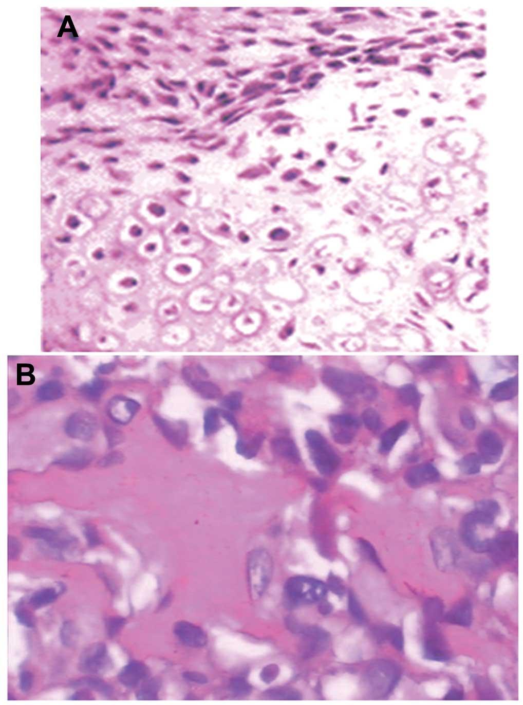
established. HE staining was used to determine the extent of tumor
metastasis. As revealed in Fig. 1,
pale-blue Mallory's trichrome staining of the tumor tissues
indicated evident osteoid growth. These results suggested that the
tumor tissues exhibited distinctive osteogenesis
characteristics.
Effects of different doses of X-ray
radiation on the proliferation of cells
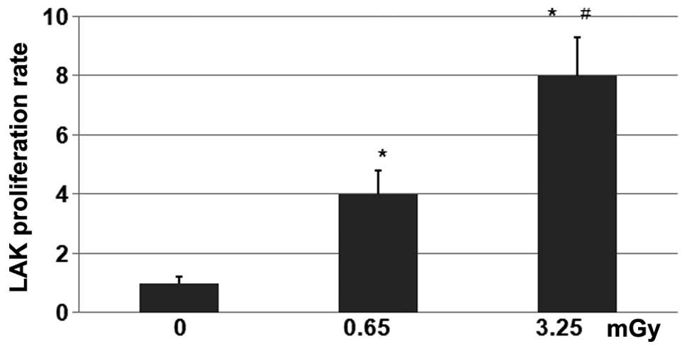
The LAK cells from the 24 mice were exposed to doses
of 0 (control), 0.65 or 3.25 mGy X-ray radiation. The number and
survival rates of the LAK cells cultured in a CO2
incubator were determined every three days. The results
demonstrated that the LAK cells began to grow from day three
post-radiation, and subsequent to attaining a higher level on day
six, the cell proliferation rate was maintained at a stable level.
As revealed in Fig. 2, the cells that
received 3.25 mGy X-ray radiation demonstrated a higher
proliferation rate than those receiving 0.65 mGy. Compared with the
control group that received 0 mGy X-ray radiation, the
proliferation rates in the 0.65- and 3.25-mGy groups were
significantly higher (P<0.01).
Therapeutic effect of LAK cells
treated with low-dose ionizing radiation on osteosarcoma
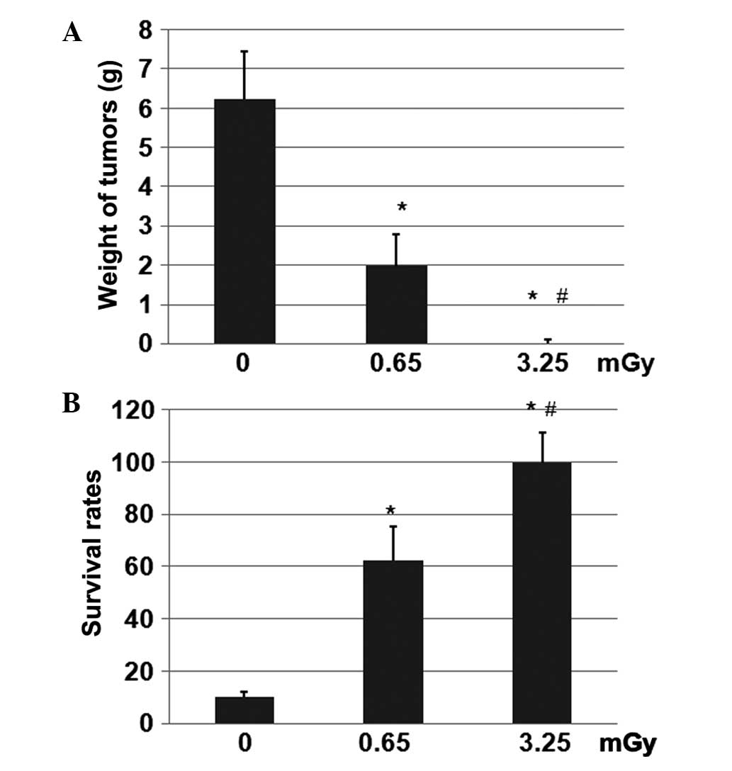
Subsequent to exposure to 0, 0.65 or 3.25 mGy X-ray
radiation, the LAK cells were inoculated into the tumor-bearing
rats. Following tumor cell inoculation, alkaline phosphatase levels
in the blood serum of the tumor-bearing rats were elevated compared
with those of rats without tumors. After 30 days, the rats were
euthanized and the tumors were collected and weighed (Fig. 3A). The mean weight of the tumors in
the control and 0.65-mGy groups was 6.25 and 2.0 g, respectively.
However, the weights of the tumors in the rats inoculated with 3.25
mGy radiation-treated LAK cells were undetectable after 30 days.
Furthermore, the rats in the group treated with 3.25 mGy radiation
demonstrated the longest survival rate (Fig. 3B), followed by those in the 0.65-mGy
group. The differences between these two groups were revealed to be
statistically significant (P<0.01). In addition, the differences
between the 0.65- and 3.25-mGy groups and the control 0-mGy group
were statistically significant (P<0.01).
Discussion
The results of the present study demonstrated that
the optimal dose of X-ray radiation for the stimulation of LAK cell
activity was 3.25 mGy. Tomuleasa et al (14) reported that low doses of ionizing
radiation were able to enhance the biological activity of stem and
bone cells. The results of the present study demonstrated that the
viability and proliferation of the LAK cells that received low
doses of X-ray radiation were higher than those of the cells that
received no radiation (P<0.01). This indicates that low doses of
ionizing radiation may promote the proliferation of certain cells.
This may be due to low doses of ionizing radiation simultaneously
accelerating cellular mitosis and increasing IL-2-mediated
proliferation. The LAK cell proliferation rate reached a peak on
days one and two, prior to gradually decreasing until day 21, where
the rate returned to the initial level. This indicated that the
proliferative ability of the cells was progressively weakened over
time.
In the present study, the viability of the
freshly-isolated cells was 98%, which gradually decreased over the
incubation period. However, the viability of the cells treated with
0.65 and 3.25 mGy X-ray radiation was 20% higher than that of the
cells in the control group. The differences were statistically
significant (P<0.01). The results of the present study indicated
that low doses of radiation may not only increase the proliferation
rate, but also decrease the mortality of LAK cells. Therefore,
radiation provides a method to shorten the culture time and
increase the proliferation of cells. The reduced mortality may be
due to the ability of low doses of radiation to specifically
stimulate certain dormant cells, but not affect the internal or
external environment of the cells.
The effect of low doses of radiation may provide a
novel approach in increasing the proliferation of cells for
clinical applications. A previous study (15) revealed that osteosarcoma cells had the
ability to produce cancer stem cells (CSCs), which are recognized
to lead to tumor recurrence. Therefore, osteosarcoma CSCs were
considered to be a major target for cancer therapy, as they were
believed to contribute to chemotherapy resistance (13,16–18).
Another previous study identified that low doses of radiation were
able to enhance immunity, stimulate the differentiation and
proliferation of T lymphocytes and natural killer (NK) cells, and
promote IL-2 secretion of spleen cells and peripheral lymphocytes
(19). In addition, previous studies
have demonstrated that low doses of ionizing radiation can
stimulate LAK cell killing activity (20). This may be due to the ability of
radiation to promote the secretion of IL-2 and stimulate NK cell
activity.
LAK cells are derived from a group of heterogeneous
large particles of lymphocytes that are cytotoxic to either
autologous or allogeneic tumor cells. IL-2 is necessary for LAK
cell activation, and activated LAK cells can directly or indirectly
kill tumor cells by the release of cytotoxic granules and the
secretion of cytokines (21). As a
biological form of tumor treatment, the clinical application of LAK
cells attracts a large amount of attention.
LAK cell therapy is an adoptive immunotherapy
(22), and is administered to
patients by the injection of IL-2-activated LAK cells. This leads
to immunity and an improvement in the body's anti-tumor
ability.
Subsequent to a certain amount of IL-2-mediated
proliferation, LAK cells demonstrate antitumor activity (23). However, the clinical culture and
proliferation of LAK cells is difficult to achieve. Therefore, to
enhance the clinical antitumor effects of LAK cells, a dose of IL-2
is usually co-administered (24).
However, high doses of IL-2 have certain side-effects that may
affect treatment. Tumor necrosis factor (TNF) can significantly
enhance LAK cell activity in vitro and reduce the
concentration of IL-2, which is necessary for the induction of LAK
activity (25). A prior study
revealed that IL-2 could induce peripheral blood mononuclear cells
to produce TNF, which improves resultant antitumor effects
(26). The injection of solasodine
hydrochloride to tumor-bearing mice may increase the antitumor
activity of LAK cells (27). The
treatment of malignant tumors by the in vivo activation of
LAK cells from lymphocytes, the expansion of a LAK cell population
and the activation of macrophages to produce large amounts of TNF,
can inhibit the growth of tumor cells and prevent tumor invasion
and metastasis (28). Consequently,
this approach can significantly improve the quality of life and
prolong the survival of cancer patients.
In order to investigate the impact of low doses of
radiation on LAK cells, the effects of different doses of radiation
on the culture and anti-tumor activity of the cells were studied in
the present study. This may provide basic information for the
further study of LAK cell therapy.
References
|
1
|
Heymann D and Rédini F: Targeted therapies
for bone sarcomas. Bonekey Rep. 2:3782013. View Article : Google Scholar : PubMed/NCBI
|
|
2
|
Hoch M, Ali S, Agrawal S, Wang C and
Khurana JS: Extraskeletal osteosarcoma: a case report and review of
the literature. J Radiol Case Rep. 7:15–23. 2013.PubMed/NCBI
|
|
3
|
Vikram S, Salih S, Krishnan A, et al:
Radiation-induced extra-osseous osteosarcoma - A case report and
review of literature. Indian J Surg Oncol. 4:374–377. 2013.
View Article : Google Scholar : PubMed/NCBI
|
|
4
|
Puri A, Pruthi M and Gulia A: Outcomes
after limb sparing resection in primary malignant pelvic tumors.
Eur J Surg Oncol. 40:27–33. 2014. View Article : Google Scholar : PubMed/NCBI
|
|
5
|
Tao LJ, Zhou XD, Shen CC, et al:
Tetrandrine induces apoptosis and triggers a caspase cascade in
U2-OS and MG-63 cells through the intrinsic and extrinsic pathways.
Mol Med Rep. 9:345–349. 2014.PubMed/NCBI
|
|
6
|
Zhao Z, Tao L, Shen C, Liu B, Yang Z and
Tao H: Silencing of Barkor/ATG14 sensitizes osteosarcoma cells to
cisplatin-induced apoptosis. Int J Mol Med. 33:271–276.
2014.PubMed/NCBI
|
|
7
|
Uno K, Tsukuda M, Kushida K, et al:
Clinical experience with In-111 labeled LAK cells and TILS for
tumor localization. Prog Clin Biol Res. 355:239–245.
1990.PubMed/NCBI
|
|
8
|
Luksch R, Perotti D, Cefalo G, et al:
Immunomodulation in a treatment program including pre- and
post-operative interleukin-2 and chemotherapy for childhood
osteosarcoma. Tumori. 89:263–268. 2003.PubMed/NCBI
|
|
9
|
Herrero-Martín D, Osuna D, Ordóñez JL, et
al: Stable interference of EWS-FLI1 in an Ewing sarcoma cell line
impairs IGF-1/IGF-1R signalling and reveals TOPK as a new target.
Br J Cancer. 101:80–90. 2009. View Article : Google Scholar : PubMed/NCBI
|
|
10
|
Park JH, Yoon DS, Choi HJ, Hahm DH and Oh
SM: Phosphorylation of IκBα at serine 32 by T-lymphokine-activated
killer cell-originated protein kinase is essential for
chemoresistance against doxorubicin in cervical cancer cells. J
Biol Chem. 288:3585–3593. 2013. View Article : Google Scholar : PubMed/NCBI
|
|
11
|
Jennings VA, Ilett EJ, Scott KJ, et al:
Lymphokine-activated killer and dendritic cell carriage enhances
oncolytic reovirus therapy for ovarian cancer by overcoming
antibody neutralization in ascites. Int J Cancer. 134:1091–1101.
2014. View Article : Google Scholar : PubMed/NCBI
|
|
12
|
Tjota A, Zhang YQ, Piedmonte MR and Lee
CL: Adoptive immunotherapy using lymphokine-activated killer cells
and recombinant interleukin-2 in preventing and treating
spontaneous pulmonary metastases of syngeneic Dunning rat prostate
tumor. J Urol. 146:177–183. 1991.PubMed/NCBI
|
|
13
|
Gillette J and Nielsen-Preiss S: Cancer
stem cells: seeds of growth in osteosarcoma. Cancer Biol Ther.
8:553–554. 2009. View Article : Google Scholar : PubMed/NCBI
|
|
14
|
Tomuleasa C, Soritau O, Brie I, et al:
Mesenchymal stem cell irradiation in culture engages differential
effect of hyper-fractionated radiotherapy for head and neck
cancers. J BUON. 15:348–356. 2010.PubMed/NCBI
|
|
15
|
Siclari VA and Qin L: Targeting the
osteosarcoma cancer stem cell. J Orthop Surg Res. 5:782010.
View Article : Google Scholar : PubMed/NCBI
|
|
16
|
Longhi A, Errani C, De Paolis M, Mercuri M
and Bacci G: Primary bone osteosarcoma in the pediatric age: state
of the art. Cancer Treat Rev. 32:423–436. 2006. View Article : Google Scholar : PubMed/NCBI
|
|
17
|
Fujii H, Honoki K, Tsujiuchi T, Kido A,
Yoshitani K and Takakura Y: Sphere-forming stem-like cell
populations with drug resistance in human sarcoma cell lines. Int J
Oncol. 34:1381–1386. 2009.PubMed/NCBI
|
|
18
|
Ta HT, Dass CR, Choong PF and Dunstan DE:
Osteosarcoma treatment: state of the art. Cancer Metastasis Rev.
28:247–263. 2009. View Article : Google Scholar : PubMed/NCBI
|
|
19
|
Zhang SY and Shi Y: Reaction of low dose
ionizing radiation on amplification in vitro and antitumor effect
of TIL cells. Chinese Journal of Cancer Prevention and Treatment.
16:1552–1553. 2009.
|
|
20
|
Fu Q, Zhang LS, Zhu SP and Liu XH: Effects
of low-dose radiation on LAK cells to kill tumor target cells.
Chinese Journal of Radiological Medicine and Protection.
15:3211995.
|
|
21
|
Wang YC, Shi EX and Ding XL: Killing
effect on lung cancer cell line A549 by lymphokine-activated killer
cells activated by dendritic cells. Northern China National Defense
Medicine. 21:11–12. 2009.
|
|
22
|
Yanagawa H, Sone S, Fukuta K, Nishioka Y
and Ogura T: Local adoptive immunotherapy using
lymphokine-activated killer cells and interleukin-2 against
malignant pleural mesothelioma: Report of two cases. Jpn J Clin
Oncol. 21:377–383. 1991.PubMed/NCBI
|
|
23
|
Mou QJ, Wang J, Cui WF and Wang ZJ:
Proliferation and cytotoxic activity comparison of three sources of
CIK cells. Shandong Medicine. 50:7–9. 2010.
|
|
24
|
Barba D, Saris SC, Holder C, Rosenberg SA
and Oldfield EH: Intratumoral LAK cell and interleukin-2 therapy of
human gliomas. J Neurosurg. 70:175–182. 1989. View Article : Google Scholar : PubMed/NCBI
|
|
25
|
Balasa B, Yun R, Belmar NA, et al:
Elotuzumab enhances natural killer cell activation and myeloma cell
killing through interleukin-2 and TNF-α pathways. Cancer Immunol
Immunother. 64:61–73. 2015. View Article : Google Scholar : PubMed/NCBI
|
|
26
|
Jin P, Wang E, Provenzano M, et al:
Molecular signatures induced by interleukin-2 on peripheral blood
mononuclear cells and T cell subsets. J Transl Med. 4:262006.
View Article : Google Scholar : PubMed/NCBI
|
|
27
|
Jinxia Ai and Huanbo Cui: The effect of
SBHL on the tumor growth and immune functions in S180-bearing mice.
Zhong Guo Mian Yi Xue Za Zhi. 26:26–29. 2010.(In Chinese).
|
|
28
|
Parhar RS, Ernst P, Sheth KV and
al-Sedairy ST: Anti-tumor cytotoxic potential and effect on human
bone marrow GM-CFU of human LAK cells generated in response to
various cytokines. Eur Cytokine Netw. 3:299–306. 1992.PubMed/NCBI
|