|
1
|
Toker C: Trabecular carcinoma of the skin.
Arch Dermatol. 105:107–110. 1972. View Article : Google Scholar : PubMed/NCBI
|
|
2
|
Merkel F: Tastzellen und Tastkörperchen
bei den Hausthieren und beim Menschen. Archiv für Mikroskopische
Anatomie. 11:636–652. 1875. View Article : Google Scholar
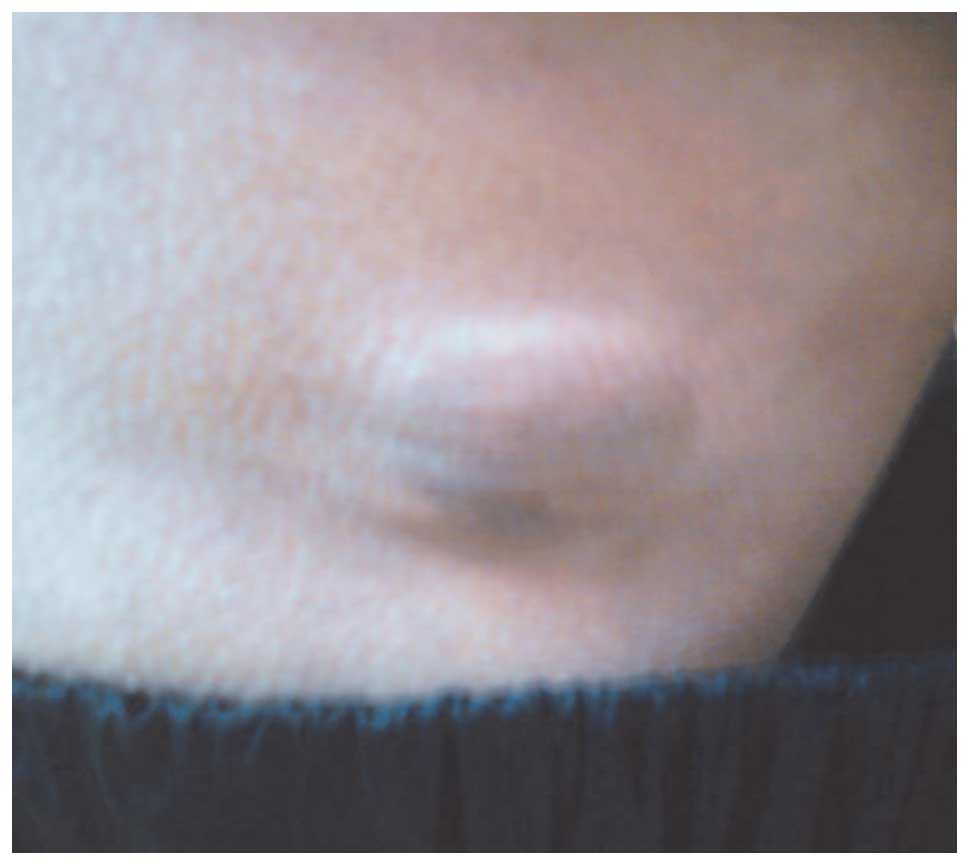
|
|
3
|
Erovic I and Erovic BM: Merkel cell
carcinoma: The past, the present and the future. J Skin Cancer.
2013:9293642013. View Article : Google Scholar : PubMed/NCBI
|
|
4
|
Tang CK and Toker C: Trabecular carcinoma
of the skin: An ultrastructural study. Cancer. 42:2311–2321. 1978.
View Article : Google Scholar : PubMed/NCBI
|
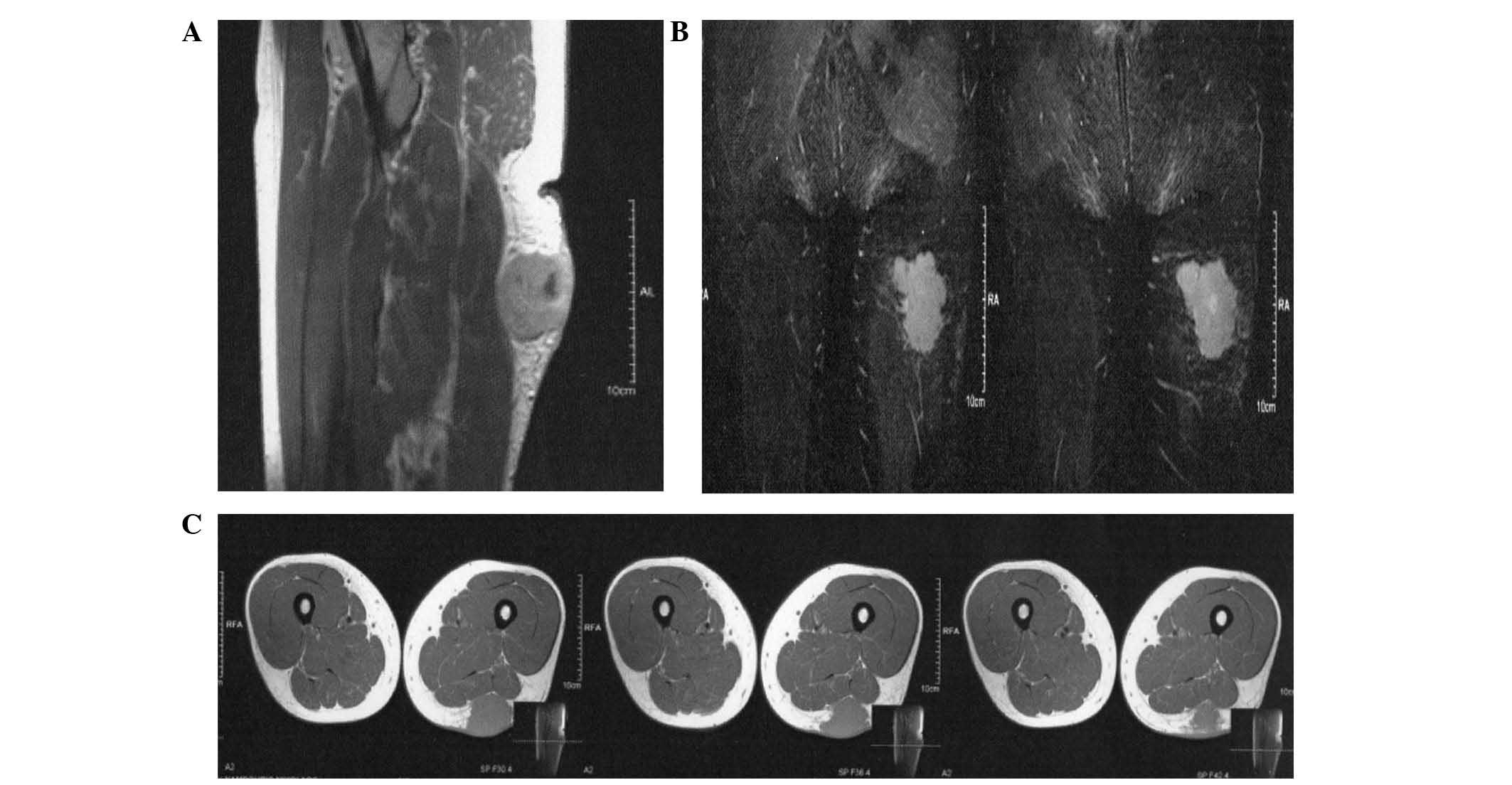
|
5
|
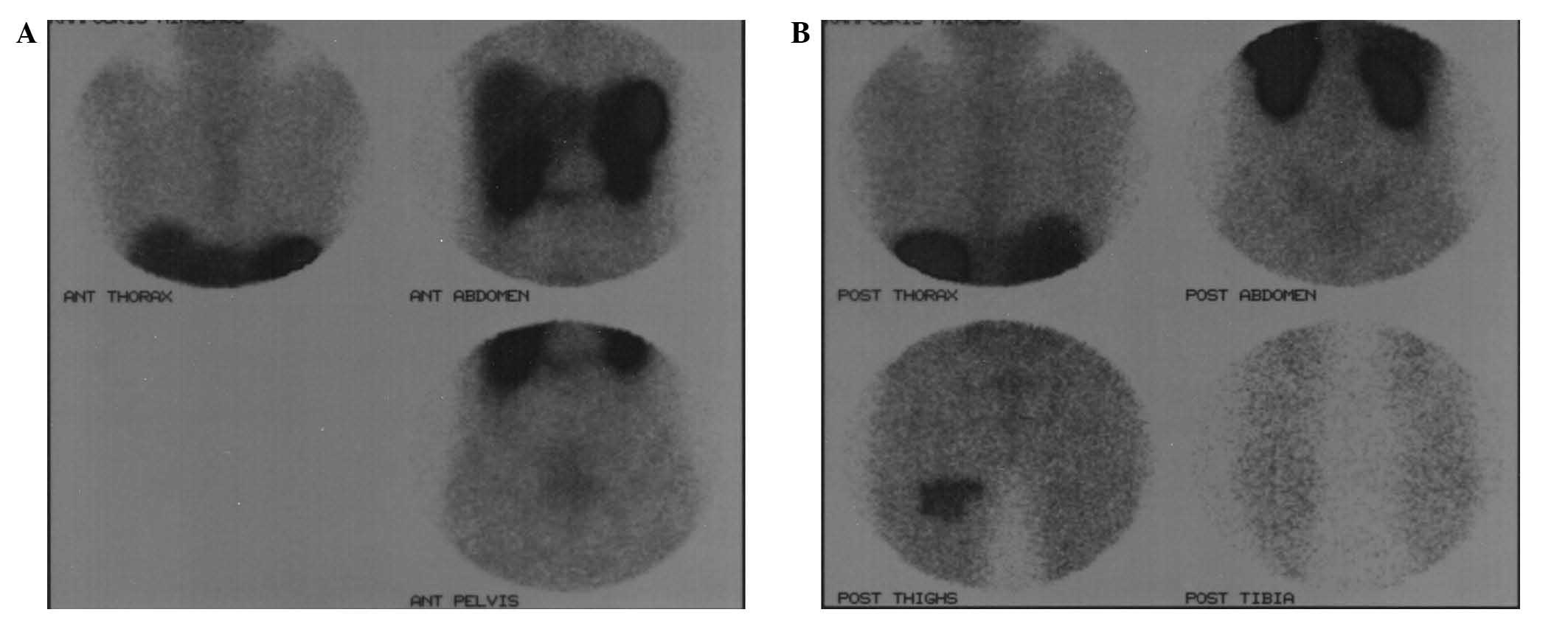
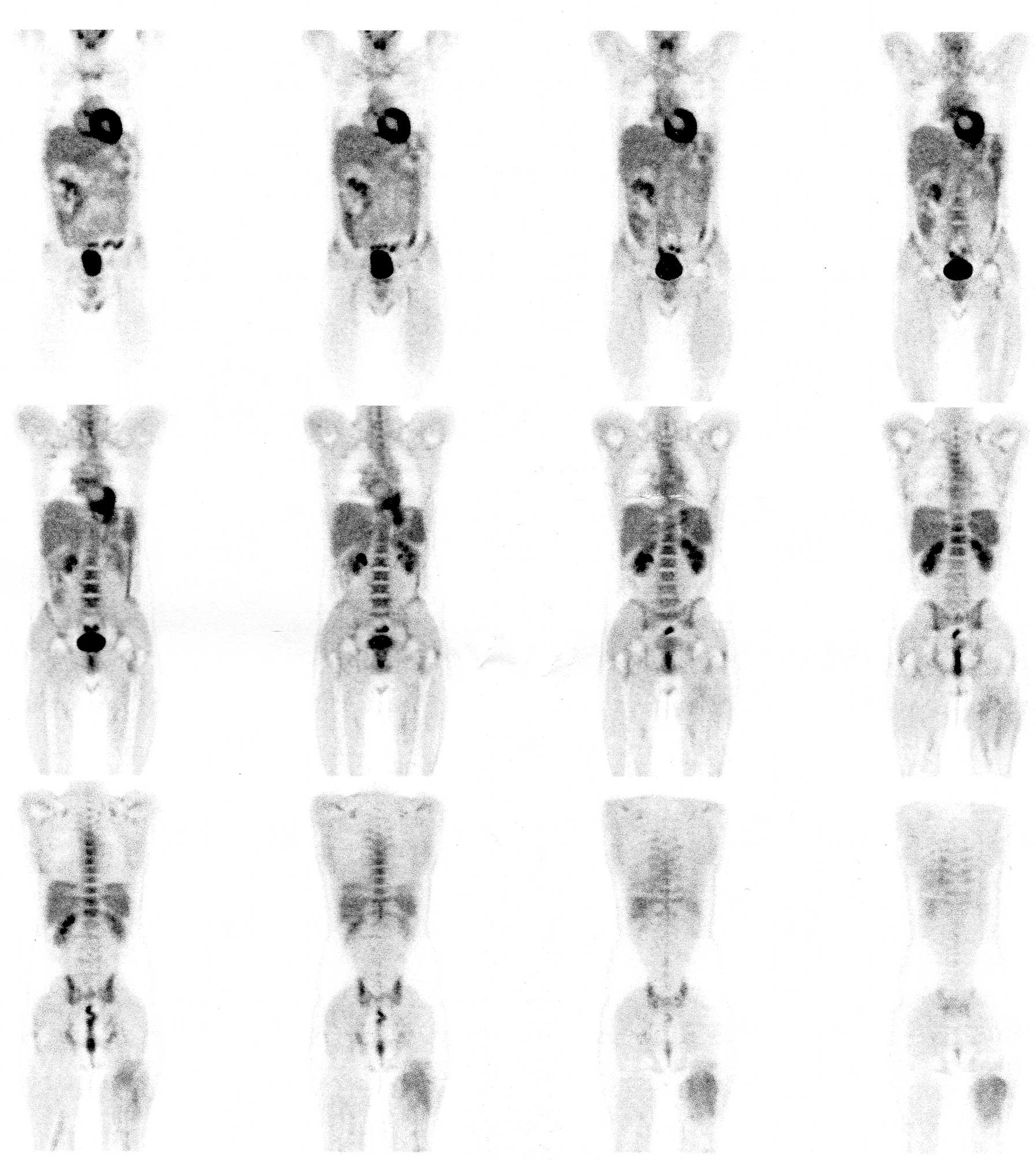
Han SY, North JP, Canavan T, et al: Merkel
cell carcinoma. Hematol Oncol Clin North Am. 26:1351–1374. 2012.
View Article : Google Scholar : PubMed/NCBI
|
|
6
|
Duprat JP, Landman G, Salvajoli JV and
Brechtbühl ER: A review of the epidemiology and treatment of Merkel
cell carcinoma. Clinics (Sao Paulo). 66:1817–1823. 2011. View Article : Google Scholar : PubMed/NCBI
|
|
7
|
Becker JC: Merkel cell carcinoma. Ann
Oncol. 21:(Sul 7). vii81–vii85. 2010. View Article : Google Scholar : PubMed/NCBI
|
|
8
|
Feng H, Shuda M, Chang Y and Moore PS:
Clonal integration of a polyomavirus in human Merkel cell
carcinoma. Science. 319:1096–1100. 2008. View Article : Google Scholar : PubMed/NCBI
|
|
9
|
Fukumoto H, Sato Y, Hasegawa H and Katano
H: Frequent detection of Merkel cell polyomavirus DNA in sera of
HIV-1-positive patients. Virol J. 10:842013. View Article : Google Scholar : PubMed/NCBI
|
|
10
|
Engels EA, Frisch M, Goedert JJ, et al:
Merkel cell carcinoma and HIV infection. Lancet. 359:497–498. 2002.
View Article : Google Scholar : PubMed/NCBI
|
|
11
|
Agelli M and Clegg LX: Epidemiology of
primary Merkel cell carcinoma in the United States. J Am Acad
Dermatol. 49:832–841. 2003. View Article : Google Scholar : PubMed/NCBI
|
|
12
|
Heath M, Jaimes N, Lemos B, et al:
Clinical characteristics of Merkel cell carcinoma at diagnosis in
195 patients: The AEIOU features. J Am Acad Dermatol. 58:375–381.
2008. View Article : Google Scholar : PubMed/NCBI
|
|
13
|
Moll R, Löwe A, Laufer J and Franke WW:
Cytokeratin 20 in human carcinomas. A new histodiagnostic marker
detected by monoclonal antibodies. Am J Pathol. 140:427–447.
1992.PubMed/NCBI
|
|
14
|
Jaeger T, Ring J and Andres C:
Histological, immunohistological and clinical features of merkel
cell carcinoma in correlation to merkel cell polyomavirus status. J
Skin Cancer. 2012:9834212012. View Article : Google Scholar : PubMed/NCBI
|
|
15
|
Mojica P, Smith D and Ellenhorn JD:
Adjuvant radiation therapy is associated with improved survival in
Merkel cell carcinoma of the skin. J Clin Oncol. 25:1043–1047.
2007. View Article : Google Scholar : PubMed/NCBI
|
|
16
|
Teunissen JJ, Kwekkeboom DJ, Valkema R and
Krenning EP: Nuclear medicine techniques for the imaging and
treatment of neuroendocrine tumours. Endocr Relat Cancer. 18:(Sul
1). S27–S51. 2011. View Article : Google Scholar : PubMed/NCBI
|
|
17
|
Lemos BD, Storer BE, Iyer JG, et al:
Pathologic nodal evaluation improves prognostic accuracy in Merkel
cell carcinoma: Analysis of 5823 cases as the basis of the first
consensus staging system. J Am Acad Dermatol. 63:751–761. 2010.
View Article : Google Scholar : PubMed/NCBI
|
|
18
|
Nguyen BD and McCullough AE: Imaging of
Merkel cell carcinoma. Radiographics. 22:367–376. 2002. View Article : Google Scholar : PubMed/NCBI
|
|
19
|
Arruda EP and Higgins KM: Role of sentinel
lymph node biopsy in the management of merkel cell carcinoma. J
Skin Cancer. 2012:1761732012. View Article : Google Scholar : PubMed/NCBI
|
|
20
|
Enzenhofer E, Ubl P, Czerny C and Erovic
BM: Imaging in patients with merkel cell carcinoma. J Skin Cancer.
2013:9731232013. View Article : Google Scholar : PubMed/NCBI
|
|
21
|
Panagiotidis E and Bomanji J: Role of
18 F-fluorodeoxyglucose PET in the study of
neuroendocrine tumors. PET Clin. 9:43–55. 2014. View Article : Google Scholar : PubMed/NCBI
|
|
22
|
Yao M, Smith RB, Hoffman HT, et al: Merkel
cell carcinoma: Two case reports focusing on the role of
fluorodeoxyglucose positron emission tomography imaging in staging
and surveillance. Am J Clin Oncol. 28:205–210. 2005. View Article : Google Scholar : PubMed/NCBI
|
|
23
|
Lin O, Thomas A, Singh A and Greenspan B:
Complementary role of positron emission tomography in merkel cell
carcinoma. South Med J. 97:1110–1112. 2004. View Article : Google Scholar : PubMed/NCBI
|
|
24
|
Belhocine T, Pierard GE, Frühling J, et
al: Clinical added-value of 18 FDG PET in
neuroendocrine-merkel cell carcinoma. Oncol Rep. 16:347–352.
2006.PubMed/NCBI
|
|
25
|
Concannon R, Larcos GS and Veness M: The
impact of (18)F-FDG PET-CT scanning for staging and management of
Merkel cell carcinoma: Results from Westmead Hospital, Sydney,
Australia. J Am Acad Dermatol. 62:76–84. 2010. View Article : Google Scholar : PubMed/NCBI
|
|
26
|
Shintani SA, Foote RL, Lowe VJ, et al:
Utility of PET/CT imaging performed early after surgical resection
in the adjuvant treatment planning for head and neck cancer. Int J
Radiat Oncol Biol Phys. 70:322–329. 2008. View Article : Google Scholar : PubMed/NCBI
|
|
27
|
Peloschek P, Novotny C, Mueller-Mang C, et
al: Diagnostic imaging in Merkel cell carcinoma: Lessons to learn
from 16 cases with correlation of sonography, CT, MRI and PET. Eur
J Radiol. 73:317–323. 2010. View Article : Google Scholar : PubMed/NCBI
|
|
28
|
Ziessman H, O'Malley JP and Thrall JH:
Nuclear Medicine: The Requisites. 3rd. Mosby; Maryland Heights, MO,
USA: pp. 279–281. 2006
|
|
29
|
Pepe G, Bombardieri E, Lorenzoni A and
Chiti A: Single-photon emission computed tomography tracers in the
diagnostics of neuroendocrine tumors. PET Clin. 9:11–26. 2014.
View Article : Google Scholar : PubMed/NCBI
|
|
30
|
Kwekkeboom DJ, Hoff AM, Lamberts SW, et
al: Somatostatin analogue scintigraphy: A simple and sensitive
method for the in vivo visualization of Merkel cell tumors and
their metastases. Arch Dermatol. 128:818–821. 1992. View Article : Google Scholar : PubMed/NCBI
|
|
31
|
Guitera-Rovel P, Lumbroso J,
Gautier-Gougis MS, et al: Indium-111 octreotide scintigraphy of
Merkel cell carcinomas and their metastases. Ann Oncol. 12:807–811.
2001. View Article : Google Scholar : PubMed/NCBI
|
|
32
|
Ambrosini V and Fanti S:
68Ga-DOTA-peptides in the diagnosis of NET. PET Clin. 9:37–42.
2014. View Article : Google Scholar : PubMed/NCBI
|
|
33
|
Schmidt MC, Uhrhan K, Markiefka B, et al:
(68)Ga-DotaTATE PET-CT followed by Peptide Receptor Radiotherapy in
combination with capecitabine in two patients with Merkel Cell
Carcinoma. Int J Clin Exp Med. 5:363–366. 2012.PubMed/NCBI
|
|
34
|
Fakiha M, Letertre P, Vuillez JP and
Lebeau J: Remission of Merkel cell tumor after somatostatin analog
treatment. J Cancer Res Ther. 6:382–384. 2010. View Article : Google Scholar : PubMed/NCBI
|