Introduction
Activation of the vascular epidermal growth factor
(VEGF) signaling pathway via interaction between VEGF and VEGF
receptors (VEGFRs) has been demonstrated to promote a network of
signaling processes that support endothelial cell-mediated
angiogenesis (1). Thus, the VEGF
pathway is activated via a paracrine action; tumor cells
overexpress the ligand, VEGFA, to bind to the receptors, such as
fms-related tyrosine kinase 1 (FLT1; also known as VEGFR1) and
kinase insert domain receptor (KDR; also known as VEGFR2), which
are expressed in the endothelial cells, in order to promote
endothelial cell growth and survival, thereby initiating
angiogenesis. However, an increasing body of evidence indicates
that the VEGF signaling pathway also functions in an autocrine
manner, in which tumor cells express VEGF ligand and receptors, and
drive intrinsic processes, including tumor cell cycle progression,
survival and chemoresistance, ultimately promoting cancer
development (2–4).
VEGFA is known to promote angiogenesis through
binding to FLT1 and KDR, thus triggering the downstream signaling
pathway. Among various pro-angiogenic factors, VEGFA has been
identified to be a predominant regulator of tumor angiogenesis
(5). High expression of these three
genes has been identified in non-small cell lung cancer xenografts
compared with xenografts originating from other sites (5), indicating that these three factors may
be significant in lung cancer progression. Indeed, VEGFA (6–8), FLT1
(9,10)
and KDR (11,12) have all been demonstrated to promote
lung cancer progression. However, their utility as prognostic
markers remains unknown owing to the conflicting findings of
previous studies (12,13).
The aim of the present study was to investigate the
prognostic significance of VEGFA, FLT1 and KDR mRNA expression,
alone or in combination, in four independent lung cancer patient
cohorts from different institutions and of different ethnicities,
with a combined patient cohort of 583 patients.
Materials and methods
Extraction of clinical and microarray
gene expression data from lung cancer patient datasets
The GSE3141 (14),
GSE4573 (15), GSE8894 (16) and GSE31210 (17) lung cancer patient datasets were
identified in the NCBI Gene Expression Omnibus (GEO) database
(http://www.ncbi.nlm.nih.gov/geo/).
Datasets using the HG-U133 microarray platform and comprising of
>100 patients for whom survival data were available, were
included in this study. Microarray gene expression data were
retrieved from the data matrixes deposited to the GEO database by
the original authors. R scripting was used to extract the
expression values from probesets of VEGFA, FLT1 and KDR, and the
clinical data was obtained from the data matrixes downloaded from
the GEO database, as previously described (18).
Correlation between gene expression
levels and clinical data
All statistical analyses were performed using SPSS
software (version 19.0; IBM SPSS, Armonk, NY, USA) and P<0.05
was considered to indicate a statistically significant difference.
The associations between mRNA expression levels were analyzed by
performing Spearman's rank tests. Expression levels were divided
into high and low levels using the median expression level as the
cut-off point for Kaplan-Meier survival analysis. Results were
compared using a log-rank test. Univariate Cox regression analysis
was performed to analyze the correlation between mRNA expression
levels and patient survival. Median level of gene expression was
used as a cut-off to divide patients into high or low expression
groups. Patients presenting a target gene expression that was over
the median value were classified into the high expression group,
while patients with a target gene expression at or below the median
value were classified into the low expression group. In addition,
patients were divided into 4 groups based on the expression levels
of VEGFa, FLT1 and KDR: Patients with a high expression of 0, 1, 2
or 3 target genes were classified as 0, 1, 2 and 3, respectively,
for statistical analysis. The survival time of patients stratified
by this grouping method were analyzed by Kaplan-Meier and Cox
regression analysis, as aforementioned.
Results
Association between high VEGFA and
FLT1 mRNA expression and the survival of patients with lung
cancer
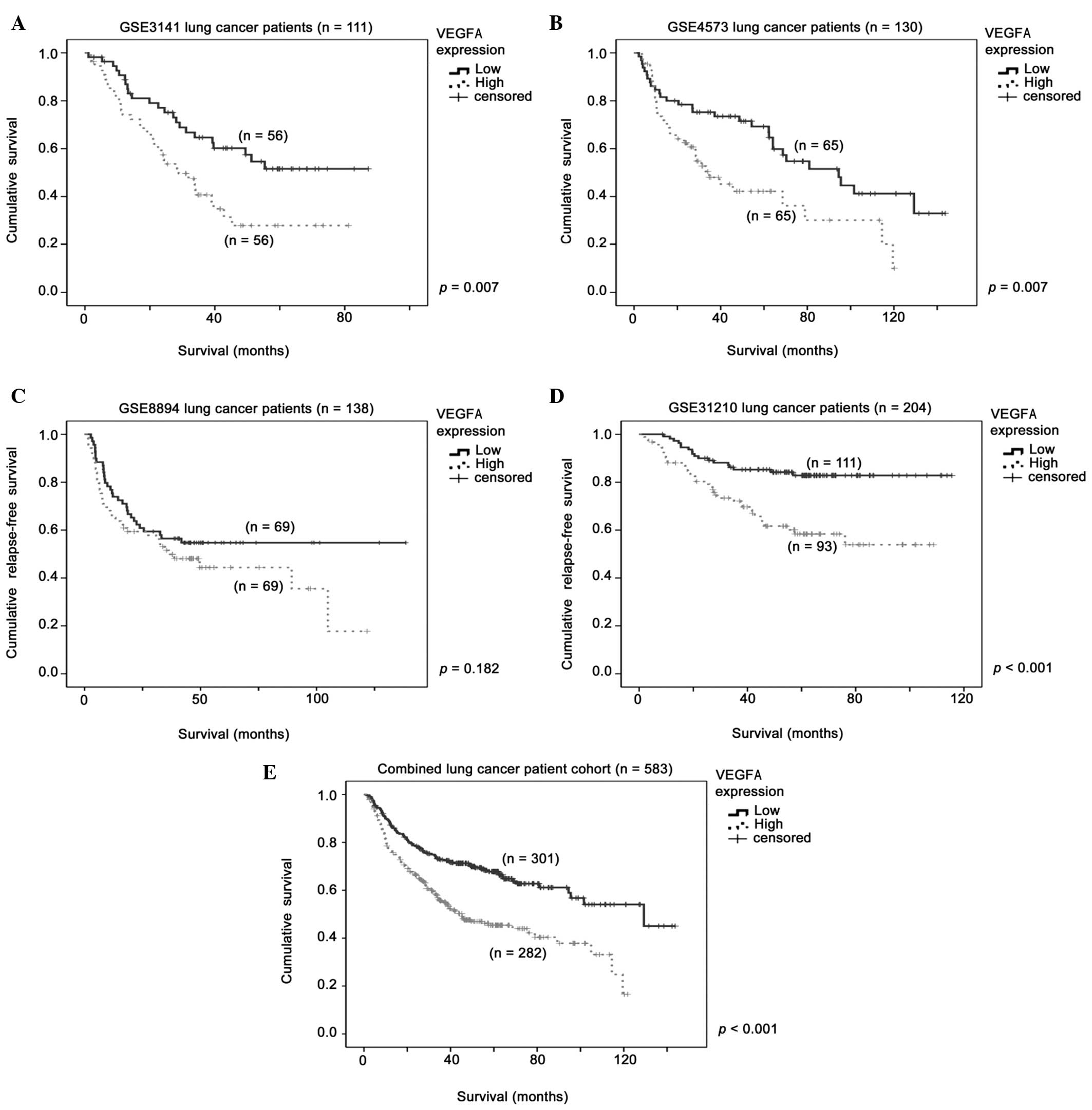
As indicated in Table
I, Cox regression identified that the VEGFA mRNA expression
level was significantly associated with a higher risk of
progression or mortality in all four datasets analyzed, with hazard
ratios (HRs) ranging between 1.43 and 2.02. Using median VEGFA mRNA
expression as the cut-off point, patients with a high level of
VEGFA mRNA expression were associated with a shorter survival time.
Kaplan-Meier analysis identified a significant association between
survival and VEGFA expression in the GSE3141 (P=0.007; Fig. 1A), GSE4573 (P=0.007; Fig. 1B) and GSE31210 (P<0.001; Fig. 1D) datasets, but not in the GSE8894
dataset (P=0.182; Fig. 1C). In the
combined datasets (n=583), a high level of VEGFA mRNA expression
was identified to be significantly associated with a shorter
survival time, according to Kaplan-Meier (log-rank test,
P<0.001; Fig. 1e) and Cox
regression (HR, 1.94; 95% CI, 1.50–2.50; P<0.001; Table I) analyses.
 | Table I.Cox regression analysis for high VEGFA
mRNA expression in lung cancer cohorts. |
Table I.
Cox regression analysis for high VEGFA
mRNA expression in lung cancer cohorts.
| Dataset | n | HR | 95% CI | P-value |
|---|
| GSE3141 | 111 | 1.68 | 1.18–2.39 |
0.004 |
| GSE4573 | 130 | 1.73 | 1.06–2.82 |
0.028 |
| GSE8894 | 138 | 1.43 | 1.11–1.84 |
0.006 |
| GSE31210 | 204 | 2.02 | 1.41–2.91 | <0.001 |
| Combined | 583 | 1.94 | 1.50–2.50 | <0.001 |
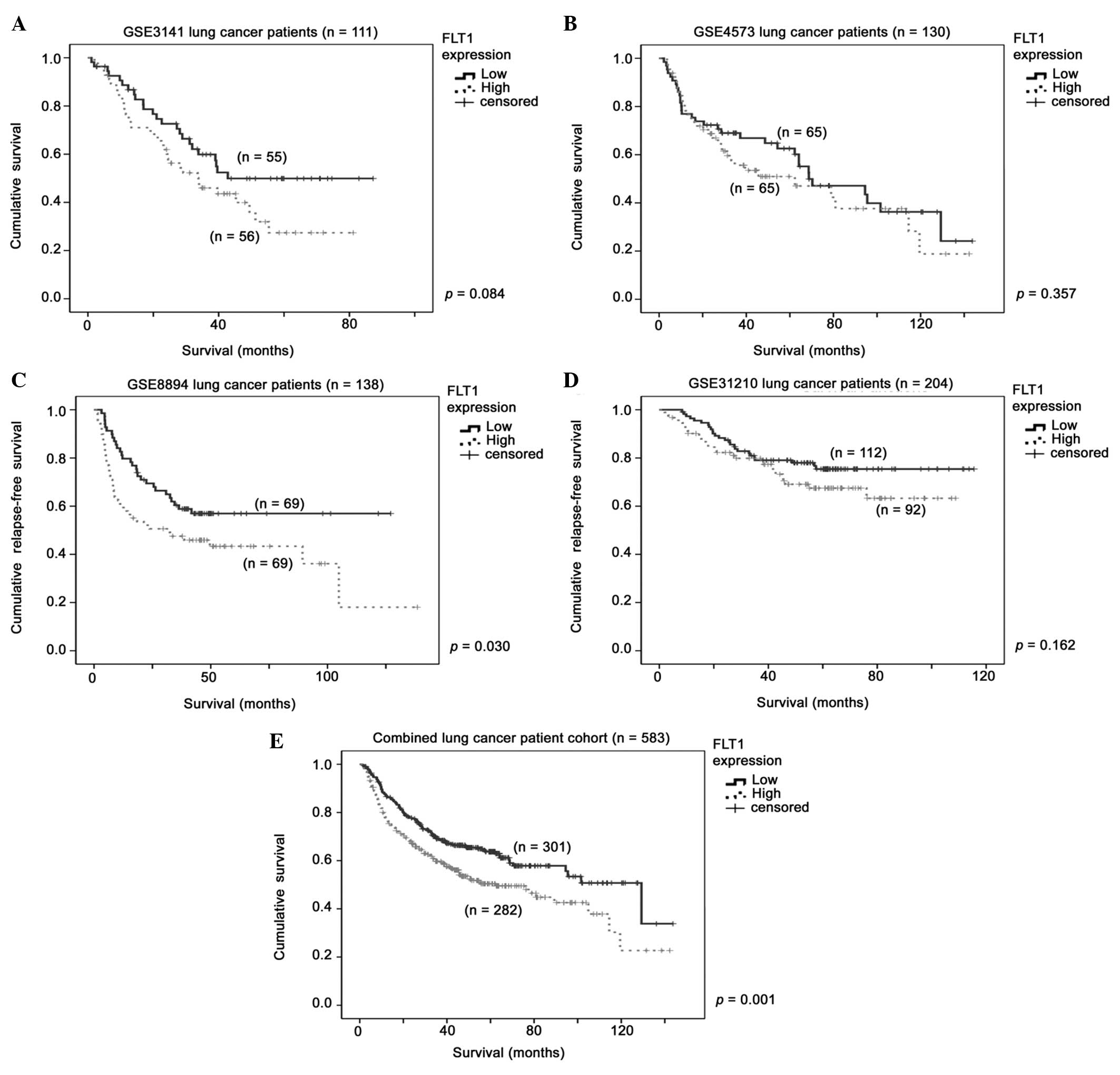
For FLT1 mRNA expression, Cox regression analysis
identified that high levels of FLT1 mRNA expression were
significantly associated with a shorter survival time in two
datasets (GSE3141 and GSE8894; P<0.05), but not in the other two
(GSE4573 and GSE31210; P>0.05; Table
II). Additionally, a high level of FLT1 expression was only
significantly associated with a shorter survival time in the
GSE8894 dataset (P=0.03; Fig. 2C),
but not in the other three independent datasets (Fig. 2A, B and D). When the four datasets
were combined, a high level of FLT1 mRNA expression was identified
to be significantly associated with a shorter patient survival time
according to Kaplan-Meier (log-rank test, P=0.001; Fig. 2E) and Cox regression (HR, 1.50; 95%
CI, 1.17–1.93; P=0.002; Table II)
analyses.
| Table II.Cox regression analysis for high FLT1
mRNA expression in lung cancer cohorts. |
Table II.
Cox regression analysis for high FLT1
mRNA expression in lung cancer cohorts.
| Dataset | n | HR | 95% CI | P-value |
|---|
| GSE3141 | 111 | 1.66 | 1.01–2.73 | 0.046 |
| GSE4573 | 130 | 1.11 | 0.63–1.95 | 0.729 |
| GSE8894 | 138 | 1.69 | 1.02–2.08 | 0.041 |
| GSE31210 | 204 | 1.30 | 0.84–2.01 | 0.234 |
| Combined | 583 | 1.50 | 1.17–1.93 | 0.002 |
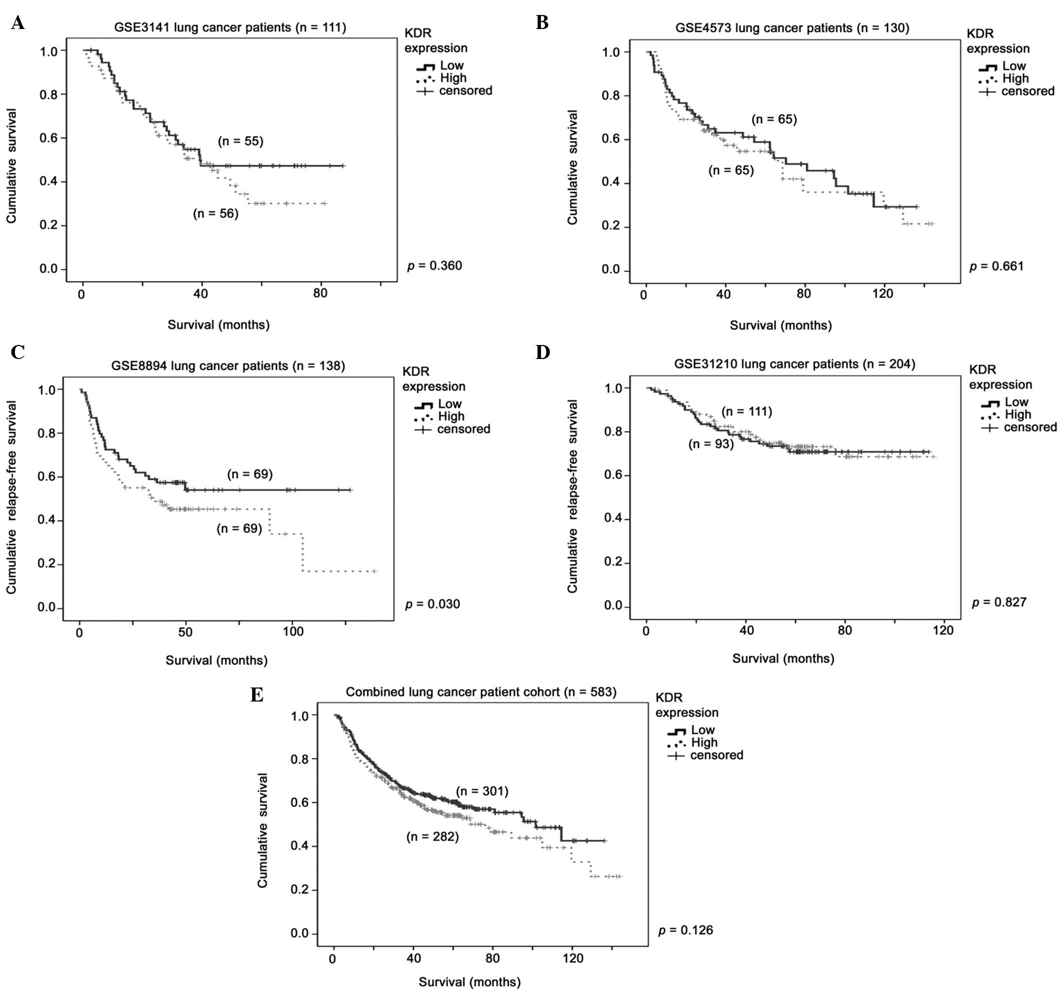
With respect to KDR mRNA expression (Table III; Fig.
3), a significant association with survival was only detected
in the GSE8894 dataset, but not in the other three datasets nor in
the combined cohort. Taken together, the current data indicate that
VEGFA mRNA expression is a consistent prognostic marker in patients
with lung cancer, while FLT1 mRNA expression is a marginal
prognostic marker and KDR mRNA expression is not a prognostic
marker.
| Table III.Cox regression analysis for high KDR
mRNA expression in lung cancer cohorts. |
Table III.
Cox regression analysis for high KDR
mRNA expression in lung cancer cohorts.
| Dataset | n | HR | 95% CI | P-value |
|---|
| GSE3141 | 111 | 1.01 | 0.76–1.30 | 0.972 |
| GSE4573 | 130 | 0.96 | 0.66–1.39 | 0.812 |
| GSE8894 | 138 | 1.23 | 1.01–1.49 | 0.040 |
| GSE31210 | 204 | 0.92 | 0.68–1.25 | 0.598 |
| Combined | 583 | 1.22 | 0.95–1.56 | 0.127 |
Positive correlation between VEGFA,
FLT1 and VDR expression and lung cancer specimens
As VEGFA must bind to its receptors to generate
downstream signaling, the overexpression of VEGFA alone may not be
sufficient to drive tumor progression (19). Therefore, the present study
investigated whether the expression of the ligand and its receptors
are correlated. As indicated in Table
IV, VEGFA mRNA expression was significantly positively
correlated with FLT1 mRNA expression in lung cancer specimens from
all four independent cohorts investigated in the current study.
Significant correlations between the expression of VEGFA and KDR,
and FLT1 and KDR, were also observed in three out of four
independent datasets (Table IV). The
current results indicate that VEGFA and its receptors, FLT1 and
KDR, may be co-regulated in tumors or in the tumor
microenvironment.
| Table IV.Correlation between the mRNA
expression levels of VEGFA, FLT1 and KDR. |
Table IV.
Correlation between the mRNA
expression levels of VEGFA, FLT1 and KDR.
| Dataset | n | VEGFA vs. KDR | VEGFA vs. FLT1 | KDR vs. FLT1 |
|---|
| GSE3141 | 111 | r=0.063,
P=0.512 | r=0.218,
P=0.022a |
r=0.678,
P<0.001a |
| GSE4573 | 130 |
r=0.242, P=0.005a | r=0.372,
P<0.001a |
r=0.318,
P<0.001a |
| GSE8894 | 138 |
r=0.371,
P<0.001a | r=0.361,
P<0.001a | r=0.143,
P=0.094 |
| GSE31210 | 204 |
r=0.394,
P<0.001a | r=0.684,
P<0.001a |
r=0.469,
P<0.001a |
High level of VEGFA and FLT1 mRNA
expression in the same patients predicts a shorter survival
outcome
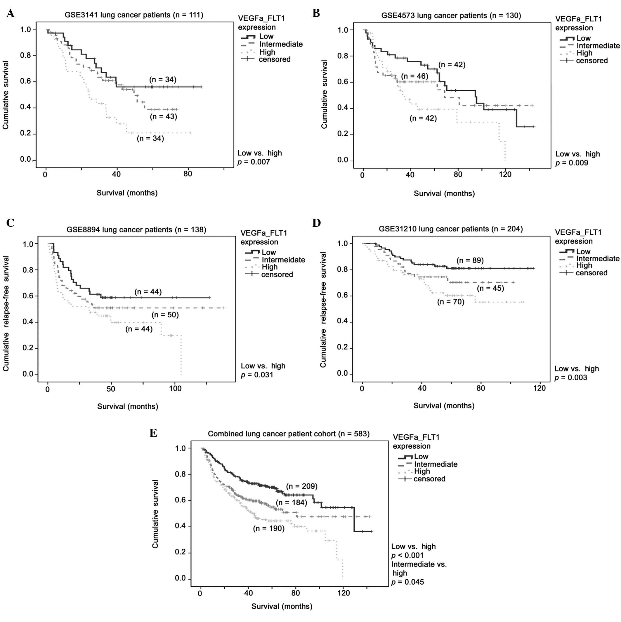
As VEGFA and FLT1 mRNA expression were significantly
correlated in the four independent lung cancer patient cohorts
(Table IV), the prognostic
significance of the co-upregulation of these two factors was
further investigated. Patients were stratified based on whether
their expression of VEGFA and FLT1 mRNAs was high (above median)
for both, high for one and low for the other (intermediate), or low
for both. As indicated in Fig. 4, a
high level expression of VEGFA and FLT1a expression was
significantly associated with a shorter survival time in the
GSE3141 (P=0.007; Fig. 4A), GSE4573
(P=0.009; Fig. 4B), GSE8894 (P=0.031;
Fig. 4C) and GSE31210 (P=0.003;
Fig. 4D) datasets. In the combined
cohort, a high level of VEGFA mRNA expression was associated with a
poor patient prognosis compared with high expression of a single
factor (P<0.001; Fig. 4E) or low
expression of both factors (P=0.045; Fig.
4E).
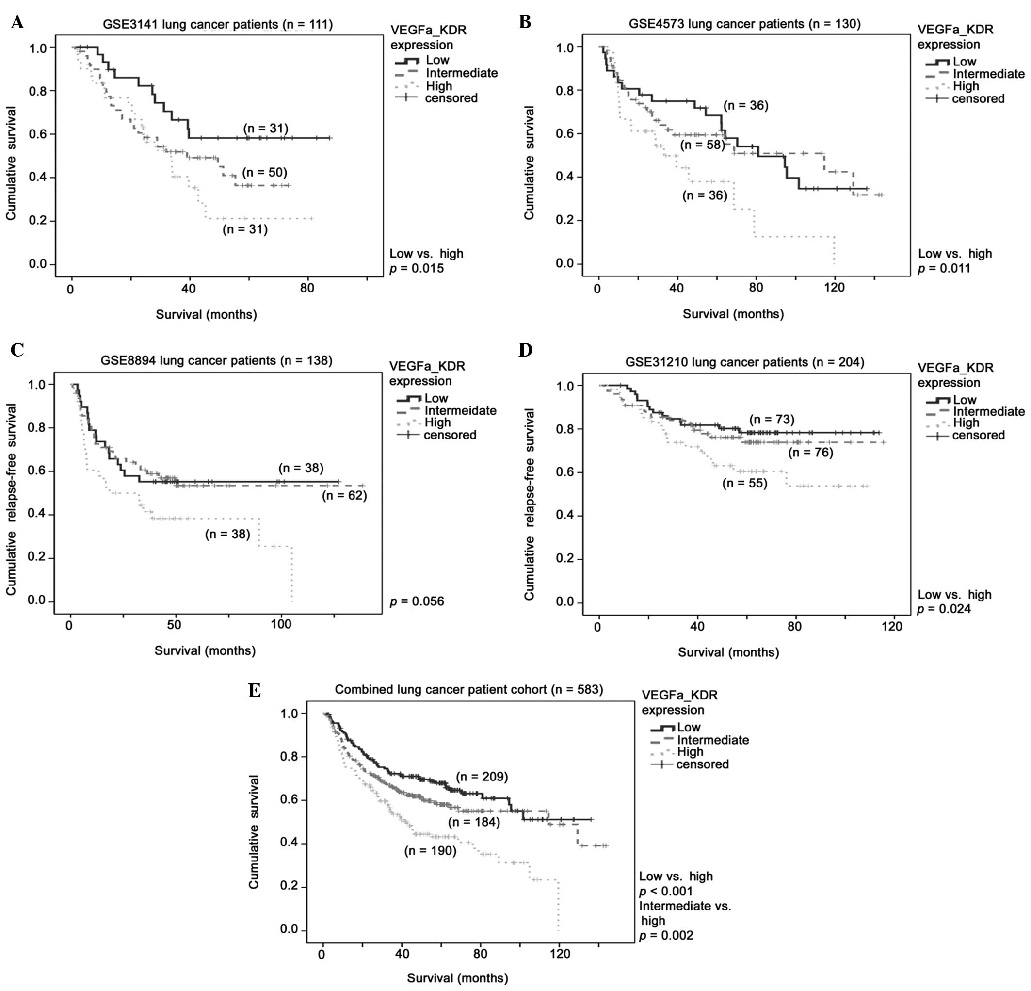
High level of VEGFA and KDR mRNA
expression in the same patients predicts a shorter survival
outcome
Comparable results were obtained when VEGFA and KDR
were similarly interrogated. A high level expression of the two
factors was significantly associated with a poor prognosis in all
four independent lung cancer patient cohorts (P<0.05; Fig. 5A–D). Notably, in the combined cohort
with a larger sample size, the patients with a high level of
expression of the two factors were associated with a significantly
shorter survival time compared with those exhibiting a low level of
expression of either one of the two factors (P<0.001; Fig. 5E) or a low level of expression of both
factors (P=0.002).
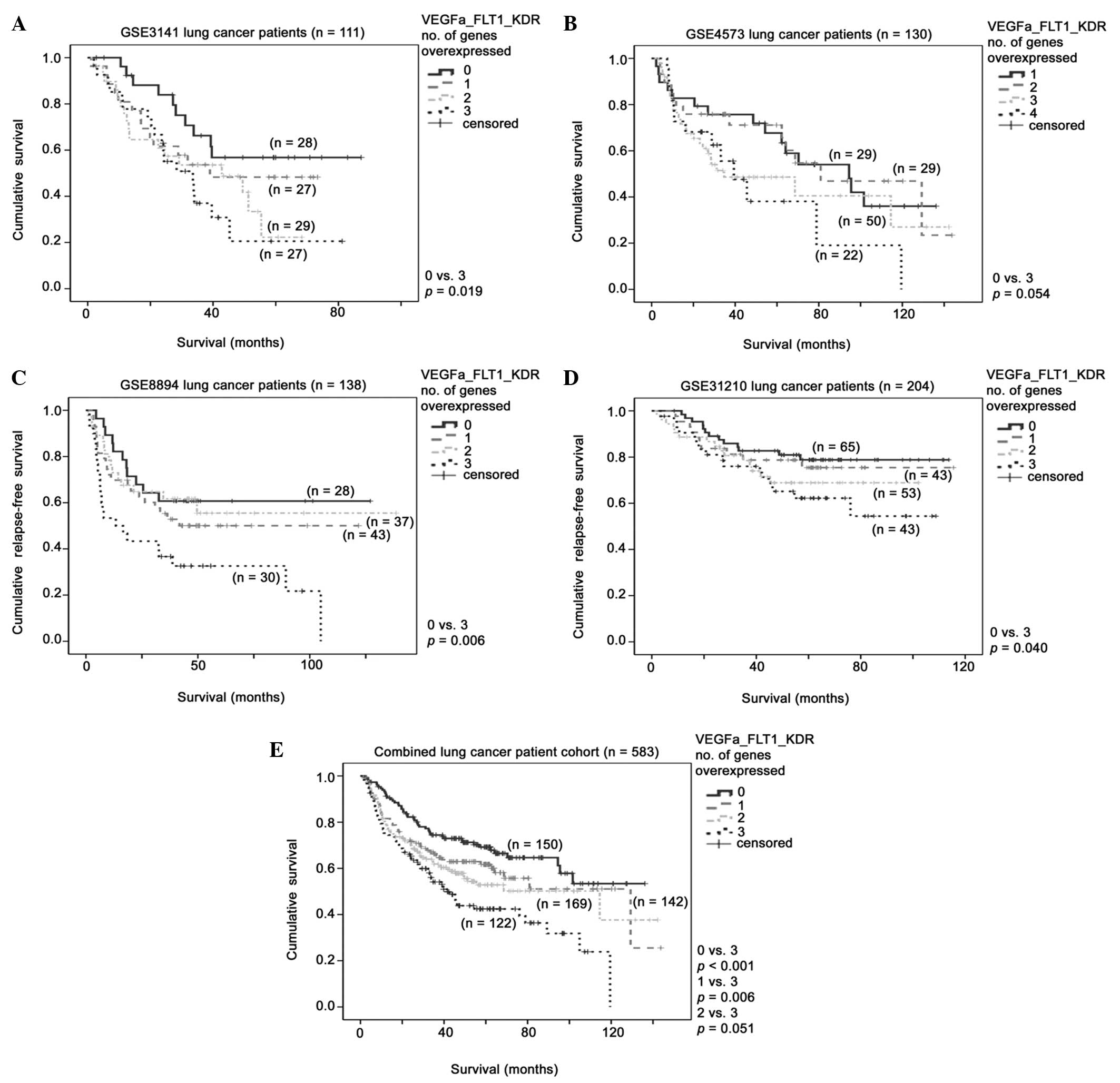
High level of VEGFA, FLT1 and KDR mRNA
expression in the same patients predicts a shorter survival
outcome
The present study investigated whether VEGFA, FLT1
and KDR, which are involved in the same angiogenesis promoting
signaling pathway, could be combined as a more effective prognostic
indicator for patients with lung cancer. Notably, in the four
independent cohorts, patients whose tumor specimens expressed a
high level of all three factors exhibited a significantly shorter
survival compared with those expressing a low level of all these
three factors (P<0.05; Fig. 6A–D).
In the combined cohort of 583 patients, the results were more
pronounced. As indicated in Fig. 6E,
the patients whose tumors overexpressed VEGFA, FLT1 and KDR
exhibited significantly shorter survival times compared with those
whose tumors overexpressed two (P=0.051), one (P=0.006) and none
(P<0.001) of these factors. Furthermore, in the patients with
lung cancer overexpressing all three, two or one of these factors,
Cox regression analysis was used to determine the HRs of 2.27, 1.65
and 1.40, respectively, for a shorter relapse-free or overall
survival time (Table V).
| Table V.Cox regression analysis for
VEGFA/FLT1/KDR mRNA overexpression in the combined cohort
(n=583). |
Table V.
Cox regression analysis for
VEGFA/FLT1/KDR mRNA overexpression in the combined cohort
(n=583).
| Overexpressed
genes, n | HR | 95% CI | P-value |
|---|
| 0 | Reference |
|
|
| 1 | 1.40 | 0.96–2.05 | 0.083 |
| 2 | 1.65 | 1.15–2.37 | 0.007 |
| 3 | 2.27 | 1.57–3.28 | <0.001 |
Discussion
A previous study attempted to combine angiogenic
factors together for improved prognostification in patients with
non-small cell lung cancer. In the study, a high level of VEGFA
mRNA expression, and a low level of VEGFB and VEGFD mRNA expression
was found to be associated with the poorest outcome (20). However, the functional roles and the
biological importance of VEGFB and VEGFD suppression are largely
unknown. By contrast, the co-upregulation of VEGFA, FLT1 and KDR
mRNA expression in the tumor environment identified in the present
study is supported by the well-known interaction between VEGFA and
the two receptors, FLT1 and KDR, though which downstream signaling
for angiogenesis is activated (5,21).
Previous studies have produced contradicting results
regarding the prognostic significance of VEGFA, FLT1 and KDR
expression in lung cancer (12,13). In
the present analysis, it was identified that only VEGFA mRNA
expression, but not FLT1 and KDR expression, was consistently
significantly associated with patient survival. Notably, and in
contrast to previous studies (12,13), a
high level of FLT1 mRNA expression was significantly associated
with poorer survival in the combined cohort of 583 patients. By
combining VEGFA and FLT1 mRNA expression, it was identified that
tumors overexpressing these two factors had a poorer prognosis
compared with tumors overexpressing only one or neither of these
two factors. By contrast, KDR mRNA expression itself was not
significantly associated with survival in the 583 patients with
lung cancer. However, combined VEGFA, FLT1 and KDR mRNA expression
was a consistent and statistically significant prognostic indicator
for lung cancer in the four independent lung cancer patient cohorts
examined, as well as in the combined cohort. Therefore, the current
results prime future investigations into whether this combination
could predict the sensitivity of the patients to agents that target
angiogenesis, including bevacizumab or nintedanib, which have
demonstrated promising results in phase III clinical trials in
patients with lung cancer (22,23).
Various studies have demonstrated that the
expression of VEGFA in fibroblasts and immune cells surrounding the
tumor mass may also be important in cancer progression, in addition
to VEGFA in the tumor cells themselves (24,25). The
results from these studies indicate that it may not be sufficient
to analyze only the tumor expression of VEGFA, FLT1 and KDR.
Rather, their expression levels at the tumor site as a whole may be
more important, as this widespread expression accounts for the
autocrine and paracrine actions of the VEGF signaling pathway,
which drive angiogenesis, and tumor growth and survival. Similar
results have been observed in colon cancer, suggesting that
co-consideration of these factors may have prognostic implication
for multiple cancer types that required angiogenesis for tumor
progression (26).
Although the cohorts included in the present study
were highly heterogeneous in terms of patient characteristics,
specimen procurement, RNA extraction, treatment and data
collection, the observations of VEGFA, and combined VEGFA, FLT1 and
KDR mRNA expression, were highly consistent between these four
independent cohorts, indicating that this combination may be highly
relevant and readily applicable in clinical practice as a more
effective prognostic indicator for patients with lung cancer.
However, additional investigations are required to demonstrate the
clinical applicability of combined VEGFA/FLT1/KDR expression, as
well as its predictive value as an anti-angiogenic agent, for
example, by conducting a prospective trial with or without
application of an agent targeting angiogenesis.
Acknowledgements
The authors would like to thank Omic Science and
Technology Ltd. (Hong Kong, SAR, China) for performing the initial
gene analysis and for their support in editing of the original
manuscript. The present study was supported by the University of
Macau Start-Up Research Grant (no. SRG2014-00006-FHS) and
Multi-Year Research Grant (no. MYRG2015-00065-FHS).
References
|
1
|
Hicklin DJ and Ellis LM: Role of the
vascular endothelial growth factor pathway in tumor growth and
angiogenesis. J Clin Oncol. 23:1011–1027. 2005. View Article : Google Scholar : PubMed/NCBI
|
|
2
|
Knizetova P, Ehrmann J, Hlobilkova A,
Vancova I, Kalita O, Kolar Z and Bartek J: Autocrine regulation of
glioblastoma cell cycle progression, viability and radioresistance
through the VEGF-VEGFR2 (KDR) interplay. Cell Cycle. 7:2553–2561.
2008. View Article : Google Scholar : PubMed/NCBI
|
|
3
|
Lichtenberger BM, Tan PK, Niederleithner
H, Ferrara N, Petzelbauer P and Sibilia M: Autocrine VEGF signaling
synergizes with EGFR in tumor cells to promote epithelial cancer
development. Cell. 140:268–279. 2010. View Article : Google Scholar : PubMed/NCBI
|
|
4
|
Hamerlik P, Lathia JD, Rasmussen R, Wu Q,
Bartkova J, Lee M, Moudry P, Bartek J Jr, Fischer W, Lukas J, et
al: Autocrine VEGF-VEGFR2-Neuropilin-1 signaling promotes glioma
stem-like cell viability and tumor growth. J Exp Med. 209:507–520.
2012. View Article : Google Scholar : PubMed/NCBI
|
|
5
|
Bieche I, Vacher S, Vallerand D, Richon S,
Hatem R, De Plater L, Dahmani A, Némati F, Angevin E, Marangoni E,
et al: Vasculature analysis of patient derived tumor xenografts
using species-specific PCR assays: Evidence of tumor endothelial
cells and atypical VEGFA-VEGFR1/2 signalings. BMC Cancer.
14:1782014. View Article : Google Scholar : PubMed/NCBI
|
|
6
|
Andersen S, Donnem T, Al-Shibli K, Al-Saad
S, Stenvold H, Busund LT and Bremnes RM: Prognostic impacts of
angiopoietins in NSCLC tumor cells and stroma: VEGF-A impact is
strongly associated with Ang-2. PLoS One. 6:e197732011. View Article : Google Scholar : PubMed/NCBI
|
|
7
|
Jantus-Lewintre E, Sanmartín E, Sirera R,
Blasco A, Sanchez JJ, Tarón M, Rosell R and Camps C: Combined
VEGF-A and VEGFR-2 concentrations in plasma: Diagnostic and
prognostic implications in patients with advanced NSCLC. Lung
Cancer. 74:326–331. 2011. View Article : Google Scholar : PubMed/NCBI
|
|
8
|
Donnem T, Al-Shibli K, Andersen S, Al-Saad
S, Busund LT and Bremnes RM: Combination of low vascular
endothelial growth factor A (VEGF-A)/VEGF receptor 2 expression and
high lymphocyte infiltration is a strong and independent favorable
prognostic factor in patients with nonsmall cell lung cancer.
Cancer. 116:4318–4325. 2010. View Article : Google Scholar : PubMed/NCBI
|
|
9
|
Takenaka K, Katakura H, Chen F, Ogawa E,
Adachi M, Wada H and Tanaka F: The ratio of membrane-bound form
Flt-1 mRNA to VEGF mRNA correlates with tumor angiogenesis and
prognosis in non-small cell lung cancer. Cancer Lett. 246:34–40.
2007. View Article : Google Scholar : PubMed/NCBI
|
|
10
|
Roybal JD, Zang Y, Ahn YH, Yang Y, Gibbons
DL, Baird BN, Alvarez C, Thilaganathan N, Liu DD, Saintigny P, et
al: miR-200 Inhibits lung adenocarcinoma cell invasion and
metastasis by targeting Flt1/VEGFR1. Mol Cancer Res. 9:25–35. 2011.
View Article : Google Scholar : PubMed/NCBI
|
|
11
|
An SJ, Nie Q, Chen ZH, Lin QX, Wang Z, Xie
Z, Chen SL, Huang Y, Zhang AY, Yan JF, et al: KDR expression is
associated with the stage and cigarette smoking of the patients
with lung cancer. J Cancer Res Clin Oncol. 133:635–642. 2007.
View Article : Google Scholar : PubMed/NCBI
|
|
12
|
Seto T, Higashiyama M, Funai H, Imamura F,
Uematsu K, Seki N, Eguchi K, Yamanaka T and Ichinose Y: Prognostic
value of expression of vascular endothelial growth factor and its
flt-1 and KDR receptors in stage I non-small-cell lung cancer. Lung
Cancer. 53:91–96. 2006. View Article : Google Scholar : PubMed/NCBI
|
|
13
|
Volm M, Koomägi R and Mattern J:
Prognostic value of vascular endothelial growth factor and its
receptor Flt-1 in squamous cell lung cancer. Int J Cancer.
74:64–68. 1997. View Article : Google Scholar : PubMed/NCBI
|
|
14
|
Bild AH, Yao G, Chang JT, Wang Q, Potti A,
Chasse D, Joshi MB, Harpole D, Lancaster JM, Berchuck A, et al:
Oncogenic pathway signatures in human cancers as a guide to
targeted therapies. Nature. 439:353–357. 2006. View Article : Google Scholar : PubMed/NCBI
|
|
15
|
Raponi M, Zhang Y, Yu J, Chen G, Lee G,
Taylor JM, Macdonald J, Thomas D, Moskaluk C, Wang Y, et al: Gene
expression signatures for predicting prognosis of squamous cell and
adenocarcinomas of the lung. Cancer Res. 66:7466–7472. 2006.
View Article : Google Scholar : PubMed/NCBI
|
|
16
|
Lee ES, Son DS, Kim SH, Lee J, Jo J, Han
J, Kim H, Lee HJ, Choi HY, Jung Y, et al: Prediction of
recurrence-free survival in postoperative non-small cell lung
cancer patients by using an integrated model of clinical
information and gene expression. Clin Cancer Res. 14:7397–7404.
2008. View Article : Google Scholar : PubMed/NCBI
|
|
17
|
Okayama H, Kohno T, Ishii Y, Shimada Y,
Shiraishi K, Iwakawa R, Furuta K, Tsuta K, Shibata T, Yamamoto S,
et al: Identification of genes upregulated in ALK-positive and
EGFR/KRAS/ALK-negative lung adenocarcinomas. Cancer Res.
72:100–111. 2012. View Article : Google Scholar : PubMed/NCBI
|
|
18
|
Yuen HF, Gunasekharan VK, Chan KK, Zhang
SD, Platt-Higgins A, Gately K, O'Byrne K, Fennell DA, Johnston PG,
Rudland PS, et al: RanGTPase: A candidate for Myc-mediated cancer
progression. J Natl Cancer Inst. 105:475–488. 2013. View Article : Google Scholar : PubMed/NCBI
|
|
19
|
Ferrara N: VEGF and the quest for tumour
angiogenesis factors. Nat Rev Cancer. 2:795–803. 2002. View Article : Google Scholar : PubMed/NCBI
|
|
20
|
Sanmartín E, Sirera R, Usó M, Blasco A,
Gallach S, Figueroa S, Martínez N, Hernando C, Honguero A,
Martorell M, et al: A gene signature combining the tissue
expression of three angiogenic factors is a prognostic marker in
early-stage non-small cell lung cancer. Ann Surg Oncol. 21:612–620.
2014. View Article : Google Scholar : PubMed/NCBI
|
|
21
|
Vieira JM, Ruhrberg C and Schwarz Q: VEGF
receptor signaling in vertebrate development. Organogenesis.
6:97–106. 2010. View Article : Google Scholar : PubMed/NCBI
|
|
22
|
Sandler A, Gray R, Perry MC, Brahmer J,
Schiller JH, Dowlati A, Lilenbaum R and Johnson DH:
Paclitaxel-carboplatin alone or with bevacizumab for non-small-cell
lung cancer. N Engl J Med. 55:2542–2550. 2006. View Article : Google Scholar
|
|
23
|
Reck M, Kaiser R, Mellemgaard A, Douillard
JY, Orlov S, Krzakowski M, von Pawel J, Gottfried M, Bondarenko I,
Liao M, et al: LUME-Lung 1 Study Group: Docetaxel plus nintedanib
versus docetaxel plus placebo in patients with previously treated
non-small-cell lung cancer (LUME-Lung 1): A phase 3, double-blind,
randomised controlled trial. Lancet Oncol. 15:143–155. 2014.
View Article : Google Scholar : PubMed/NCBI
|
|
24
|
Fukumura D, Xavier R, Sugiura T, Chen Y,
Park EC, Lu N, Selig M, Nielsen G, Taksir T, Jain RK, et al: Tumor
induction of VEGF promoter activity in stromal cells. Cell.
94:715–725. 1998. View Article : Google Scholar : PubMed/NCBI
|
|
25
|
Liang WC, Wu X, Peale FV, Lee CV, Meng YG,
Gutierrez J, Fu L, Malik AK, Gerber HP, Ferrara N, et al:
Cross-species vascular endothelial growth factor (VEGF)-blocking
antibodies completely inhibit the growth of human tumor xenografts
and measure the contribution of stromal VEGF. J Biol Chem.
281:951–961. 2006. View Article : Google Scholar : PubMed/NCBI
|
|
26
|
Zhang SD, McCrudden CM, Meng C, Lin Y and
Kwok HF: The signifiance of combining VEGFA, FLT1 and KDR
expressions in colon cancer patient prognosis and predicting
response to bevacizumab. Onco Targets Ther. 8:835–843.
2015.PubMed/NCBI
|