Introduction
Malignant melanoma is a potentially fatal form of
skin cancer, with a strong capacity for invasion and metastasis,
and high rates of recurrence and mortality (1,2), as well
as a limited response to currently available treatments, such as
chemotherapy and radiotherapy. The incidence and rate of mortality
of malignant melanoma have been increasing in the USA and Europe
faster than any those of other type of cancer (3). The median survival following the onset
of distant metastases is just 6–9 months and the five-year survival
rate is <5% (4).
The progression of melanoma is necessarily a complex
multistep process, as a cancer cell must acquire the ability to
survive under attachment-free conditions; migrate and invade
through the surrounding stroma; intravasate into the vascular
system; extravasate into, and subsequently endure a disadvantageous
distant environment; adhere to the local tissue; and proliferate
(5). The initiation of metastasis has
been attributed to the process of epithelial to mesenchymal
transition (EMT), in which a differentiated tumor cell transforms
into a more invasive, motile and resistant cell (6). EMT is characterized as a downregulation
of epithelial markers, in particular E-cadherin, and an
upregulation of mesenchymal markers, particularly vimentin or
fibronectin (7). E-cadherin is a key
mediator of cell-cell adhesions in epithelial tissues, and loss of
E-cadherin may enhance the invasive and metastatic behavior of
melanoma cells (8). During malignant
transformation, the transition from radial growth phase (RGP) to
vertical growth phase (VGP) is characterized by reduced E-cadherin
expression that results in the loss of keratinocyte-mediated growth
and motility control (9).
Metformin, a biguanide, is the most widely
prescribed drug for type 2 diabetes, worldwide (10). Metformin exerts its effects by
repressing hepatic gluconeogenesis, and increasing insulin
sensitivity and glucose uptake. Studies have indicated that
metformin efficiently suppresses the growth of various tumors, such
as prostate carcinoma, and breast, lung and pancreatic cancer
(11,12), which is in accordance with the results
of a retrospective epidemiological study, showing a reduction in
cancer risk in patients with diabetes who were receiving metformin
(13). Furthermore, a number of
studies have reported that metformin inhibits melanoma cell
proliferation (14–17).
Although, Cerezo et al (18) reported that metformin inhibited
melanoma invasion, and that this was associated with reduced
expression of proteins involved in EMT, such as fibronectin,
N-cadherin, Vimentin and SPARC, the effect of metformin on the
expression of E-cadherin was not demonstrated. In the present
study, the effect of metformin on the protein and mRNA expression
of E-cadherin was measured, in addition to the impact of metformin
on the migration and invasion of B16F10 melanoma cells.
Materials and methods
Chemicals
Metformin was obtained from Sigma Chemical Co. (St.
Louis, MO, USA). RPMI 1640 medium was obtained from Gibco BRL
(Gaithersburg, MD, USA). Penicillin and streptomycin were obtained
from North China Pharmaceutical Group Corp (Shijiazhuang, China)).
TRIzol reagent and crystal violet were obtained from Solarbio
(Beijing, China). 0.05% trypsine-EDTA and fetal bovine serum (FBS)
were obtained from Invitrogen Life Technologies (Carlsbad, CA,
USA).
Cell culture
The B16F10 melanoma cell line was obtained from
KeyGen Biotech (Nanjing, China) and cultured in RPMI 1640 medium,
containing 10% FBS, 100 g/ml streptomycin and 100 U/ml penicillin
at 37°C in a humidified atmosphere of 5% CO2. The B16F10
cells were cultured in a 6-well plate (1×10⁶ cells/well)
for RNA isolation and western blot analysis, and incubated in a
96-well flat-bottom plate (1×10⁴ cells/well) for cell
viability analysis. Trypsin/EDTA was used to prepare cells for the
experiments and cells were rested for 24 h in FBS-free medium prior
to treatment with the indicated concentrations of metformin
(specified for each experiment) for 24 h.
Analysis of cell proliferation
A
3-(4,5-dimethylthiazol-2-yl)-2,5-diphenyl-tetrazolium bromide (MTT)
assay was used to determine whether metformin altered cell
proliferation. Cells were seeded at a density of 1×10⁴
cells/well in a 96-well plate. The cells were treated with
metformin at various concentrations (0, 1, 2.5 or 5 mM) for 24 h.
Following incubation, cells were incubated with the MTT solution
(Sigma-Aldrich, St. Louis, MO, USA) at a final concentration of 0.5
mg/ml for 4 h, 37°C in a humidified atmosphere of 5%
CO2. Subsequently, the cells were lysed in dimethyl
sulfoxide and the absorbance at 570 nm was measured using a
microplate reader (Bio-Rad Laboratories, Hercules, CA, USA).
Wound healing assay
The wound healing assay was conducted as described
previously, by Liang et al (19). B16F10 cells were seeded in a 6-well
plate and grown to confluent monolayers. Next, the B16F10 cells
were cultured in RPMI-1640 medium without FBS for 12 h. A 200 µl
pipette tip was drawn across the center of the well in order to
produce a wound area and washed twice with serum-free RPMI 1640 to
remove loose cells. The cells were then treated with a medium
containing different concentrations of metformin (2.5 or 5 mM) and
1% FBS (1% FBS permits cell survival but not cell proliferation).
Subsequently, images of the wound healing process were photographed
digitally (x100) at 0, 12 and 24 h. Each value was derived from
three randomly selected fields.
Matrigel invasion assay
B16F10 melanoma cells were incubated in RPMI 1640
with 10% FBS and collected by trypsinization. A cell suspension
(200 µl of 5×10⁵ cells/ml with 1% FBS), containing
various concentrations of metformin (1, 2.5 or 5 mM) was added into
the inner cup of a 24-well Transwell chamber, which had been coated
with 50 µl of Matrigel™ (BD Biosciences, Franklin Lakes, NJ, USA;
1:4 dilution in serum-free medium). The medium, supplemented with
10% serum, was added to the outer cup. After 24 h, non-invading
cells were removed from the upper surface by gently rubbing with a
cotton-tipped swab. Cells that had migrated through the Matrigel
and the 8-µm pore membrane, were fixed with 3% paraformaldehyde for
15 min, stained with crystal violet for 30 min and then counted in
five random microscopic fields of the lower filter surface. Each
experiment was performed in triplicate.
Migration assay
A 24-well Transwell chamber (Corning Life Sciences,
Corning, New York, NY, USA) with an 8-µm-pore PET membrane, was
used to conduct the migration assay. The lower chamber was filled
with 600 µl RPMI 1640, containing 10% FBS. Subsequently, 200 µl
B16F10 melanoma cells suspension (5×10⁵ cells/ml with 1%
FBS), containing various concentrations of metformin (1, 2.5 or 5
mM) were added into the insert. The cells were allowed to migrate
at 37°C with 5% CO2 over 24 h. Non-migrating cells were
removed from the upper surface of the inserts by gently rubbing
with a cotton-tipped swab. Cells that had migrated to the lower
surface of the inserts were washed, fixed and stained with crystal
violet. Quantitative OD at a wavelength of 570 nm of crystal violet
staining dissolved in 33% acetic acid, represented migrated cells.
Experiments were performed at least in triplicate.
Immunocytochemistry
Immunocytochemistry was performed in order to
determine the expression of E-cadherin in B16F10 melanoma cells.
Briefly, sterilized slides were placed into a 24-well flat-bottomed
plate. B16F10 melanoma cells with 1% FBS, containing various
concentrations of metformin (0, 1, 2.5 and 5 mM), were added into
the 24-well plate (1×10⁵ cells/well); the total volume
of each well was 500 ml. Following a 24 h incubation, slides were
fixed with 3% paraformaldehyde for 15 min and incubated in 3%
hydrogen peroxide to inhibit endogenous peroxidase activity. After
blocking with 5% normal goat serum, a rabbit polyclonal primary
antibody against E-cadherin (cat. no. ZS-7870; ZSGB-BIO, Beijing,
China) was used at a 1:50 dilution and incubated overnight at 4°C.
Subsequently, a polyclonal peroxidase-conjugated goat anti-rabbit
antibody (cat. no. ZDR-5306, ZSGB-BIO) was added at a 1:500
dilution and incubated for 1 h at room temperature (25°C). Finally,
slides were developed in a substrate solution of DAB (Vector
Laboratories, Burlingame, CA, USA) and counter-stained with
hematoxylin. All Slides were examined and photographed under a
light microscope.
Reverse transcription-quantitative
polymerase chain reaction (RT-qPCR) analysis
Total RNA was extracted from B16F10 cells that had
been treated with various concentrations of metformin (0,1, 2.5 or
5 mM) for 24 h. An RNA extraction kit (Solarbio,) was used for the
extraction of total RNA. The concentration of total RNA was
quantified by measuring the optical density (OD) at 260 nm and RNA
integrity was confirmed using denaturing agarose gel
electrophoresis. Reverse transcription was conducted using a
PrimeScript™ RT Master Mix kit (Takara, Dalian, China), according
to the manufacturer's instructions. RT-qPCR was conducted in the
Stratagene Mx3000P (Stratagene, La Jolla, CA, USA) using a SYBR®
Premix DimerEraser™ kit (Takara). By detecting the increase in the
level of the reporter dye (SYBR), the PCR product was monitored
continuously during the reaction using MxProTMQPCR software
(Stratagene, La Jolla, CA, USA). The cycling conditions were in two
stages. An initial denaturation step at 95°C for 30 sec for one
cycle. The PCR amplification step was 40 cycles of 95°C for 30 sec,
58°C for 30 sec and 72°C for 30 sec. The expression of E-cadherin
mRNA in the treated cells was compared to that in the control cells
at each timepoint, using the comparative cycle threshold (Ct)
method. The following primers were used: Forward,
5′-ATTGCAAGTTCCTGCCATCCTC-3′ and reverse,
5′-CACATTGTCCCGGGTATCATCA-3′ for E-cadherin and forward,
5′-CATCCGTAAAGACCTCTATGCCAAC-3′ and reverse,
5′-ATGGAGCCACCGATCCACA-3′ for β-actin. According to the
manufacturer's instructions, the quantity of each transcript was
calculated and normalized to the quantity of β-actin, as an
internal standard.
Western blot analysis
B16F10 melanoma cells (1×10⁶) in the
exponential phase of growth, were plated in a 6-well plate and
treated with metformin (0, 1, 2.5 or 5 mM). Following treatment,
cells were collected and washed with phosphate-buffered saline. The
total protein was extracted using a RIPA lysis buffer kit (BestBio,
shanghai, China), on ice for 30 min. The lysates were collected and
centrifuged at 12,000 × g for 30 min at 4°C. Protein concentrations
were detected using a bicinchoninic acid protein assay kit
(Multisciences, Hangzhou, China). Aliquots of the lysates were
boiled for 5 min, electrophoresed on 10% SDS-PAGE gels and
transferred to a PVDF membrane (Merck Millipore, Darmstadt,
Germany). The membrane was blocked with 1% BSA at room temperature
for 1 h and then probed with primary polyclonal antibodies to
E-cadherin at a 1:50 dilution (cat. no.ZS-7870; ZSGB-BIO) and
β-actin at a 1:1,000 dilution (cat. no. sc-7210; Santa Cruz
Biotechnology, Inc., Dallas, TX, USA) overnight at 4°C, followed by
incubation with the HRP-labeled goat-anti-rabbit secondary
antibodies at a 1:5,000 dilution (KPL, Gaithersburg, MD, USA) for 1
h at 25°C. Finally, protein bands were detected using an enhanced
chemiluminescence Western blotting detection kit (Santa Cruz
Biotechnology, Inc.). The bands were quantified with a Gel Doc 2000
system and Quantity One software, version 4.6 (Bio-Rad
Laboratories), and expressed as a ratio of the quantity of
E-cadherin to β-actin, followed by standardization, with the ratio
of the normal control set as 1.
Statistical analysis
All experiments were repeated at least three times.
The mean ± standard deviation was calculated for each group, and
data were analyzed using one-way analysis of variance followed by a
post hoc least significant differences multiple comparison test.
Statistical analysis was performed using SPSS software, version
17.0 (SPSS, Inc., Chicago, IL, USA). P≤0.05 was considered to
indicate a statistically significant difference.
Results
Effect of metformin on the
proliferation of B16F10 melanoma cells
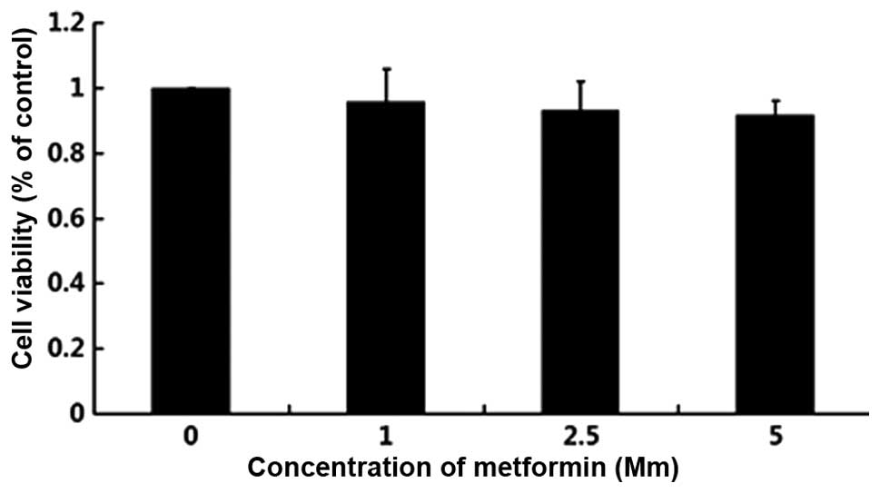
The cytotoxicity of metformin in the B16F10 murine
melanoma cell line was evaluated by treating the cells with
metformin, at concentrations of 0, 1, 2.5 or 5 mM for 24 h. As
shown in Fig. 1, no significant
cytotoxicity was observed. Since metformin was not cytotoxic in
B16F10 cells at 5 mM, metformin were used at concentrations of 5 mM
or lower in subsequent experiments.
Metformin inhibits B16F10 cell
motility, invasion and migration
Increased cell motility is a characteristic that is
associated with malignancy. The ‘wound’ repair model was used to
evaluate the effect of metformin on the motility of B16F10 cells.
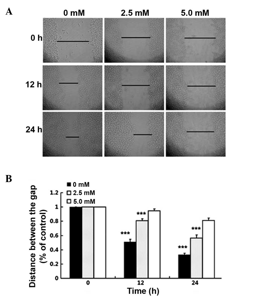
As shown by the wound healing assay (Fig.
2), in the group treated with metformin the rate of cell
migration into the wounded area was significantly reduced compared
with that in the control group (control group, 51.2 and 33%; 2.5 mM
group, 80.8 and 56.6%; and 5 mM group, 94.7 and 81.6% at 12 and 24
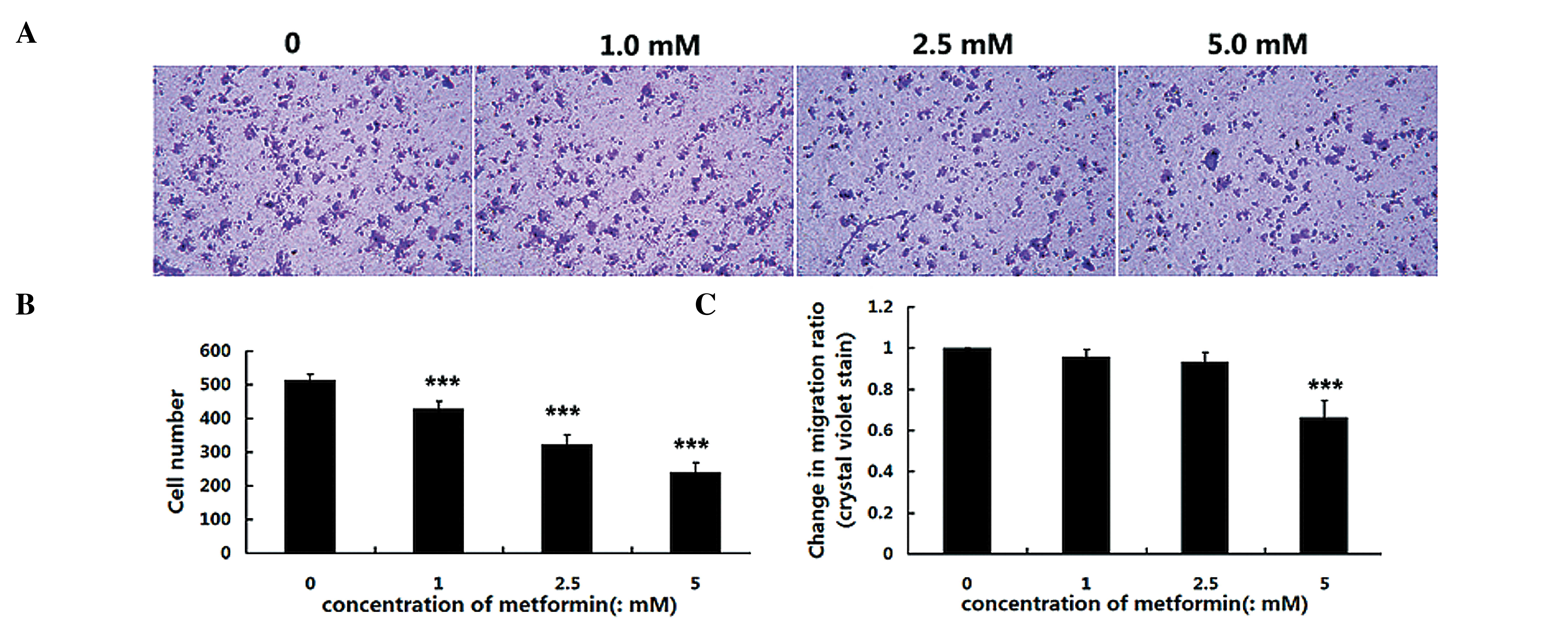
h, respectively). Furthermore, Transwell migration and Matrigel
invasion assays were used to investigate the inhibitory effects of
metformin on the invasive potency of cells. The Matrigel invasion
assay, a three dimensional model, showed that metformin
significantly suppressed B16F10 cell invasion, while 1 mM metformin
inhibited the invasion of cells by 17%, 2.5 mM inhibited invasion
by 36% and 5 mM by 47% (Fig. 3A and
B). In the Transwell migration assay, a significantly reduced
number of migrating cells was observed when cells were treated with
metformin for 24 h, compared with that in the control group
(Fig. 3C). These results suggested
that non-toxic concentrations of metformin inhibit the motility,
invasion and migration of B16F10 melanoma cells in a dose-dependent
manner.
Metformin upregulates the expression
of E-cadherin
E-cadherin is involved in the progression of cancer.
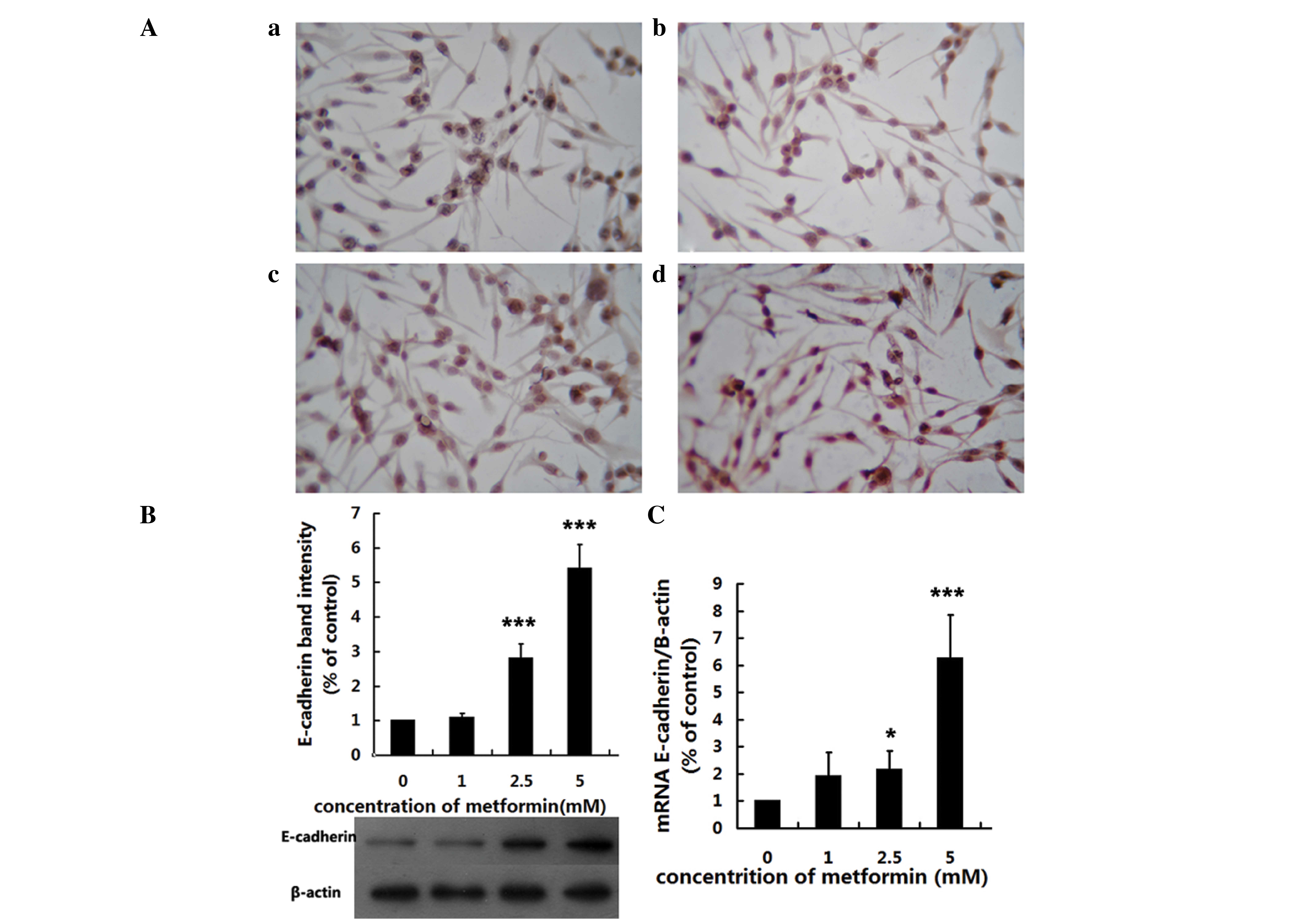
The results of the immunocytochemistry experiment demonstrated that
the level E-cadherin was markedly reduced in B16F10 cells following
24 h treatment with metformin, compared with that in the control
group (Fig. 4A). Western blot
analysis showed that E-cadherin expression was significantly
suppressed by metformin at concentrations of 1, 2.5 and 5 mM,
compared with the control group (all P<0.05; Fig. 4B). RT-qPCR demonstrated that the level
of E-cadherin mRNA in B16F10 cells was significantly reduced in the
groups treated with metformin at concentrations of 1, 2.5 and 5 mM,
compared with that in the control group (all P<0.05; Fig. 4C).
Discussion
Metformin, the most widely prescribed antidiabetic
drug worldwide, is well tolerated and has the major advantage of
not inducing hypoglycemia. Metformin reduces hepatic glucose
production through a mechanism requiring liver kinase B1, which is
important in the control of the metabolic checkpoint, AMP-activated
protein kinase-mammalian target of rapamycin, and neoglucogenic
genes, resulting in inhibition of protein synthesis and cell
proliferation (20). Observations
have shown that metformin affects the regulation of tumor cell
proliferation, cell-cycle regulation, apoptosis and autophagy
(21). It has been reported that
metformin may be effective as an anticancer drug in a variety of
types of tumors, such as prostate (22), breast (23), lung (24) and pancreatic cancer (25), and melanoma (15). A retrospective epidemiological study
demonstrated a reduction in cancer risk in diabetic patients
treated with metformin (13). The
present study focused on the effects of metformin on the invasion
and migration of B16F10 melanoma cells in order to determine its
effects on the metastasis of tumor cells.
Invasiveness and metastasis are the most important
characteristics of malignant tumors, including melanoma. Cell
adhesion is closely associated with tumor invasiveness and
metastasis, and the cadherin superfamily is a group of
transmembrane glycoproteins that mediate calcium-dependent
homophilic intercellular adhesion. Perturbations in cadherins have
been shown to be associated with the development of cancer,
particularly invasion and metastasis (26). E-cadherin, expressed by the majority
of normal epithelial tissues, is a prototypic member of the
cadherin superfamily, and reduced expression and abnormal cellular
distribution of E-cadherin have been observed to be associated with
dedifferentiation and invasiveness in various human malignancies
(27). Of the events occurring during
the complex metastasis process, EMT, the initial step, is the most
important (28). EMT promotes tumor
progression, by endowing cells with migratory and invasive
properties, inducing stem cell properties, suppressing apoptosis
and senescence, and contributing to immunosuppression (8). Previous studies have shown a link
between EMT markers in primary tumor cells and aggressive clinical
behavior (29,30). The majority of evidence indicates that
E-cadherin is associated with EMT, and that the loss of E-cadherin
may promote invasive and metastatic behavior in many types of
epithelial tumors (31–33). Therefore, downregulation of E-cadherin
is one of the essential hallmarks of EMT (34). During melanoma progression, the
transition from RGP to VGP is characterized by reduced E-cadherin
expression, accompanied by loss of keratinocyte-mediated growth and
motility control (9). Tang et
al (35) reported that E-cadherin
expression decreased in a number of human malignant melanoma cell
lines. The current study showed that motility, migration and
invasion of B16F10 melanoma cells are markedly inhibited by
metformin, and that E-cadherin mRNA transcription and E-cadherin
protein production were upregulated by metformin in a
dose-dependent manner, implying that it may be beneficial in the
treatment of cancer.
Although, the E-cadherin expression in B16F10 cells,
as measured by western blotting, was not entirely consistent with
the results of the RT-qPCR analysis at a dose of 2.5 mM metformin,
the overall trend was similar, in that there was a dose-dependent
increase in E-cadherin expression. This inconsistency may have been
due to the use of different assays with different sensitivities,
and different kinetics between the protein and mRNA.
In conclusion, the present data demonstrated that
metformin inhibits the migration and invasion of B16F10 melanoma
cells, via the upregulation of E-cadherin in a dose-dependent
manner. Further in vivo studies are required to determine
the inhibitory effects of metformin on tumor cell invasion. The
findings in the present study indicate that metformin has the
potential to be an efficient anticancer drug in the treatment of
metastatic malignant melanoma.
References
|
1
|
Shore RN, Shore P, Monahan NM and Sundeen
J: Serial screening for melanoma: Measures and strategies that have
consistently achieved early detection and cure. J Drugs Dermatol.
10:244–252. 2011.PubMed/NCBI
|
|
2
|
Boyle GM: Therapy for metastatic melanoma:
An overview and update. Expert Rev Anticancer Ther. 11:725–737.
2011. View Article : Google Scholar : PubMed/NCBI
|
|
3
|
MacKie RM, Hauschild A and Eggermont AM:
Epidemiology of invasive cutaneous melanoma. Ann Oncol. 20 (Suppl
6):vi1–vi7. 2009. View Article : Google Scholar : PubMed/NCBI
|
|
4
|
Houghton AN and Polsky D: Focus on
melanoma. Cancer Cell. 2:275–278. 2002. View Article : Google Scholar : PubMed/NCBI
|
|
5
|
Chaffer CL and Weinberg RA: A perspective
on cancer cell metastasis. Science. 331:1559–1564. 2011. View Article : Google Scholar : PubMed/NCBI
|
|
6
|
Hollier BG, Evans K and Mani SA: The
epithelial-to-mesenchymal transition and cancer stem cells: A
coalition against cancer therapies. J Mammary Gland Biol Neoplasia.
14:29–43. 2009. View Article : Google Scholar : PubMed/NCBI
|
|
7
|
Zeisberg M and Neilson EG: Biomarkers for
epithelial-mesenchymal transitions. J Clin Invest. 119:1429–1437.
2009. View
Article : Google Scholar : PubMed/NCBI
|
|
8
|
Thiery JP, Acloque H, Huang RY and Nieto
MA: Epithelial-mesenchymal transitions in development and disease.
Cell. 139:871–890. 2009. View Article : Google Scholar : PubMed/NCBI
|
|
9
|
Hsu MY, Meier FE, Nesbit M, Hsu JY, Van
Belle P, Elder DE and Herlyn M: E-cadherin expression in melanoma
cells restores keratinocyte-mediated growth control and
down-regulates expression of invasion-related adhesion receptors.
Am J Pathol. 156:1515–1525. 2000. View Article : Google Scholar : PubMed/NCBI
|
|
10
|
Kourelis TV and Siegel RD: Metformin and
cancer: New applications for an old drug. Med Oncol. 29:1314–1327.
2012. View Article : Google Scholar : PubMed/NCBI
|
|
11
|
Ben Sahra I, Le Marchand-brustel Y, Tanti
JF and Bost F: Metformin in cancer therapy: A new perspective for
an old antidiabetic drug? Mol Cancer Ther. 9:1092–1099. 2010.
View Article : Google Scholar : PubMed/NCBI
|
|
12
|
Pollak M: Metformin and other biguanides
in oncology: Advancing the research agenda. Cancer Prev Res
(Phila). 3:1060–1065. 2010. View Article : Google Scholar : PubMed/NCBI
|
|
13
|
Noto H, Goto A, Tsujimoto T and Noda M:
Cancer risk in diabetic patients treated with metformin: A
systematic review and meta-analysis. PLoS One. 7:e334112012.
View Article : Google Scholar
|
|
14
|
Woodard J and Platanias LC: AMP-activated
kinase (AMPK)-generated signals in malignant melanoma cell growth
and survival. Biochem Biophys Res Commun. 398:135–139. 2010.
View Article : Google Scholar : PubMed/NCBI
|
|
15
|
Janjetovic K, Harhaji-Trajkovic L,
Misirkic-Marjanovic M, Vucicevic L, Stevanovic D, Zogovic N,
Sumarac-Dumanovic M, Micic D and Trajkovic V: In vitro and
in vivo anti-melanoma action of metformin. Eur J Pharmacol.
668:373–382. 2011. View Article : Google Scholar : PubMed/NCBI
|
|
16
|
Niehr F, von EE, Attar N, Guo D, Matsunaga
D, Sazegar H, Ng C, Glaspy JA, Recio JA, Lo RS, et al: Combination
therapy with vemurafenib (PLX4032/RG7204) and metformin in melanoma
cell lines with distinct driver mutations. J Transl Med. 9:762011.
View Article : Google Scholar : PubMed/NCBI
|
|
17
|
Tomic T, Botton T, Cerezo M, Robert G,
Luciano F, Puissant A, Gounon P, Allegra M, Bertolotto C, Bereder
JM, et al: Metformin inhibits melanoma development through
autophagy and apoptosis mechanisms. Cell Death Dis. 2:e1992011.
View Article : Google Scholar : PubMed/NCBI
|
|
18
|
Cerezo M, Tichet M, Abbe P, Ohanna M,
Lehraiki A, Rouaud F, Allegra M, Giacchero D, Bahadoran P,
Bertolotto C, et al: Metformin blocks melanoma invasion and
metastasis development in AMPK/p53-dependent manner. Mol Cancer
Ther. 12:1605–1615. 2013. View Article : Google Scholar : PubMed/NCBI
|
|
19
|
Liang CC, Park AY and Guan JL: In vitro
scratch assay: A convenient and inexpensive method for analysis of
cell migration in vitro. Nat Protoc. 2:329–333. 2007. View Article : Google Scholar : PubMed/NCBI
|
|
20
|
Andújar-Plata P, Pi-Sunyer X and Laferrère
B: Metformin effects revisited. Diabetes Res Clin Pract. 95:1–9.
2012. View Article : Google Scholar : PubMed/NCBI
|
|
21
|
Cerezo M, Tomic T, Ballotti R and Rocchi
S: Is it time to test biguanide metformin in the treatment of
melanoma? Pigment Cell Melanoma Res. 28:8–20. 2014. View Article : Google Scholar : PubMed/NCBI
|
|
22
|
Ben Sahra I, Laurent K, Loubat A,
Giorgetti-Peraldi S, Colosetti P, Auberger P, Tanti JF, Le
Marchand-Brustel Y and Bost F: The antidiabetic drug metformin
exerts an antitumoral effect in vitro and in vivo
through a decrease of cyclin D1 level. Oncogene. 27:3576–3586.
2008. View Article : Google Scholar : PubMed/NCBI
|
|
23
|
Zakikhani M, Dowling R, Fantus IG,
Sonenberg N and Pollak M: Metformin is an AMP kinase-dependent
growth inhibitor for breast cancer cells. Cancer Res.
66:10269–10273. 2006. View Article : Google Scholar : PubMed/NCBI
|
|
24
|
Algire C, Zakikhani M, Blouin MJ, Shuai JH
and Pollak M: Metformin attenuates the stimulatory effect of a
high-energy diet on in vivo LLC1 carcinoma growth. Endocr
Relat Cancer. 15:833–839. 2008. View Article : Google Scholar : PubMed/NCBI
|
|
25
|
Wang LW, Li ZS, Zou DW, Jin ZD, Gao J and
Xu GM: Metformin induces apoptosis of pancreatic cancer cells.
World J Gastroenterol. 14:7192–7198. 2008. View Article : Google Scholar : PubMed/NCBI
|
|
26
|
Takeichi M: Cadherins in cancer:
Implications for invasion and metastasis. Curr Opin Cell Biol.
5:806–811. 1993. View Article : Google Scholar : PubMed/NCBI
|
|
27
|
Christofori G and Semb H: The role of the
cell-adhesion molecule E-cadherin as a tumour-suppressor gene.
Trends Biochem Sci. 24:73–76. 1999. View Article : Google Scholar : PubMed/NCBI
|
|
28
|
Rattan R, Ali Fehmi R and Munkarah A:
Metformin: An emerging new therapeutic option for targeting cancer
stem cells and metastasis. J Oncol. 2012:9281272012. View Article : Google Scholar : PubMed/NCBI
|
|
29
|
Sarrió D, Rodriguez-Pinilla SM, Hardisson
D, Cano A, Moreno-Bueno G and Palacios J: Epithelial-mesenchymal
transition in breast cancer relates to the basal-like phenotype.
Cancer Res. 68:989–997. 2008. View Article : Google Scholar : PubMed/NCBI
|
|
30
|
Yang J, Mani SA, Donaher JL, Ramaswamy S,
Itzykson RA, Come C, Savagner P, Gitelman I, Richardson A and
Weinberg RA: Twist, a master regulator of morphogenesis, plays an
essential role in tumor metastasis. Cell. 117:927–939. 2004.
View Article : Google Scholar : PubMed/NCBI
|
|
31
|
Birchmeier W and Behrens J: Cadherin
expression in carcinomas: role in the formation of cell junctions
and the prevention of invasiveness. Biochim Biophys Acta.
1198:11–26. 1994.PubMed/NCBI
|
|
32
|
Kalluri R and Weinberg RA: The basics of
epithelial-mesenchymal transition. J Clin Invest. 119:1420–148.
2009. View
Article : Google Scholar : PubMed/NCBI
|
|
33
|
Perl AK, Wilgenbus P, Dahl U, Semb H and
Christofori G: A causal role for E-cadherin in the transition from
adenoma to carcinoma. Nature. 392:190–193. 1998. View Article : Google Scholar : PubMed/NCBI
|
|
34
|
Nakamura M and Tokura Y:
Epithelial-mesenchymal transition in the skin. J Dermatol Sci.
61:7–13. 2011. View Article : Google Scholar : PubMed/NCBI
|
|
35
|
Tang A, Eller MS, Hara M, Yaar M,
Hirohashi S and Gilchrest BA: E-cadherin is the major mediator of
human melanocyte adhesion to keratinocytes in vitro. J Cell
Sci. 107:983–992. 1994.PubMed/NCBI
|


