Introduction
Tongue squamous cell carcinoma (TSCC) is the most
common type of oral cancer. According to data released by the
American Cancer Society, an estimated 13,590 novel TSCC cases were
predicted to occur in the USA in 2014, accounting for one-third of
all oral cavity and pharynx cancers (1). TSCC tends to demonstrate more aggressive
behavior, including a high frequency of local invasion and regional
lymph node metastasis (2).
Consequently, the mortality rate for TSCC continues to be high,
with a typical overall five-year survival rate of <50% (3). In order to delineate the clinical
behavior of TSCC and to personalize therapy, the identification of
novel biomarkers for tumor aggressiveness is required.
Raf kinase inhibitor protein (RKIP) is a member of
the phosphatidylethanolamine-binding protein family. RKIP was
original identified as a physiological inhibitor of the
Raf/mitogen-activated protein/extracellular signal-regulated kinase
(ERK) kinase (MEK)/ERK pathway (4,5) and
appears to be implicated in a variety of intracellular signaling
pathways (6–8) that control cell proliferation (9), differentiation (10), adhesion (11) and epithelial-mesenchymal transition
(12). RKIP has also been implicated
in tumor progression. It was suggested that RKIP acts as a
metastasis suppressor in a variety of malignancies, including
prostate (13), breast (14), hepatocellular (15), colorectal (16) and gastric cancers (17). These previous findings prompted the
elucidation of the clinicopathological features and implications of
RKIP expression in patients with TSCC. Therefore, the present study
investigated the protein levels of RKIP in non-cancerous and tumor
tissues, and assessed the role of RKIP in the clinical outcome of
the patients with TSCC.
Materials and methods
Patients and tissue samples
All tissue samples were obtained from the Affiliated
Hospital of Stomatology, Sun Yat-Sen University (Guangzhou, China)
between January 2007 and December 2012. In total, 85 pairs of
paraffin-embedded tissue samples from patients with TSCC,
consisting of TSCC tissue and adjacent non-cancerous tissues, were
assessed. An additional 32 oral leukoplakia lesions were also
analyzed (Table I). In patients that
demonstrated lymph node involvement (n=30), the corresponding lymph
node metastases were also examined. The specimens were obtained
from the patients subsequent to radical surgery, with informed
consent being obtained from all patients for the use of the
surgically-resected specimens for research purposes, according to
the guidelines for research on human tissues and samples set by the
Institution Review Board of Sun Yat-Sen University. No patients
received any form of adjuvant therapy prior to surgery. All
histopathological diagnoses were based on the Union for
International Cancer Control or the 2002 American Joint Committee
on Cancer criteria (18), and the
diagnoses were reviewed by two experienced pathologists.
 | Table I.Association between RKIP expression
and clinicopathological features in patients with tongue squamous
cell carcinoma. |
Table I.
Association between RKIP expression
and clinicopathological features in patients with tongue squamous
cell carcinoma.
|
|
| RKIP expression, n
(%) |
|
|---|
|
|
|
|
|
|---|
| Clinicopathological
features | Patients, n | High | Low | P-value |
|---|
| Gender |
|
|
|
|
| Male | 51 | 19 (37.3) | 32 (62.7) | 0.153 |
|
Female | 34 | 18 (52.9) | 16 (47.1) |
|
| Age, years |
|
|
|
|
| ≥55 | 40 | 16 (40.0) | 24 (60.0) | 0.536 |
|
<55 | 45 | 21 (46.7) | 24 (53.3) |
|
| Differentiation |
|
|
|
|
| Well | 49 | 23 (46.9) | 26 (53.1) | 0.460 |
|
Moderately or poorly | 36 | 14 (38.9) | 22 (61.1) |
|
| pT stage |
|
|
|
|
| T1-2 | 66 | 31 (47.0) | 35 (53.0) | 0.233 |
| T3-4 | 19 | 6
(31.6) | 13 (68.4) |
|
| pN stage |
|
|
|
|
| N0 | 55 | 29 (52.7) | 26 (47.3) | 0.021 |
| N+ | 30 | 8
(26.7) | 22 (73.3) |
|
| pTNM stage |
|
|
|
|
| I–II | 45 | 25 (55.6) | 20 (44.4) | 0.018 |
|
III–IV | 40 | 12 (30.0) | 28 (70.0) |
|
Immunohistochemistry
Formalin-fixed, paraffin-embedded tissue sections
were subjected to immunohistochemical analysis. The 4-µm tissue
sections were routinely dewaxed, rehydrated and blocked using 0.3%
hydrogen peroxide in methanol for 30 min at room temperature.
Antigen retrieval was performed using 10 mM sodium citrate buffer
(pH 6.0) at 95°C for 20 min. To reduce non-specific binding, the
slides were blocked with 10% goat serum for 30 min at room
temperature. The tissue sections were incubated in a humidified
chamber at 4°C overnight with a primary polyclonal IgG rabbit
anti-human RKIP antibody (dilution, 1:100; catalog no. sc-28837;
Santa Cruz Biotechnology, Inc., Santa Cruz, CA, USA). Subsequent to
washing three times in phosphate-buffered saline (PBS), the slides
were incubated with horseradish peroxidase and visualized with
diaminobenzidine (DAKO Deutschland GmbH, Hamburg, Germany), and the
slides were counterstained with hematoxylin. To confirm the
specificity of the immunostaining, a negative control was included
in each run by substituting PBS for the primary antibody.
Evaluation of immunohistochemical
staining
Five random fields of each section were viewed under
a light microscope (Axioskop 40; Zeiss, Oberkochen, Germany) at
x400 magnification. The sections were independently examined and
scored by three investigators, who were blind to the clinical
features and outcomes of the cases. The expression of RKIP was
scored by multiplication of the average signal intensity, which was
scored on a scale of 0–3, and percentage of positively stained
tumor cells, which was scored on a scale of 0–4, as previously
described (18). The final
immunoreactive score reported is the average of the scores from the
three investigators. Receiver operating characteristic (ROC)
analysis resulted in tissues with a final score >4 being
classified as demonstrating high RKIP expression (sensitivity,
81.5%; specificity, 93.7%). The tissues that obtained a score ≤4
were classified as demonstrating low RKIP expression.
Statistical analysis
All statistical analyses were performed using SPSS
18.0 software (SPSS, Inc., Chicago, IL, USA). The association
between RKIP expression and clinicopathological parameters was
determined using the χ2 test. Survival was evaluated
using the Kaplan-Meier method, and the statistical significance of
the differences between the survival of patients was examined using
the Log-rank test. Analysis of the association between the survival
time and the clinicopathological variables was performed by
univariate and multivariate analyses using a Cox regression.
P<0.05 was considered to indicate a statistically significant
difference.
Results
Patient characteristics
The clinicopathological characteristics of the
patients are summarized in Table I.
The patients consisted of 51 males and 34 females, with a median
age of 54 years (range, 22–82-years). Post-operative pathological
tumor-node-metastasis (pTNM) staging evaluation revealed stage I
disease in 15 patients, stage II disease in 30 patients, stage III
disease in 22 patients and stage IV disease in 18 patients. The
histopathological diagnoses consisted of well-differentiated TSCC
in 49 patients (57.6%), moderately differentiated TSCC in 30
patients (35.3%) and poorly differentiated TSCC in 6 patients
(7.1%). The median follow-up time was 39 months (range, 4–83
months) and 37.6% (32/85) of patients had succumbed to TSCC at the
time of the final follow-up.
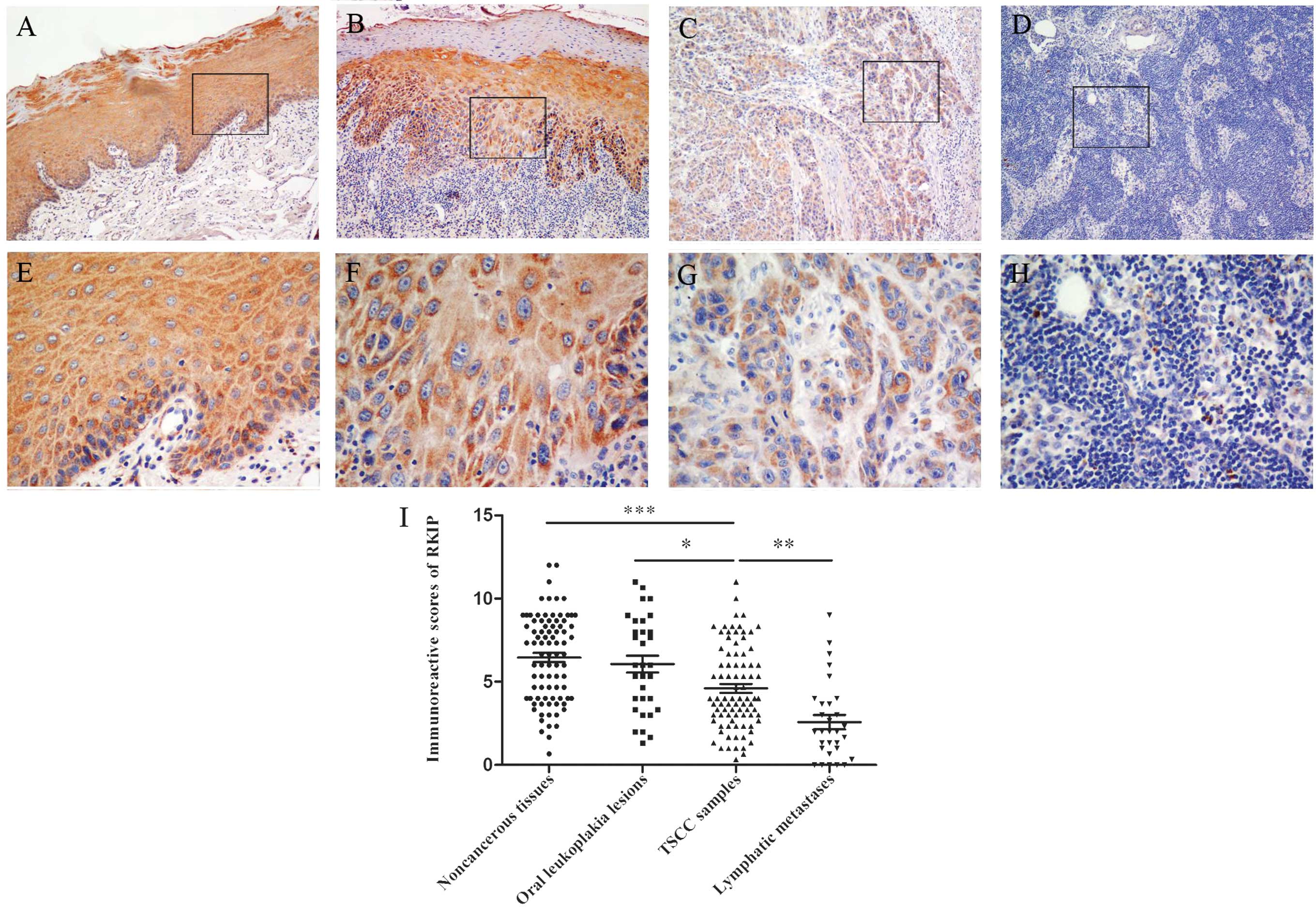
Pattern of RKIP expression in TSCC and
association with clinicopathological parameters
In non-cancerous tissues adjacent to TSCC and oral
leukoplakia lesions, RKIP was detected in almost all the layers of
the epithelia, and was mainly expressed in the cytoplasm. In TSCC
samples, RKIP was also predominantly detected in the cytoplasm, and
sporadic nuclear staining was also found. Overall, 43.5% (37/85) of
the primary tumor tissues demonstrated high RKIP expression, while
71.8% (61/85) of the corresponding adjacent non-cancerous tissues
and 65.6% (21/32) of the oral leukoplakia lesions demonstrated high
RKIP expression (Table II). There
was no statistically significant difference between RKIP expression
in adjacent non-cancerous tissues and oral leukoplakia lesions
(P=0.518). However, a significant decrease in RKIP expression was
noted in TSCC samples compared with either adjacent non-cancerous
tissues (P=0.000) or oral leukoplakia lesions (P=0.033).
| Table II.RKIP expression level in TSCCs and
related tissues. |
Table II.
RKIP expression level in TSCCs and
related tissues.
|
|
| RKIP expression, n
(%) |
|---|
|
|
|
|
|---|
| Tissue type | Patients, n | High | Low |
|---|
| Adjacent
non-cancerous tissue | 85 | 61 (71.8) | 24 (28.2) |
| TSCC tissue | 85 | 37 (43.5) | 48 (56.5) |
| Oral leukoplakia
lesions | 32 | 21 (65.6) | 11 (34.4) |
| Lymph node
metastases | 30 | 5
(16.7) | 25 (83.3) |
In order to evaluate the role of RKIP in TSCC, the
association between RKIP expression and any of the
clinicopathological parameters was investigated (Table I). The results revealed that RKIP
expression was significantly associated with the pN stage (P=0.021)
and pTNM stage (P=0.018). No significant association was identified
between RKIP expression and gender (P=0.153), age (P=0.536), tumor
differentiation (P=0.460) and pT stage (P=0.233).
Loss of RKIP in lymph node
metastases
RKIP expression was considerably less frequent in
patients with lymph node involvement (26.7%; 8/30) compared with
patients without node involvement (52.7%; 29/55). In addition, RKIP
expression was less frequent in the corresponding lymph node
metastases (16.7%; 5/30) compared with the primary TSCC tissues
(P=0.009). Notably, 5 of the metastases demonstrating low RKIP
expression completely lacked RKIP expression, obtaining an
immunoreactive score of 0 (Fig. 1D and
H).
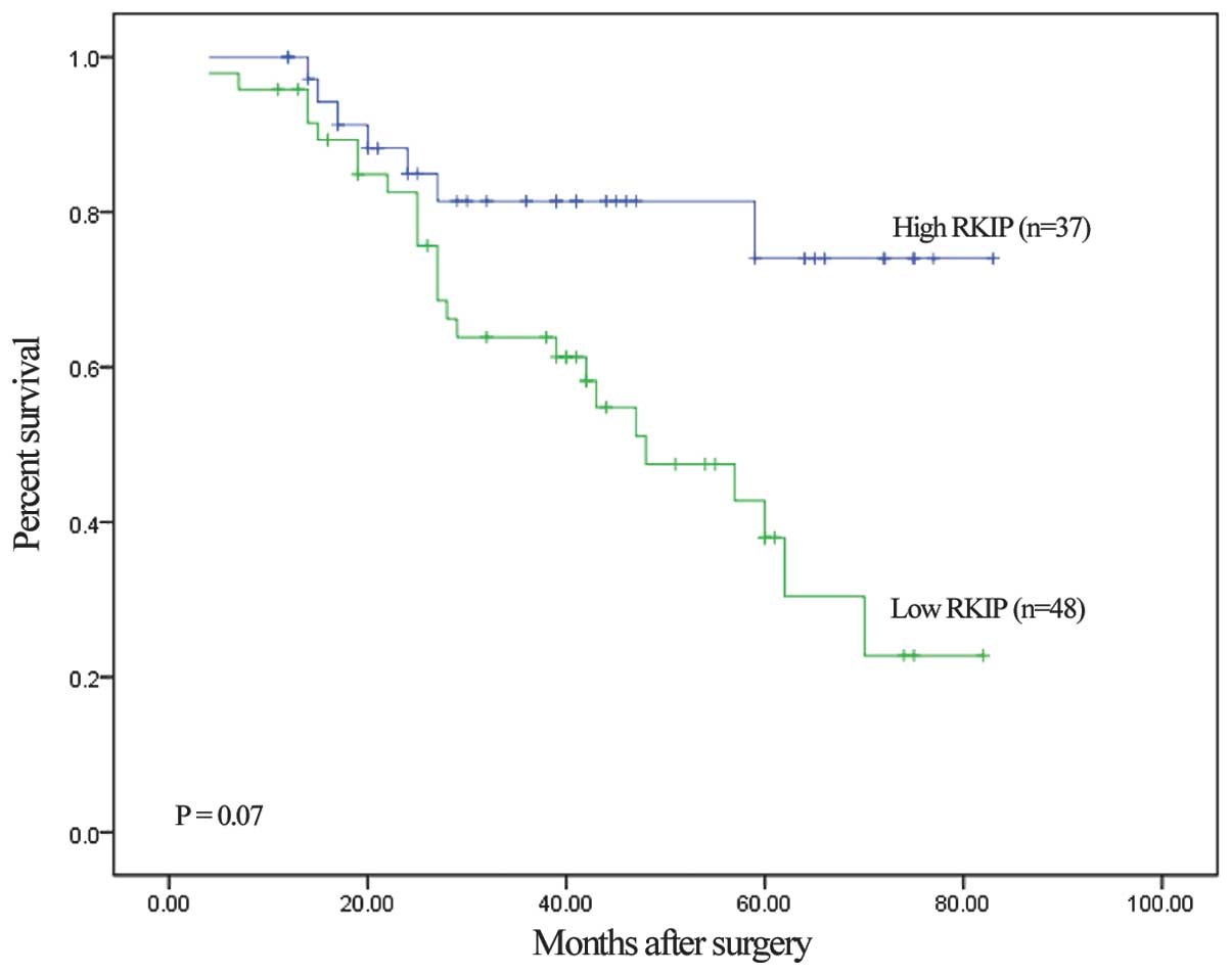
Association between RKIP expression
and survival in TSCC patients
To investigate the association between RKIP
expression and the clinical outcome of TSCC patients, the
association between the survival status and RKIP expression of
patients was analyzed. The results revealed that patients
possessing a TSCC tumor with low RKIP expression demonstrated a
significantly worse prognosis compared with patients that possessed
a tumor with high RKIP expression (P=0.007; Fig. 2). The mean survival time of patients
with low RKIP expression (n=37) was 44.1 months, while the mean
survival time of the patients with high RKIP expression (n=48) was
30.3 months.
To further assess whether RKIP expression is a
prognostic parameter in patients with TSCC, regression analysis
using a Cox proportional hazards model was performed. The covariate
parameters included several clinicopathological variables in
addition to RKIP, as reported in Table
III. In univariate analysis, low RKIP expression, lymph node
involvement and pTNM stage III or IV lesions demonstrated a
significantly increased hazard ratio (HR) for poor prognosis. In
addition, multivariate analysis was performed using variables that
demonstrated a significantly increased HR in univariate analysis.
The results of multivariate analysis revealed that RKIP expression
was the only independent prognostic predictor of TSCC outcome
(P=0.032; Table III). These results
strongly indicated that the downregulated RKIP expression in TSCC
patients is closely associated with a poor prognosis.
| Table III.Cox proportional hazards model
analysis of variables affecting survival in patients with tongue
squamous cell carcinoma. |
Table III.
Cox proportional hazards model
analysis of variables affecting survival in patients with tongue
squamous cell carcinoma.
|
|
| Univariate
analysis | Multivariate
analysis |
|---|
|
|
|
|
|
|---|
| Variable | Comparison | HR (95% CI) | P-value | HR (95% CI) | P-value |
|---|
| Gender | Male vs.
female | 0.693
(0.333–1.445) | 0.328 |
|
|
| Age | ≥55 years vs.
<55 years | 1.104
(0.549–2.219) | 0.781 |
|
|
|
Differentiation | Well vs. moderately
or poorly | 1.727
(0.858–3.475) | 0.126 |
|
|
| pT stage | T1-2 vs. T3-4 | 0.811
(0.350–1.890) | 0.625 |
|
|
| pN stage | N0 vs.
N+ | 2.261
(1.127–4.539) | 0.022 | 1.512
(0.503–4.546) | 0.462 |
| pTNM stage | I–II vs.
III–IV | 2.135
(1.041–4.380) | 0.038 | 1.286
(0.412–4.011) | 0.665 |
| RKIP
expression | High vs. low | 3.015
(1.299–6.999) | 0.010 | 2.567
(1.083–6.083) | 0.032 |
Discussion
RKIP plays an important role in cell growth
(9), mitosis (19), motility (20) and apoptosis (21) by regulating multiple intracellular
signaling pathways, including the Raf/MEK/ERK pathway (4,5), nuclear
factor-κB pathway (6), G-protein
coupled receptor signaling cascade (7,8) and
glycogen synthase kinase-3β/β-catenin pathway (22). Previous studies have revealed that
RKIP acts as a tumor suppressor and suppressor of metastasis in a
variety of malignancies (13–17). However, little is known about the
function of RKIP in oral cancer, and particularly in TSCC. The
present study observed that RKIP was intensively expressed in
non-cancerous and pre-cancerous tongue tissues. This result is
consistent with a previous study that reported a high rate (85.7%)
of RKIP immunostaining in normal tissue of the head and neck,
consisting of tissues from the tongue, lip, mouth, larynx and
pharynx (16). By contrast, the
present study found that RKIP expression is markedly reduced in
human TSCC tissues. Notably, the level of RKIP expression was found
to be even lower in the matched tissues from lymph node metastasis.
Loss of RKIP expression was also found to be significantly
associated with the presence of lymphatic metastasis and advanced
clinical stage. This finding is consistent with the reported
association between the reduction of RKIP and tumor progression and
metastasis in numerous other cancers. However, the present study
found no significant association between RKIP expression and tumor
size or histological grade in patients with TSCC. These results
indicate that although RKIP affects progression and metastasis, it
does not have an influence on the tumorigenic properties of TSCC.
Similarly, previous studies have reported that RKIP expression is
not associated with tumor size or histological grade in breast
cancer (14). In addition, the
restoration of RKIP expression has been reported to suppress
invasion and lung metastasis in prostate cancer, but not tumor
growth (13). Thus, the present
results suggest that RKIP may function as a suppressor of
metastasis in TSCC.
Furthermore, the loss of RKIP expression is
associated with a poor prognosis in prostate (23), gastric (24), pancreatic (25), and bladder cancers (26) and glioma (27). In the present study, well-established
prognostic markers, such as lymph node metastasis and an advanced
pTNM stage, were significantly associated with patient survival
time. The present study revealed that reduced cytoplasmic
expression of RKIP, which was found in 56.5% of the tissues, is
significantly associated with a shorter overall survival time in
patients with TSCC. In addition, a trend was observed between the
loss of RKIP expression and the presence of metastasis.
Accordingly, patients with positive lymphatic metastases
demonstrated relatively lower RKIP expression compared with the
patients without metastases. Although the mechanistic basis for
these subset differences is not known, the identification of these
differences may aid in future tailored therapy and early prediction
of the survival time of patients with TSCC. Additional studies with
larger cohorts are required to validate the role of RKIP as a
prognostic marker in TSCC.
To the best of our knowledge, the current study is
the first study that has analyzed RKIP expression in TSCC to be
reported in the literature. The expression level of RKIP was also
assessed in the metastatic lymph node lesions of TSCC. The present
results revealed that loss of RKIP expression was significantly
associated with tumor progression and metastasis. Despite the
importance of RKIP in tumor progression and the development of
metastasis, the mechanism of RKIP downregulation remains largely
unknown. Previous studies have investigated the CpG methylation
status of the RKIP promoter in human cancers as a possible
mechanism for the downregulation (28,29), but
the results are inconsistent and may not completely explain the
loss of RKIP expression in malignancies. Future studies evaluating
the possible mechanisms of RKIP downregulation in TSCC may be
required.
The present study reports that RKIP expression is
lost during TSCC progression, and in particular, is absent in lymph
node metastases. The current study presents evidence that the loss
of RKIP expression in TSCC is associated with the
clinicopathological characteristics of cancer aggressiveness.
Notably, loss of RKIP expression is associated with poor survival
time and may act as a potential biomarker of poor prognosis in TSCC
patients. Additional studies in larger series and with in
vitro and in vivo models are required to assess the role
of RKIP expression in the tumor progression, metastasis and
survival of TSCC patients.
Acknowledgements
This study was supported by the National Natural
Science Foundation of China (grant nos. 81172567, 81272949,
81202136 and 81372885), Program for New Century Excellent Talent in
University, Ministry of Education, China (grant no. NCET-11-0535)
and the Fundamental Research Funds for the Central Universities
(grant no. 12ykpy69).
References
|
1
|
Siegel R, Ma J, Zou Z and Jemal A: Cancer
statistics, 2014. CA Cancer J Clin. 64:9–29. 2014. View Article : Google Scholar : PubMed/NCBI
|
|
2
|
Yuasa-Nakagawa K, Shibuya H, Yoshimura R,
Miura M, Watanabe H, Kishimoto S and Omura K: Cervical lymph node
metastasis from early-stage squamous cell carcinoma of the oral
tongue. Acta Otolaryngol. 133:544–551. 2013. View Article : Google Scholar : PubMed/NCBI
|
|
3
|
Hauswald H, Zwicker F, Rochet N, Jensen
AD, Debus J and Lindel K: Treatment of squamous cell carcinoma of
the mobile tongue or tongue margins: An interdisciplinary
challenge. Acta Oncol. 52:1017–1021. 2013. View Article : Google Scholar : PubMed/NCBI
|
|
4
|
Yeung K, Seitz T, Li S, Janosch P,
McFerran B, Kaiser C, Fee F, Katsanakis KD, Rose DW, Mischak H, et
al: Suppression of Raf-1 kinase activity and MAP kinase signalling
by RKIP. Nature. 401:173–177. 1999. View
Article : Google Scholar : PubMed/NCBI
|
|
5
|
Yeung K, Janosch P, McFerran B, Rose DW,
Mischak H, Sedivy JM and Kolch W: Mechanism of suppression of the
Raf/MEK/extracellular signal-regulated kinase pathway by the raf
kinase inhibitor protein. Mol Cell Biol. 20:3079–3085. 2000.
View Article : Google Scholar : PubMed/NCBI
|
|
6
|
Yeung KC, Rose DW, Dhillon AS, Yaros D,
Gustafsson M, Chatterjee D, McFerran B, Wyche J, Kolch W and Sedivy
JM: Raf kinase inhibitor protein interacts with NF-kappaB-inducing
kinase and TAK1 and inhibits NF-kappaB activation. Mol Cell Biol.
21:7207–7217. 2001. View Article : Google Scholar : PubMed/NCBI
|
|
7
|
Corbit KC, Trakul N, Eves EM, Diaz B,
Marshall M and Rosner MR: Activation of Raf-1 signaling by protein
kinase C through a mechanism involving Raf kinase inhibitory
protein. J Biol Chem. 278:13061–13068. 2003. View Article : Google Scholar : PubMed/NCBI
|
|
8
|
Lorenz K, Lohse MJ and Quitterer U:
Protein kinase C switches the Raf kinase inhibitor from Raf-1 to
GRK-2. Nature. 426:574–579. 2003. View Article : Google Scholar : PubMed/NCBI
|
|
9
|
Zhang L, Fu Z, Binkley C, Giordano T,
Burant CF, Logsdon CD and Simeone DM: Raf kinase inhibitory protein
inhibits beta-cell proliferation. Surgery. 136:708–715. 2004.
View Article : Google Scholar : PubMed/NCBI
|
|
10
|
Schuierer MM, Heilmeier U, Boettcher A,
Ugocsai P, Bosserhoff AK, Schmitz G and Langmann T: Induction of
Raf kinase inhibitor protein contributes to macrophage
differentiation. Biochem Biophys Res Commun. 342:1083–1087. 2006.
View Article : Google Scholar : PubMed/NCBI
|
|
11
|
McHenry KT, Montesano R, Zhu S, Beshir AB,
Tang HH, Yeung KC and Fenteany G: Raf kinase inhibitor protein
positively regulates cell-substratum adhesion while negatively
regulating cell-cell adhesion. J Cell Biochem. 103:972–985. 2008.
View Article : Google Scholar : PubMed/NCBI
|
|
12
|
Shin SY, Rath O, Zebisch A, Choo SM, Kolch
W and Cho KH: Functional roles of multiple feedback loops in
extracellular signal-regulated kinase and Wnt signaling pathways
that regulate epithelial-mesenchymal transition. Cancer Res.
70:6715–6724. 2010. View Article : Google Scholar : PubMed/NCBI
|
|
13
|
Fu Z, Smith PC, Zhang L, Rubin MA, Dunn
RL, Yao Z and Keller ET: Effects of raf kinase inhibitor protein
expression on suppression of prostate cancer metastasis. J Natl
Cancer Inst. 95:878–889. 2003. View Article : Google Scholar : PubMed/NCBI
|
|
14
|
Hagan S, Al-Mulla F, Mallon E, Oien K,
Ferrier R, Gusterson B, García JJ and Kolch W: Reduction of Raf-1
kinase inhibitor protein expression correlates with breast cancer
metastasis. Clin Cancer Res. 11:7392–7397. 2005. View Article : Google Scholar : PubMed/NCBI
|
|
15
|
Schuierer MM, Bataille F, Weiss TS,
Hellerbrand C and Bosserhoff AK: Raf kinase inhibitor protein is
downregulated in hepatocellular carcinoma. Oncol Rep. 16:451–456.
2006.PubMed/NCBI
|
|
16
|
Al-Mulla F, Hagan S, Behbehani AI, Bitar
MS, George SS, Going JJ, García JJ, Scott L, Fyfe N, Murray GI and
Kolch W: Raf kinase inhibitor protein expression in a survival
analysis of colorectal cancer patients. J Clin Oncol. 24:5672–5679.
2006. View Article : Google Scholar : PubMed/NCBI
|
|
17
|
Chatterjee D, Sabo E, Tavares R and
Resnick MB: Inverse association between Raf Kinase Inhibitory
Protein and signal transducers and activators of transcription 3
expression in gastric adenocarcinoma patients: Implications for
clinical outcome. Clin Cancer Res. 14:2994–3001. 2008. View Article : Google Scholar : PubMed/NCBI
|
|
18
|
Wang C, Liu X, Huang H, Ma H, Cai W, Hou
J, Huang L, Dai Y, Yu T and Zhou X: Deregulation of Snai2 is
associated with metastasis and poor prognosis in tongue squamous
cell carcinoma. Int J Cancer. 130:2249–2258. 2012. View Article : Google Scholar : PubMed/NCBI
|
|
19
|
Eves EM, Shapiro P, Naik K, Klein UR,
Trakul N and Rosner MR: Raf kinase inhibitory protein regulates
aurora B kinase and the spindle checkpoint. Mol Cell. 23:561–574.
2006. View Article : Google Scholar : PubMed/NCBI
|
|
20
|
Bement WM: A role for RKIP in cell
motility. Chem Biol. 12:953–954. 2005. View Article : Google Scholar : PubMed/NCBI
|
|
21
|
Odabaei G, Chatterjee D, Jazirehi AR,
Goodglick L, Yeung K and Bonavida B: Raf-1 kinase inhibitor
protein: Structure, function, regulation of cell signaling and
pivotal role in apoptosis. Adv Cancer Res. 91:169–200. 2004.
View Article : Google Scholar : PubMed/NCBI
|
|
22
|
Al-Mulla F, Bitar MS, Al-Maghrebi M,
Behbehani AI, Al-Ali W, Rath O, Doyle B, Tan KY, Pitt A and Kolch
W: Raf kinase inhibitor protein RKIP enhances signaling by glycogen
synthase kinase-3β. Cancer Res. 71:1334–1343. 2011. View Article : Google Scholar : PubMed/NCBI
|
|
23
|
Fu Z, Kitagawa Y, Shen R, Shah R, Mehra R,
Rhodes D, Keller PJ, Mizokami A, Dunn R, Chinnaiyan AM, et al:
Metastasis suppressor gene Raf kinase inhibitor protein (RKIP) is a
novel prognostic marker in prostate cancer. Prostate. 66:248–256.
2006. View Article : Google Scholar : PubMed/NCBI
|
|
24
|
Martinho O, Simões K, Longatto-Filho A,
Jacob CE, Zilberstein B, Bresciani C, Gama-Rodrigues J, Cecconello
I, Alves V and Reis RM: Absence of RKIP expression is an
independent prognostic biomarker for gastric cancer patients. Oncol
Rep. 29:690–696. 2013.PubMed/NCBI
|
|
25
|
Song SP, Zhang SB, Li ZH, Zhou YS, Li B,
Bian ZW, Liao QD and Zhang YD: Reduced expression of Raf kinase
inhibitor protein correlates with poor prognosis in pancreatic
cancer. Clin Transl Oncol. 14:848–852. 2012. View Article : Google Scholar : PubMed/NCBI
|
|
26
|
Afonso J, Longatto-Filho A, Martinho O,
Lobo F, Amaro T, Reis RM and Santos LL: Low RKIP expression
associates with poor prognosis in bladder cancer patients. Virchows
Arch. 462:445–453. 2013. View Article : Google Scholar : PubMed/NCBI
|
|
27
|
Martinho O, Granja S, Jaraquemada T,
Caeiro C, Miranda-Gonçalves V, Honavar M, Costa P, Damasceno M,
Rosner MR, Lopes JM and Reis RM: Downregulation of RKIP is
associated with poor outcome and malignant progression in gliomas.
PLoS One. 7:e307692012. View Article : Google Scholar : PubMed/NCBI
|
|
28
|
Guo W, Dong Z, Guo Y, Lin X, Chen Z, Kuang
G and Yang Z: Aberrant methylation and loss expression of RKIP is
associated with tumor progression and poor prognosis in gastric
cardia adenocarcinoma. Clin Exp Metastasis. 30:265–275. 2013.
View Article : Google Scholar : PubMed/NCBI
|
|
29
|
Al-Mulla F, Hagan S, Al-Ali W, Jacob SP,
Behbehani AI, Bitar MS, Dallol A and Kolch W: Raf kinase inhibitor
protein: Mechanism of loss of expression and association with
genomic instability. J Clin Pathol. 61:524–529. 2008. View Article : Google Scholar : PubMed/NCBI
|