Introduction
Extramedullary hematopoiesis (EMH), which refers to
hematopoiesis occurring outside the medulla of the bone, is a rare
hematological disease, secondary to insufficient bone marrow
function (1,2). EMH occurs under conditions of
myelofibrosis and ineffective erythropoiesis, including that in
thalassemia, hereditary spherocytosis and sickle cell disease
(3–5).
While EMH may occur anywhere in the body, it most commonly presents
as diffuse lesions in the liver, spleen and/or lymph nodes. In rare
cases, EMH presents as a solitary mass (6), appearing as a tumor-simulating lesion in
an atypical location, and is frequently misdiagnosed.
Alpha-thalassaemia is inherited as an autosomal
recessive disorder characterized by a microcytic hypochromic
anaemia, and is a result of impaired production of 1,2,3, or 4
alpha globin chains, leading to a relative excess of beta globin
chains of haemoglobin (Hb) (7). It is
one of the most common monogenic gene disorders worldwide and is
particularly frequently observed in Mediterranean countries,
South-East Asia and Africa. As a compensatory response in various
anaemias, EMH may develop with thalassaemia (7).
The present study describes a case of EMH presenting
as multiple tumor-like lesions in the mediastinum of a patient with
α-thalassemia. The patient was diagnosed with mediastinal EMH
following a tissue biopsy via video-assisted thoracoscopic surgery
(VATS). The study protocol was approved by the Research Ethics
Committee of The Third Affiliated Hospital, Sun Yat-Sen University
(Guangzhou, China).
The present case report discusses the major
diagnostic problems of mediastinal EMH, distinct characteristics
for differentiating EMH from other mediastinal tumors and the
treatment strategy applied, as well as a literature review of this
rare clinical entity.
Case report
A 61-year-old Chinese male presented with a 3-year
history of intermittent chest pain and tightness. Physical
examination revealed elevated blood pressure (149/96 mm Hg), but no
other remarkable findings. A chest X-ray and computed tomography
(CT) scan were performed and multiple paravertebral masses were
identified (Fig. 1A). Subsequently, a
contrast-enhanced CT scan of the chest revealed multiple bilateral
mediastinal masses, observed as round or oval, smooth, soft-tissue
opacities immediately lateral to the thoracic vertebra. The mass in
the left thoracic paravertebral space at the T3-T4 vertebral level
measured 44×25 mm, and the two masses on the right of the T10
vertebra measured 52×48×17 mm and 32×22 mm (Fig. 1B). In addition, the initial CT scan
results indicated the possibility of neurogenic tumors.
The complete blood count revealed the following
results: White blood cells, 11.44×10⁹/l; hemoglobin
(Hb), 119 g/l; platelets, 176×109/l; mean corpuscular
volume, 76.4 fl; mean corpuscular Hb concentration, 258 g/l;
segmental neutrophils, 48.1%; lymphocytes, 44.5%; monocytes, 5% and
eosinophils, 1.9%. No pathological changes were detected in
erythrocyte sedimentation rate or C-reactive protein.
To determine the diagnosis of the mediastinal
masses, a surgical tissue biopsy via VATS was performed following
the provision of consent of the patient and family. A section of
the dark-red mass in the left thoracic cavity was excised and
electrocoagulation was used for hemostasis. Frozen section
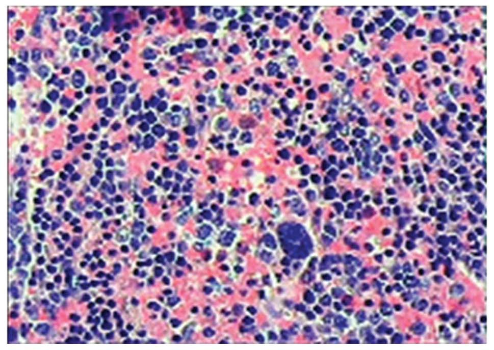
examination revealed EMH. A conventional permanent
histopathological examination was also performed; the biopsied
tissue was stained (hematoxylin and eosin) and examined under a
BX43 microscope (Olympus Corporation, Tokyo, Japan), revealing
hyperplasia with activity of two types of hematopoietic cell,
namely erythroid and megakaryocytic cells (Fig. 2). Furthermore, immunohistochemical
staining indicated that diffuse cells from the mediastinal lesions
were positive for CD43, CD99 and high Ki-67, while sporadic cells
were positive for myeloperoxidase, CD15, CD3, CD45RO and CD61. The
diffuse and sporadic cells were negative for creatine kinase, CD30
and Pax-5. Myeloid sarcoma, a rare extramedullary form of
myelogenous leukemia, also known as granulocytic sarcoma or
chloroma was therefore excluded. Based on these pathological and
clinical findings, the patient was diagnosed with thoracic EMH.
Postoperatively, various laboratory tests were
performed, which demonstrated low Hb (90 g/l). Subsequently,
thalassemia-associated tests were performed. A diagnosis of
α-thalassemia was made based on the increased glucose-6-phosphate
dehydrogenase activity (15919.000 mU/g), decreased HbA2 (0.9%) and
RBC-BT (erythrocyte fragility test; 29.0%), as well as positive
hemoglobin H staining with hemoglobin electrophoretic studies (HbH,
10.7%).
The patient was transferred to the Department of
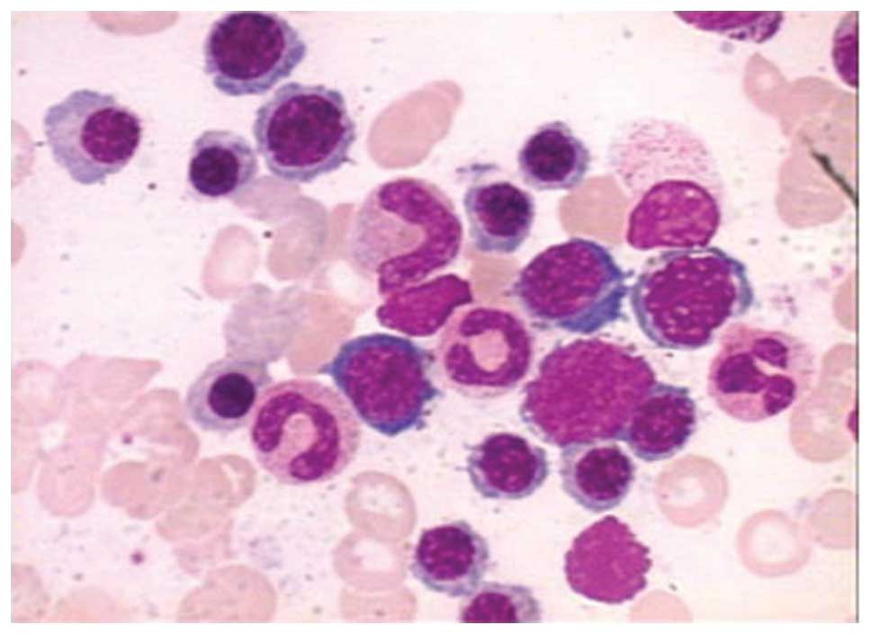
Hematology for further treatment, where bone marrow aspiration
cytology confirmed the findings of anemia and erythroid hyperplasia
(G:E, 0.82:1). In addition, the relative proportion of immature red
blood cells was also significantly increased, but was not
accompanied by malignant cell invasion (Fig. 3).
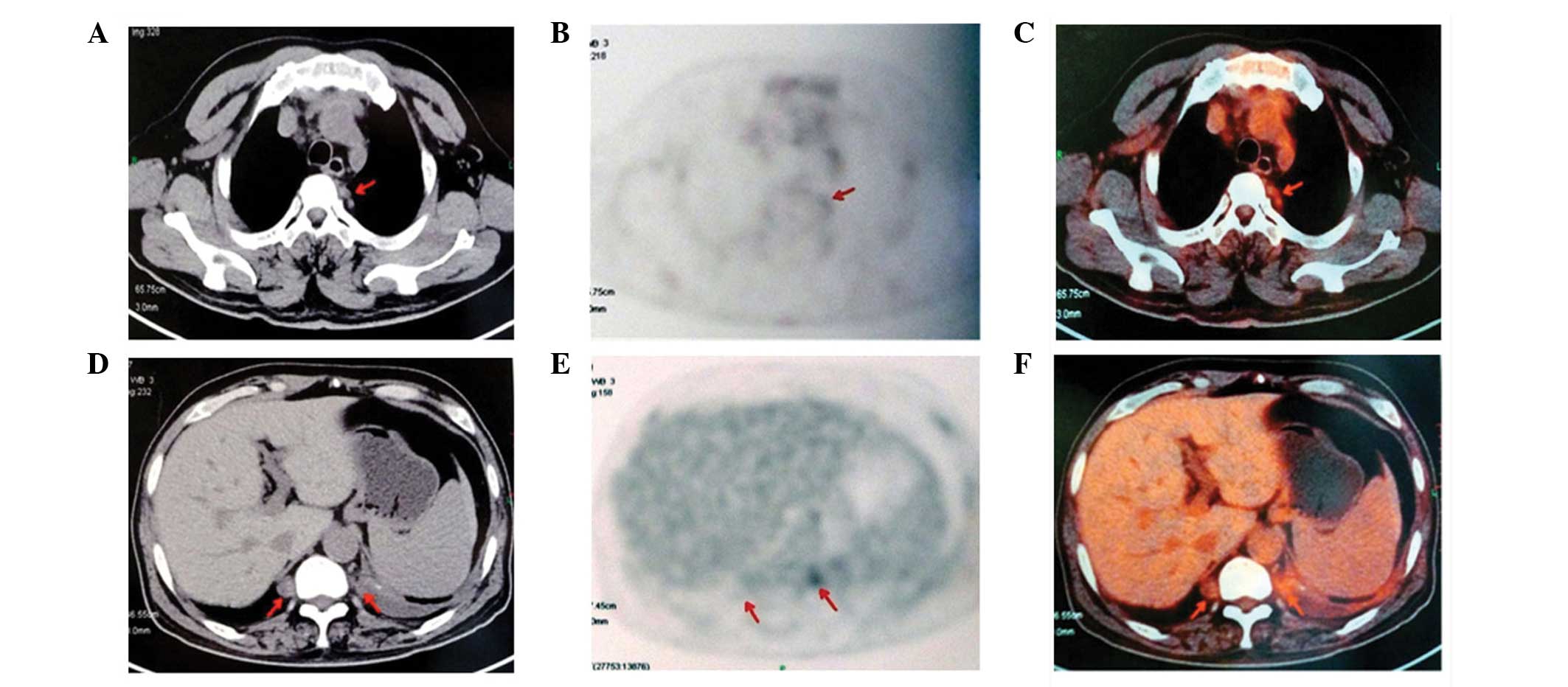
Subsequently, 18-fluorodeoxyglucose (FDG) positron
emission tomography-computed tomography (PET/CT) was performed to
evaluate the mediastinal masses and determine the presence of any
additional extramedullary masses. PET/CT identified remnant tissue
in the left thoracic T3-T4 paravertebral space following biopsy,
and the maximum standardized uptake value (SUVmax) was
5.2. The remaining two paravertebral masses, at the
posterior-inferior mediastinum at the T10 level, demonstrated no
increased glucose metabolism, with SUVmax values of 3.8
(Fig. 4).
Since the postoperative course following VATS was
uneventful and the patient exhibited no clinical symptoms, regular
outpatient follow-up observation was selected, rather than
treatment. At 8 months postoperatively, at the most recent
follow-up visit, the patient exhibited no evidence of disease.
Discussion
For a primary mediastinal mass, the potential
clinical differential diagnoses are extensive. However, EMH, a rare
entity involving the formation and development of blood cells
outside of the bone marrow, is an unlikely diagnostic
candidate.
EMH occurs in response to the failure of
erythropoiesis in bone marrow and may occur in myeloproliferative
disorders, hemoglobinopathies or bone marrow infiltration. EMH most
commonly affects the spleen and liver and, occasionally, the lymph
nodes. Less commonly involved organs include the pleura, lungs,
gastrointestinal tract, breast, skin, brain, kidneys and adrenal
glands (8). The occurrence of EMH as
a tumor-simulating mass in the intrathoracic cavity, particularly a
posterior mediastinal mass, is very rare (9). A review of the literature revealed that
only three cases of EMH in patients with α-thalassemia have been
reported, to date (5,10,11).
EMH is frequently characterized by the development
of soft tissue masses in the paravertebral thoracic regions. These
masses rarely induce significant symptoms, but may result in
hemothorax and pleural effusion (5,12). In the
present case, the patient was asymptomatic and EMH was identified
by chance.
Patients with no hematologic disorder, presenting
with such masses induced by EMH, are frequently misdiagnosed
(9,13). EMH is often incidentally diagnosed,
when patients undergo evaluation for unrelated symptoms, as in the
present case. While EMH diagnosis requires a biopsy, a needle
biopsy carries the risk of hemothorax. In the presence of anemia, a
diagnosis of EMH may be considered preoperatively; however, in the
present case, the primary blood tests identified Hb as 119 g/l;
therefore, EMH was initially overlooked as a potential
diagnosis.
Notably, extramedullary myeloid sarcoma (EMS) is
also a rare extra medullary tumor mass consisting of myeloid
blasts. EMS is also known as granulocytic sarcoma or chloroma,
since it is diagnosed based on the presence of a green color caused
by myeloperoxidase. EMS occurs in ~5% of patients with acute
myeloid leukemia (AML) (14). Rarely,
EMS may predate AML, presenting as an isolated mass in various
sites of the body. However, EMS in the mediastinal region has
rarely been reported (15,16). EMS is primarily diagnosed based on
immunohistochemical staining of tissue biopsy. Although EMH and EMS
arise from distinct hematological diseases, the two conditions
should be considered as differential diagnoses for a mediastinal
tumor.
A PET/CT scan may be diagnostically useful in such
cases. To date, only a few cases regarding the diagnosis of EMH
using PET/CT have been reported (17,18), with
EMH detected as a benign mass with low SUVmax values and
normal appearance of the tissue. The concurrent presence of an
underlying hematopoietic disorder may suggest a diagnosis of EMH.
In EMS, a PET/CT scan identifies intense FDG uptake (16).
As a result of its rarity, there are no
evidence-based guidelines for the treatment of thoracic
pseudotumors induced by EMH. Essentially, EMH is a compensatory
mechanism for chronic anemia; therefore, recurrent blood
transfusions to correct such anemia may result in shrinkage of the
mass. However, blood transfusions are not entirely harmless or
risk-free, and this therapeutic effect is temporary, slow to occur
and may be insufficient (19–21). Hematopoietic tissue is notably
radiosensitive and undergoes shrinkage with radiotherapy; thus,
excellent results have been obtained in cases of EMH with a few
days of radiotherapy alone (22,23).
Surgical excision is required in patients with EMH associated with
complications, including bleeding from the mass (9), spinal cord compression or spinal canal
invasion (13). Such excision does
not only staunch the bleeding and/or allow decompression but also
provides a histological diagnosis. Following surgical excision,
radiotherapy may aid the prevention of recurrence of EMH or
facilitate complete remission (24,25).
Based on the literature review, it was hypothesized
that close follow-up of the patient was important. Considering the
excellent results obtained with low-dose radiotherapy for EMH
masses, the patient will be advised to undergo radiation therapy in
the future.
EMH should be considered in the differential
diagnosis of an intrathoracic mass. Particularly, if a history of
thalassemia or chronic anemia is discovered, based on evaluation of
the patient's medical history, as well as laboratory studies.
Surgical resection and radiation therapy should be considered as
treatment options for EMH.
Acknowledgements
The present study was supported by the ‘985’ project
grant of Sun Yat-sen University (no. 82000-31101301), which was
awarded to Dr Junhang Zhang (The Third Affiliated Hospital, Sun
Yat-Sen University, Guangzhou, China).
References
|
1
|
Bolaman Z, Polatli M, Cildag O, Kadikoylu
G and Culhaci N: Intrathoracic extramedullary hematopoiesis
resembling posterior mediastinal tumor. Am J Med. 112:739–741.
2002. View Article : Google Scholar : PubMed/NCBI
|
|
2
|
Xiros N, Economopoulos T, Papageorgiou E,
Mantzios G and Raptis S: Massive hemothorax due to intrathoracic
extramedullary hematopoiesis in a patient with hereditary
spherocytosis. Ann Hematol. 80:38–40. 2001. View Article : Google Scholar : PubMed/NCBI
|
|
3
|
Gumbs R, Ford EAH, Teal JS, Kletter GG and
Castro O: Intrathoracic extramedullary hematopoiesis in sickle cell
disease. Amer J Radiol. 149:889–893. 1987.
|
|
4
|
Scott L, Blair G, Taylor G, Dimmick J and
Fraser G: Inflammatory pseudotumors in children. J Pediatr Surg.
23:755–758. 1988. View Article : Google Scholar : PubMed/NCBI
|
|
5
|
Chu KA, Lai RS, Lee CH, Lu JY, Chang HC
and Chiang HT: Intrathoracic extramedullary haematopoiesis
complicated by massive haemothorax in alpha-thalassaemia. Thorax.
54:466–468. 1999. View Article : Google Scholar : PubMed/NCBI
|
|
6
|
Haidar R, Mhaidli H and Taher AT:
Paraspinal extramedullary hematopoiesis in patients with
thalassemia intermedia. Eur Spine J. 19:871–878. 2010. View Article : Google Scholar : PubMed/NCBI
|
|
7
|
Harteveld CL and Higgs DR:
Alpha-thalassaemia. Orphanet J Rare Dis. 5:132010. View Article : Google Scholar : PubMed/NCBI
|
|
8
|
Choi H, David CL, Katz RL and Podoloff DA:
Case 69: Extramedullary hematopoiesis. Radiology. 231:52–56. 2004.
View Article : Google Scholar : PubMed/NCBI
|
|
9
|
Kubokura H, Koizumi K, Yoshino N,
Yamagishi S, Mikami I, Hirata T, Harada A, Kawamoto M and Shimizu
K: A case report: Thoracic extramedullary hematopoiesis found by
occurring spontaneous pneumothorax. Ann Thorac Cardiovasc Surg.
14:382–385. 2008.PubMed/NCBI
|
|
10
|
Bobylev D, Zhang R, Haverich A and Krueger
M: Extramedullary haematopoiesis presented as intrathoracic tumour
in a patient with alpha-thalassaemia. J Cardiothorac Surg.
8:1202013. View Article : Google Scholar : PubMed/NCBI
|
|
11
|
Wu JH, Shih LY, Kuo TT and Lan RS:
Intrathoracic extramedullary hematopoietic tumor in hemoglobin H
disease. Am J Hematol. 41:285–288. 1992. View Article : Google Scholar : PubMed/NCBI
|
|
12
|
Peng MJ, Kuo HT and Chang MC: A case of
intrathoracic extramedullary hematopoiesis with massive pleural
effusion: Successful pleurodesis with intrapleural minocycline. J
Formos Med Assoc. 93:445–447. 1994.PubMed/NCBI
|
|
13
|
Yeom SY, Lim JH, Han KN, Kang CH, Park IK
and Kim YT: Extramedullary hematopoiesis at the posterior
mediastinum in patient with hereditary spherocytosis: A case
report. Korean J Thorac Cardiovasc Surg. 46:156–158. 2013.
View Article : Google Scholar : PubMed/NCBI
|
|
14
|
Liu PI, Ishimaru T, McGregor DH, Okada H
and Steer A: Autopsy study of granulocytic sarcoma (chloroma) in
patients with myelogenous leukemia, Hiroshima-Nagasaki 1949–1969.
Cancer. 31:948–955. 1973. View Article : Google Scholar : PubMed/NCBI
|
|
15
|
Nounou R, Al Zahrani HH, Ajarim DS, Martin
J, Iqbal A, Naufal R, Stuart R, Roberts G and Gyger M:
Extramedullary myeloid cell tumours localised to the mediastinum: A
rare clinicopathological entity with unique karyotypic features. J
Clin Pathol. 55:221–225. 2002. View Article : Google Scholar : PubMed/NCBI
|
|
16
|
Makis W, Hickeson M and Derbekyan V:
Myeloid sarcoma presenting as an anterior mediastinal mass invading
the pericardium: Serial imaging with F-18 FDG PET/CT. Clin Nucl
Med. 35:706–709. 2010. View Article : Google Scholar : PubMed/NCBI
|
|
17
|
Zade A, Purandare N, Rangarajan V, Shah S,
Agrawal A, Ashish J and Kulkarni M: Noninvasive approaches to
diagnose intrathoracic extramedullary hematopoiesis: 18F-FLT PET/CT
and 99mTc-SC SPECT/CT scintigraphy. Clin Nucl Med. 37:788–789.
2012. View Article : Google Scholar : PubMed/NCBI
|
|
18
|
Mosley C, Jacene HA, Holz A, Grand DJ and
Wahl RL: Extramedullary hematopoiesis on F-18 FDG PET/CT in a
patient with metastatic colon carcinoma. Clin Nucl Med. 32:878–880.
2007. View Article : Google Scholar : PubMed/NCBI
|
|
19
|
Chehal A, Aoun E, Koussa S, Skoury H,
Koussa S and Taher A: Hypertransfusion: A successful method of
treatment in thalassemia intermedia patients with spinal cord
compression secondary to extramedullary hematopoiesis. Spine (Phila
Pa 1976). 28:E245–E249. 2003. View Article : Google Scholar : PubMed/NCBI
|
|
20
|
Tai SM, Chan JS, Ha SY, Young BW and Chan
MS: Successful treatment of spinal cord compression secondary to
extramedullary hematopoietic mass by hypertransfusion in a patient
with thalassemia major. Pediatr Hematol Oncol. 23:317–321. 2006.
View Article : Google Scholar : PubMed/NCBI
|
|
21
|
Berkmen YM and Zalta BA: Case 126:
Extramedullary hematopoiesis. Radiology. 245:905–908. 2007.
View Article : Google Scholar : PubMed/NCBI
|
|
22
|
Singhal S, Sharma S, Dixit S, De S,
Chander S, Rath GK and Mehta VS: The role of radiation therapy in
the management of spinal cord compression due to extramedullary
haematopoiesis in thalassaemia. J Neurol Neurosurg Psychiatry.
55:310–312. 1992. View Article : Google Scholar : PubMed/NCBI
|
|
23
|
Wen BC, Hussey DH, Hitchon PW, Schelper
RL, Vigliotti AP, Doornbos JF and VanGilder JC: The role of
radiation therapy in the management of ependymomas of the spinal
cord. Int J Radiat Oncol Biol Phys. 20:781–786. 1991. View Article : Google Scholar : PubMed/NCBI
|
|
24
|
Dore F, Pardini S, Gaviano E, Longinotti
M, Bonfigli S, Rovasio S, Tomiselli A and Cossu F: Recurrence of
spinal cord compression from extramedullary hematopoiesis in
thalassemia intermedia treated with low doses of radiotherapy. Am J
Hematol. 44:1481993. View Article : Google Scholar : PubMed/NCBI
|
|
25
|
NebenWittich MA, Brown PD and Tefferi A:
Successful treatment of severe extremity pain in myelofibrosis with
low-dose single-fraction radiation therapy. Am J Hematol.
85:808–810. 2010. View Article : Google Scholar : PubMed/NCBI
|


