Introduction
Hepatocellular carcinoma (HCC), which accounts for
almost 598,000 mortalities each year, is the fifth most common type
of cancer and the third leading cause of cancer-associated
mortality worldwide (1). The risk
factors for HCC include hepatitis B virus (HBV) or hepatitis C
virus (HCV) infection, alcoholic liver disease and non-alcoholic
fatty liver disease. Of these, HBV is the major risk factor for HCC
(2). The majority of HCC cases
(>80%) occur in either sub-Saharan Africa or Eastern Asia, with
China accounting for >50% of worldwide cases, demonstrating
age-standardized incidence rates of 35.2 and 13.3 cases per 100,000
men and women, respectively (3).
Tumor recurrence is common following local treatments, and there
continues to be no curative therapy once the tumor reaches an
advanced stage (4). Numerous factors,
including tumor size, number of lesions, cell differentiation,
venous invasion and degree of inflammation, have been identified as
predictors of recurrence and prognosis in patients with HCC.
However, few studies have focused on the association between
oxidative stress and the prognosis of HCC (5).
Reactive oxygen species (ROS) are generated as
byproducts of mitochondrial respiration, and include superoxide
(O2−) and hydrogen peroxide
(H2O2). In particular, the superoxide anion
(O2−) mediates oxidative damage to numerous
intracellular components, including proteins, nucleic acids and
lipid membranes (6,7). This ROS-induced damage may contribute to
carcinogenesis and numerous other diseases (8). DNA damage mediated by oxidative stress
and the ensuing genetic mutations are crucial for the pathogenesis
and progression of certain human malignancies (9,10).
Mitochondrial DNA (mtDNA) is the only extra-nuclear genetic
material in mammalian cells that is more susceptible to DNA damage
and mutation than nuclear DNA. This is due to the high levels of
ROS, a lack of protective histones and a limited capacity for DNA
repair (11,12). Damage to mtDNA due to elevated ROS
production is widely accepted as a key event in the initiation and
promotion of tumorigenesis (13).
Mitochondrial complex II, also termed
succinate:quinone oxidoreductase or succinate dehydrogenase,
oxidizes succinate to fumarate in the Krebs cycle and reduces
ubiquinone in the respiratory chain. Complex II is ubiquitous in
diverse aerobic species and contributes significantly to the
excessive production of ROS by mitochondria (14,15).
Recently, several tumors caused by complex II gene mutations have
been identified (16).
Although numerous studies have suggested the value
of oxidative stress markers, such as ROS and complex II (14–16), in
molecular cancer research, little is known regarding the potential
predictive power of oxidative stress markers for the prognosis of
HCC patients. The aim of the present study was to evaluate the
ability of ROS and complex II mtDNA levels in HCC liver tissues for
the survival of patients with HCC.
Materials and methods
Tissue specimens and mitochondrial
isolation
Histologically confirmed cancerous liver tissues
were obtained from 42 patients with HBV-HCC that underwent liver
cancer resection at the Department of Hepatobiliary Surgery of The
Fourth Hospital of Hebei University (Shijiazhuang, China) between
March 2007 and September 2008. All the tissues were stored in
liquid nitrogen immediately following surgical resection.
Mitochondria were isolated using an animal tissue mitochondria
isolation kit (GenMed Scientifics, Shanghai, China) and
mitochondrial protein was extracted using an animal tissue
mitochondria lysis kit (GenMed Scientifics).
Ethical approval
The protocol of the present study was approved by
the ethics committee of The Fourth Hospital of Hebei Medical
University. Written informed consent for the collection of samples
and subsequent analysis was obtained from all patients.
Measurement of mitochondrial ROS
activity
A quantitative colorimetric method was adopted to
evaluate the activity level of ROS, using a tissue ROS
determination kit (GenMed Scientifics). All experiments were
conducted according to the manufacturer's instructions. Each
experiment was performed three times, and the absorbance in each
well was measured at a wavelength of 420 nm, using a Synergy 2
Multi-Mode Microplate Reader (BioTek Instruments Inc., Winooski,
VT, USA). The activity was calculated using the following formula:
Specimen activity = OH− µM/L.
Measurement of the mitochondrial
complex II levels
A quantitative colorimetric method was used to
evaluate the activity level of complex II, and a tissue complex II
determination kit was used for the detection of complex II (GenMed
Scientifics). All experimental steps were performed according to
the manufacturer's instructions. Each sample was tested in
triplicate, and the absorbance in each well was measured at a
wavelength of 600 nm using a Synergy 2 Multi-Mode Microplate Reader
(BioTek Instruments, Inc.). The activity was calculated using the
following formula: Specimen activity = U/mg, where 1 U =
dichlorophenol µM/min- indophenol µM/min.
Statistical analysis
Data were expressed as inter-quartile ranges and
survival curves were constructed using the Kaplan-Meier method.
Comparisons between the curves were performed using the log-rank
test. Multivariate survival analysis was performed using the Cox
proportional hazards model. Pearson's correlation test was used to
analyze the association between the level of ROS and complex II.
All statistical analyses were performed using the SPSS 18.0
software package (SPSS, Inc., Chicago, IL, USA). P<0.05 was
considered to indicate a statistically significant difference.
Results
Clinical characteristics of patients
with HCC
A total of 42 patients with HBV-HCC were enrolled in
the present study. A review was conducted every 3 months for 3
years, and no patient received adjuvant chemotherapy or radiation
therapy following HCC resection. The association between the data
collected during the 3-year follow-up and the clinical
characteristics of the patients was calculated using the
Kaplan-Meier method and the log-rank test. Patient gender, patient
age, tumor size and number of tumors were not statistically
significant predictors for the post-operative survival time.
However, Child-Pugh classification and portal vein thrombosis were
significantly associated with the survival time (Table I).
 | Table I.Multivariate analysis of prognostic
factors associated with post-operative survival in hepatocellular
carcinoma patients, performed using the Cox proportional hazards
model. |
Table I.
Multivariate analysis of prognostic
factors associated with post-operative survival in hepatocellular
carcinoma patients, performed using the Cox proportional hazards
model.
| Factors | Relative risk | 95% CI | P-value |
|---|
| Child-Pugh
classification | 16.428 |
1.632–165.376 | 0.018 |
| Portal vein
thrombosis |
4.209 |
1.519–11.661 | 0.006 |
| Complex II
activity |
5.422 |
1.273–23.088 | 0.022 |
| ROS activity |
2.867 | 1.062–7.737 | 0.038 |
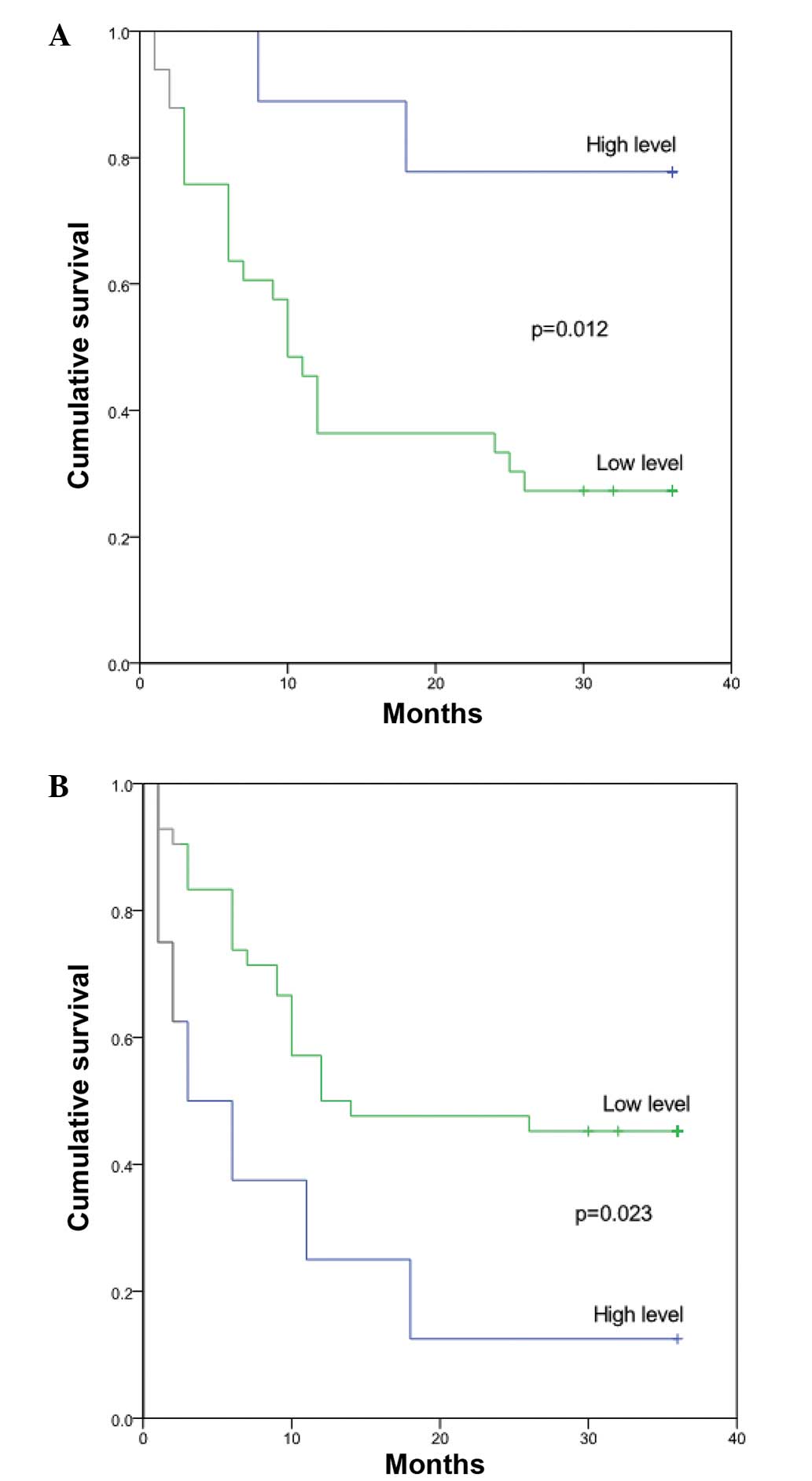
Association between the oxidative
stress parameters and HCC outcome
The association between ROS and complex II and the
outcome of HCC was assessed using the Kaplan-Meier method. The HCC
patients were divided into two groups, consisting of the upper
quartile and lower three quartiles, based on the ROS and complex II
levels. High levels of ROS and low levels of complex II were
associated with a shorter survival time, as assessed by the
log-rank test (P=0.017 and P=0.04 for ROS and complex II,
respectively) (Table II; Fig. 1). However, the levels of ROS and
complex II were not correlated when assessed by linear correlation
analysis (r=-0.110, P=0.489).
| Table II.Univariate analysis of the clinical
characteristics associated with post-operative survival in
hepatocellular carcinoma patients, performed using the log-rank
test. |
Table II.
Univariate analysis of the clinical
characteristics associated with post-operative survival in
hepatocellular carcinoma patients, performed using the log-rank
test.
| Characteristics | Patients, n | 3-year survival rate,
% | P-value |
|---|
| Gender |
|
| 0.769 |
| Male | 36 | 33.3 |
|
|
Female | 6 | 33.3 |
|
| Age |
|
| 0.629 |
| ≤55
years | 23 | 30.4 |
|
| >55
years | 19 | 36.8 |
|
| Tumor diameter |
|
| 0.165 |
| ≤5
cm | 12 | 50.0 |
|
| >5
cm | 30 | 26.7 |
|
| Number of tumors |
|
| 0.693 |
|
Single | 35 | 34.3 |
|
|
Multiple | 7 | 28.6 |
|
| Child-Pugh
classification |
|
| 0.003 |
| A | 41 | 34.1 |
|
| B | 1 | 0.0 |
|
| Portal vein
thrombosis |
|
| 0.002 |
| Yes | 6 | 0.0 |
|
| No | 36 | 38.9 |
|
| TNM
classification |
|
| 0.328 |
| I–II | 20 | 40.0 |
|
| III | 22 | 27.3 |
|
| Complex II
activity |
|
| 0.017 |
| High | 11 | 77.8 |
|
| Low | 31 | 21.2 |
|
| ROS activity |
|
| 0.040 |
| High | 11 | 16.7 |
|
| Low | 31 | 40.9 |
|
Multivariate analysis was performed to determine the
outcome predictors, which consisted of the Child-Pugh
classification, portal vein thrombosis, and complex II and ROS
activity, using a Cox proportional hazards model. The multivariate
analysis identified complex II (RR=5.422; 95% CI, 1.062–7.737;
P=0.022) and ROS (RR=2.867; 95% CI, 1.273–23.088; P=0.038) as
independent predictors for the outcome of HCC (Table I).
Discussion
Oxidative stress causes notable damage to mtDNA,
which is considered to significantly contribute to carcinogenesis
(17). The present study aimed to
identify effective predictors of HCC prognosis by examining two
parameters of oxidative stress. The present data provided initial
support for the conclusion that the levels of complex II and ROS
were associated with carcinogenesis and the outcome of HCC, using a
small sample size.
A previous study has demonstrated that oxidative
stress of mtDNA is associated with the post-operative survival of
patients with HCC (18). In the
present study, the results demonstrated that ROS levels were
associated with the carcinogenesis of HCC and the post-operative
survival of HCC patients. Endogenous ROS escape from the electron
transport chain during the oxidative phosphorylation process,
rendering nuclear and mtDNA susceptible to damage. DNA mutations
resulting from this damage may promote the malignant transformation
of cells (19). A certain amount of
ROS is required for cellular survival and adaptation (19), which may explain to some degree the
high ROS levels associated with the poor prognosis of HCC patients.
Furthermore, previous studies suggest that oxidative DNA damage is
associated with cancer progression, including cell proliferation,
apoptosis, genetic instability and chemo-resistant phenotypes
(20–22).
To the best of our knowledge, the present study is
the first to report an association between complex II levels and
the outcome of HCC. However, the detailed mechanism of the effect
of complex II on HCC progression remains to be elucidated. Complex
II contributes significantly to the production of ROS (14,15), which
may explain the strong association between complex II and HCC
development. The levels of complex II and ROS demonstrated no
association in the present analysis, suggesting that an alternative
mechanism may regulate the generation of ROS in patients with HCC.
Mitochondrial complex II has previously been used as a novel target
for anticancer agents, and several anticancer agents may induce
cancer cell apoptosis by interacting with subunits of complex II
(23). Cancer cells with the highest
levels of complex II were targeted and driven to apoptosis
(23,24).
In conclusion, the levels of ROS and complex II were
identified as independent prognostic markers for the outcome of
HCC. The analysis of ROS and complex II levels may provide a useful
research and therapeutic tool for ROS- and complex II-associated
hepatocarcinogenesis.
Acknowledgements
This study was supported by the Key Basic Research
Program of Hebei (grant no. 14967713D).
References
|
1
|
Parkin DM, Bray F, Ferlay J and Pisani P:
Global cancer statistics, 2002. CA Cancer J Clin. 55:74–108. 2005.
View Article : Google Scholar : PubMed/NCBI
|
|
2
|
El-Serag HB: Hepatocellular carcinoma. N
Engl J Med. 365:1118–1127. 2011. View Article : Google Scholar : PubMed/NCBI
|
|
3
|
El-Serag HB and Rudolph KL: Hepatocellular
carcinoma: Epidemiology and molecular carcinogenesis.
Gastroenterology. 132:2557–2576. 2007. View Article : Google Scholar : PubMed/NCBI
|
|
4
|
Hoshida Y, Villanueva A and Llovet JM:
Molecular profiling to predict hepatocellular carcinoma outcome.
Expert Rev Gastroenterol Hepatol. 3:101–103. 2009. View Article : Google Scholar : PubMed/NCBI
|
|
5
|
Okada S, Shimada K, Yamamoto J, Takayama
T, Kosuge T, Yamasaki S, Sakamoto M and Hirohashi S: Predictive
factors for postoperative recurrence of hepatocellular carcinoma.
Gastroenterology. 106:1618–1624. 1994.PubMed/NCBI
|
|
6
|
Csiszar A, Podlutsky A, Podlutskaya N,
Sonntag WE, Merlin SZ, Philipp EE, Doyle K, Davila A, Recchia FA,
Ballabh P, et al: Testing the oxidative stress hypothesis of aging
in primate fibroblasts: Is there a correlation between species
longevity and cellular ROS production? J Gerontol A Biol Sci Med
Sci. 67:841–852. 2012. View Article : Google Scholar : PubMed/NCBI
|
|
7
|
Park J, Lee J and Choi C: Mitochondrial
network determines intracellular ROS dynamics and sensitivity to
oxidative stress through switching inter-mitochondrial messengers.
PLoS One. 6:e232112011. View Article : Google Scholar : PubMed/NCBI
|
|
8
|
Kawanishi S, Hiraku Y and Oikawa S:
Mechanism of guanine-specific DNA damage by oxidative stress and
its role in carcinogenesis and aging. Mutat Res. 488:65–76. 2001.
View Article : Google Scholar : PubMed/NCBI
|
|
9
|
Valko M, Leibfritz D, Moncol J, Cronin MT,
Mazur M and Telser J: Free radicals and antioxidants in normal
physiological functions and human disease. Int J Biochem Cell Biol.
39:44–84. 2007. View Article : Google Scholar : PubMed/NCBI
|
|
10
|
DeNicola GM, Karreth FA, Humpton TJ,
Gopinathan A, Wei C, Frese K, Mangal D, Yu KH, Yeo CJ, Calhoun ES,
et al: Oncogene-induced Nrf2 transcription promotes ROS
detoxification and tumorigenesis. Nature. 475:106–109. 2011.
View Article : Google Scholar : PubMed/NCBI
|
|
11
|
DiMauro S and Schon EA: Mitochondrial DNA
mutations in human disease. Am J Med Genet. 106:18–26. 2001.
View Article : Google Scholar : PubMed/NCBI
|
|
12
|
Lightowlers RN, Chinnery PF, Turnbull DM
and Howell N: Mammalian mitochondrial genetics: Heredity,
heteroplasmy and disease. Trends Genet. 13:450–455. 1997.
View Article : Google Scholar : PubMed/NCBI
|
|
13
|
Yang L, Wang P, Wang H, Li Q, Teng H, Liu
Z, Yang W, Hou L and Zou X: Fucoidan derived from Undaria
pinnatifida induces apoptosis in human hepatocellular carcinoma
SMMC-7721 cells via the ROS-mediated mitochondrial pathway. Mar
Drugs. 11:1961–1976. 2013. View Article : Google Scholar : PubMed/NCBI
|
|
14
|
Quinlan CL, Orr AL, Perevoshchikova IV,
Treberg JR, Ackrell BA and Brand MD: Mitochondrial complex II can
generate reactive oxygen species at high rates in both the forward
and reverse reactions. J Biol Chem. 287:27255–27264. 2012.
View Article : Google Scholar : PubMed/NCBI
|
|
15
|
Moreno-Sánchez R, Hernández-Esquivel L,
Rivero-Segura NA, Marín-Hernández A, Neuzil J, Ralph SJ and
Rodríguez Enríquez S: Reactive oxygen species are generated by the
respiratory complex II - evidence for lack of contribution of the
reverse electron flow in complex I. FEBS J. 280:927–938.
2013.PubMed/NCBI
|
|
16
|
Hoekstra AS and Bayley JP: The role of
complex II in disease. Biochim Biophys Acta. 1827:543–551. 2013.
View Article : Google Scholar : PubMed/NCBI
|
|
17
|
Halliwell B: Can oxidative DNA damage be
used as a biomarker of cancer risk in humans? Problems, resolutions
and preliminary results from nutritional supplementation studies.
Free Radic Res. 29:469–486. 1998. View Article : Google Scholar : PubMed/NCBI
|
|
18
|
Li S, Wang X, Wu Y, Zhang H, Zhang L, Wang
C, Zhang R and Guo Z: 8-Hydroxy-2′-deoxyguanosine expression
predicts hepatocellular carcinoma outcome. Oncol Lett. 3:338–342.
2012.PubMed/NCBI
|
|
19
|
Cheng CW, Kuo CY, Fan CC, Fang WC, Jiang
SS, Lo YK, Wang TY, Kao MC and Lee AY: Overexpression of Lon
contributes to survival and aggressive phenotype of cancer cells
through mitochondrial complex I-mediated generation of reactive
oxygen species. Cell Death Dis. 4:e6812013. View Article : Google Scholar : PubMed/NCBI
|
|
20
|
Inoue S and Kawanishi S: Oxidative DNA
damage induced by simultaneous generation of nitric oxide and
superoxide. FEBS Lett. 371:86–88. 1995. View Article : Google Scholar : PubMed/NCBI
|
|
21
|
Erhola M, Toyokuni S, Okada K, Tanaka T,
Hiai H, Ochi H, Uchida K, Osawa T, Nieminen MM, Alho H, et al:
Biomarker evidence of DNA oxidation in lung cancer patients:
Association of urinary 8-hydroxy-2′-deoxyguanosine excretion with
radiotherapy, chemotherapy, and response to treatment. FEBS Lett.
409:287–291. 1997. View Article : Google Scholar : PubMed/NCBI
|
|
22
|
Miyake H, Hara I, Gleave ME and Eto H:
Protection of androgen-dependent human prostate cancer cells from
oxidative stress-induced DNA damage by overexpression of clusterin
and its modulation by androgen. Prostate. 61:318–323. 2004.
View Article : Google Scholar : PubMed/NCBI
|
|
23
|
Kluckova K, Bezawork-Geleta A, Rohlena J,
Dong L and Neuzil J: Mitochondrial complex II, a novel target for
anti-cancer agents. Biochim Biophys Acta. 1827:552–564. 2013.
View Article : Google Scholar : PubMed/NCBI
|
|
24
|
Albayrak T, Scherhammer V, Schoenfeld N,
Braziulis E, Mund T, Bauer MK, Scheffler IE and Grimm S: The tumor
suppressor cybL, a component of the respiratory chain, mediates
apoptosis induction. Mol Biol Cell. 14:3082–3096. 2003. View Article : Google Scholar : PubMed/NCBI
|