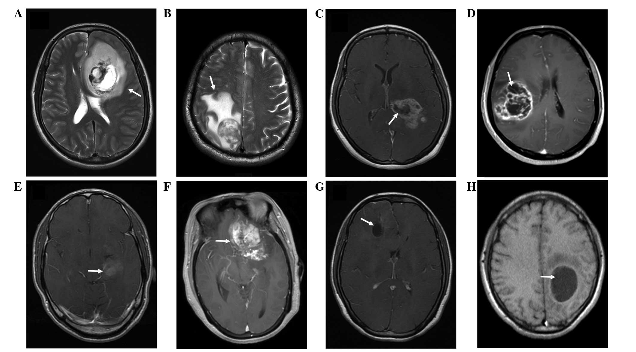
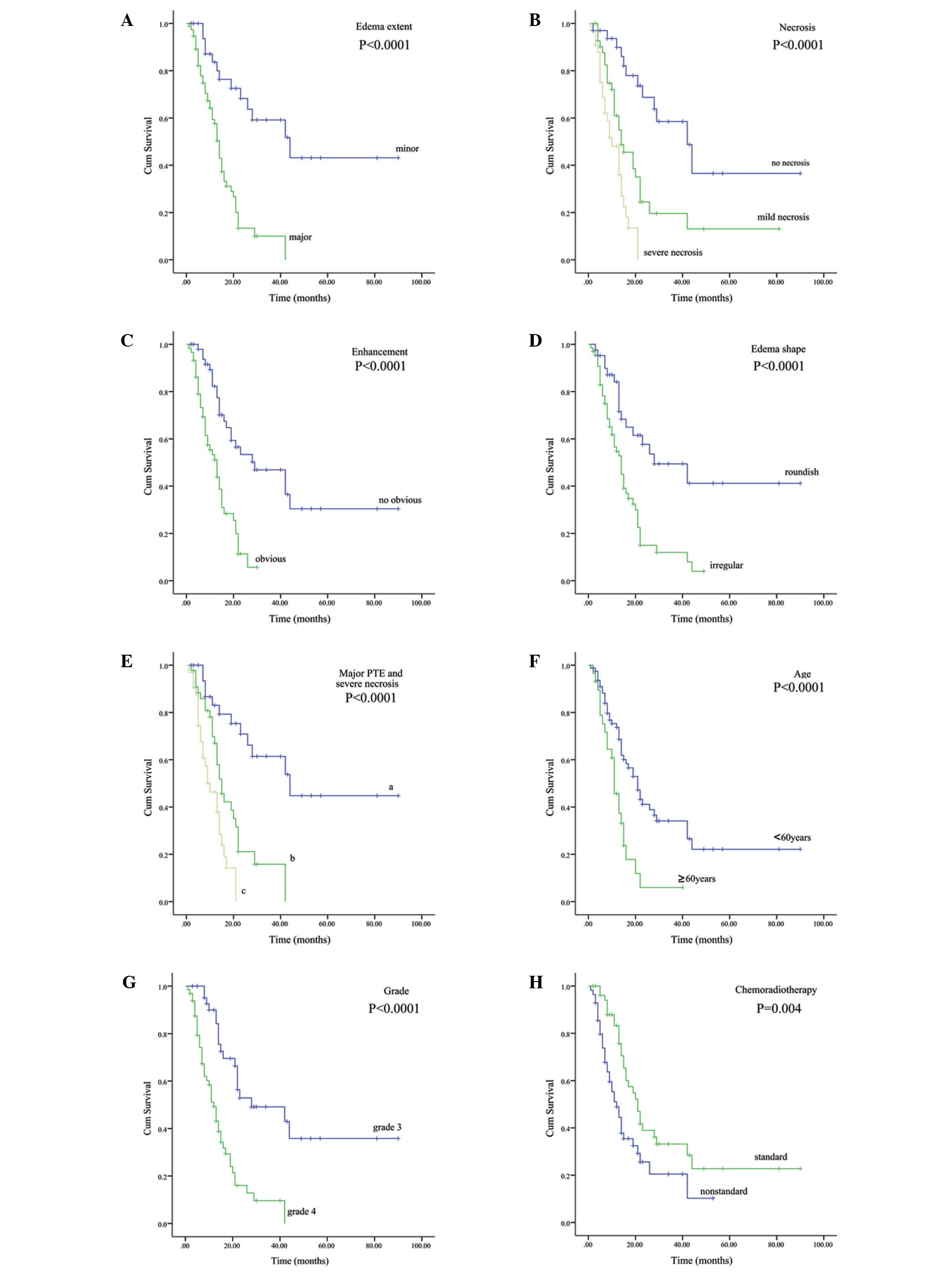
|
1
|
Zhang X, Zhang W, Cao WD, Cheng G and
Zhang YQ: Glioblastoma multiforme: Molecular characterization and
current treatment strategy (Review). Exp Ther Med. 3:9–14.
2012.PubMed/NCBI
|
|
2
|
Yang LJ, Zhou CF and Lin ZX: Temozolomide
and radiotherapy for newly diagnosed glioblastoma multiforme: A
systematic review. Cancer Invest. 32:31–36. 2014. View Article : Google Scholar : PubMed/NCBI
|
|
3
|
Buckner JC: Factors influencing survival
in high-grade gliomas. Semin Oncol. 30:10–14. 2003. View Article : Google Scholar : PubMed/NCBI
|
|
4
|
Yang LS, Huang FP, Zheng K, et al: Factors
affecting prognosis of patients with intracranial anaplastic
oligodendrogliomas: A single institutional review of 70 patients. J
Neurooncol. 100:113–120. 2010. View Article : Google Scholar : PubMed/NCBI
|
|
5
|
Schoenegger K, Oberndorfer S, Wuschitz B,
et al: Peritumoral edema on MRI at initial diagnosis: An
independent prognostic factor for glioblastoma? Eur J Neurol.
16:874–878. 2009. View Article : Google Scholar : PubMed/NCBI
|
|
6
|
Das P, Puri T, Jha P, Pathak P, Joshi N,
Suri V, Sharma MC, Sharma BS, Mahapatra AK, Suri A and Sarkar C: A
clinicopathological and molecular analysis of glioblastoma
multiforme with long-term survival. J Clin Neurosci. 18:66–70.
2011. View Article : Google Scholar : PubMed/NCBI
|
|
7
|
Arshad H, Ahmad Z and Hasan SH: Gliomas:
Correlation of histologic grade, Ki67 and p53 expression with
patient survival. Asian Pac J Cancer Prev. 11:1637–1640.
2010.PubMed/NCBI
|
|
8
|
Hammoud MA, Sawaya R, Shi W, Thall PF and
Leeds NE: Prognostic significance of preoperative MRI scans in
glioblastoma multiforme. J Neurooncol. 27:65–73. 1996. View Article : Google Scholar : PubMed/NCBI
|
|
9
|
Pope WB, Sayre J, Perlina A, Villablanca
JP, Mischel PS and Cloughesy TF: MR imaging correlates of survival
in patients with high-grade gliomas. AJNR Am J Neuroradiol.
26:2466–2474. 2005.PubMed/NCBI
|
|
10
|
Maldaun MV, Suki D, Lang FF, Prabhu S, Shi
W, Fuller GN, Wildrick DM and Sawaya R: Cystic glioblastoma
multiforme: Survival outcomes in 22 cases. J Neurosurg. 100:61–67.
2004. View Article : Google Scholar : PubMed/NCBI
|
|
11
|
Li WB, Tang K, Chen Q, Li S, Qiu G, Li SW
and Jiang T: MRI manifestions correlate with survival of
glioblastoma multiforme patients. Cancer Biol Med. 9:120–123.
2012.PubMed/NCBI
|
|
12
|
Kaur G, Bloch O, Jian BJ, Kaur R, Sughrue
ME, Aghi MK, McDermott MW, Berger MS, Chang SM and Parsa AT: A
critical evaluation of cystic features in primary glioblastoma as a
prognostic factor for survival. J Neurosurg. 115:754–759. 2011.
View Article : Google Scholar : PubMed/NCBI
|
|
13
|
Pierallini A, Bonamini M, Osti MF, Pantano
P, Palmeggiani F, Santoro A, Enrici R Maurizi and Bozzao L:
Supratentorial glioblastoma: Neuroradiological findings and
survival after surgery and radiotherapy. Neuroradiology. 38(Suppl
1): S26–S30. 1996. View Article : Google Scholar : PubMed/NCBI
|
|
14
|
Liu SY, Mei WZ and Lin ZX: Pre-operative
peritumoral edema and survival rate in glioblastoma multiforme.
Onkologie. 36:679–684. 2013.PubMed/NCBI
|
|
15
|
Lin ZX: Glioma-related edema: New insight
into molecular mechanisms and their clinical implications. Chin J
Cancer. 32:49–52. 2013. View Article : Google Scholar : PubMed/NCBI
|
|
16
|
Louis DN, Ohgaki H, Wiestler OD, Cavenee
WK, Burger PC, Jouvet A, Scheithauer BW and Kleihues P: The 2007
WHO classification of tumours of the central nervous system. Acta
Neuropathol. 114:97–109. 2007. View Article : Google Scholar : PubMed/NCBI
|
|
17
|
Lin GS, Yang LJ, Wang XF, Chen YP, Tang
WL, Chen L and Lin ZX: STAT3 Tyr705 phosphorylation affects
clinical outcome in patients with newly diagnosed supratentorial
glioblastoma. Med Oncol. 31:9242014. View Article : Google Scholar : PubMed/NCBI
|
|
18
|
Hartmann M, Jansen O, Egelhof T, Forsting
M, Albert FK and Sartor K: Effect of brain edema on the recurrence
pattern of malignant gliomas. Radiologe. 38:948–953. 1998.(In
German). View Article : Google Scholar : PubMed/NCBI
|
|
19
|
Seidel C, Dörner N, Osswald M, Wick A,
Platten M, Bendszus M and Wick W: Does age matter? A MRI study on
peritumoral edema in newly diagnosed primary glioblastoma. BMC
Cancer. 11:1272011. View Article : Google Scholar : PubMed/NCBI
|
|
20
|
Yamahara T, Numa Y, Oishi T, Kawaguchi T,
Seno T, Asai A and Kawamoto K: Morphological and flow cytometric
analysis of cell infiltration in glioblastoma: A comparison of
autopsy brain and neuroimaging. Brain Tumor Pathol. 27:81–87. 2010.
View Article : Google Scholar : PubMed/NCBI
|
|
21
|
Ruiz-Ontañon P, Orgaz JL, Aldaz B,
Elosegui-Artola A, Martino J, Berciano MT, Montero JA, Grande L,
Nogueira L, Diaz-Moralli S, et al: Cellular plasticity confers
migratory and invasive advantages to a population of
glioblastoma-initiating cells that infiltrate peritumoral tissue.
Stem Cells. 31:1075–1085. 2013. View Article : Google Scholar : PubMed/NCBI
|
|
22
|
Zhang X, Zhang W, Mao XG, Zhen HN, Cao WD
and Hu SJ: Targeting role of glioma stem cells for glioblastoma
multiforme. Curr Med Chem. 20:1974–1984. 2013. View Article : Google Scholar : PubMed/NCBI
|
|
23
|
Chen J, Li Y, Yu TS, McKay RM, Burns DK,
Kernie SG and Parada LF: A restricted cell population propagates
glioblastoma growth after chemotherapy. Nature. 488:522–526. 2012.
View Article : Google Scholar : PubMed/NCBI
|
|
24
|
Mangiola A, de Bonis P, Maira G, Balducci
M, Sica G, Lama G, Lauriola L and Anile C: Invasive tumor cells and
prognosis in a selected population of patients with glioblastoma
multiforme. Cancer. 113:841–846. 2008. View Article : Google Scholar : PubMed/NCBI
|
|
25
|
Lacroix M, Abi-Said D, Fourney DR,
Gokaslan ZL, Shi W, DeMonte F, Lang FF, McCutcheon IE, Hassenbusch
SJ, Holland E, et al: A multivariate analysis of 416 patients with
glioblastoma multiforme: Prognosis, extent of resection and
survival. J Neurosurg. 95:190–198. 2001. View Article : Google Scholar : PubMed/NCBI
|
|
26
|
Pierallini A, Bonamini M, Pantano P, et
al: Radiological assessment of necrosis in glioblastoma:
Variability and prognostic value. Neuroradiology. 40:150–153. 1998.
View Article : Google Scholar : PubMed/NCBI
|
|
27
|
Rong Y, Durden DL, Van Meir EG and Brat
DJ: 'Pseudopalisading' necrosis in glioblastoma: A familiar
morphologic feature that links vascular pathology, hypoxia and
angiogenesis. J Neuropathol Exp Neurol. 65:529–539. 2006.
View Article : Google Scholar : PubMed/NCBI
|
|
28
|
Brat DJ, Castellano-Sanchez AA, Hunter SB,
Pecot M, Cohen C, Hammond EH, Devi SN, Kaur B and Van Meir EG:
Pseudopalisades in glioblastoma are hypoxic, express extracellular
matrix proteases and are formed by an actively migrating cell
population. Cancer Res. 64:920–927. 2004. View Article : Google Scholar : PubMed/NCBI
|
|
29
|
Huang XD, Wang ZF, Dai LM and Li ZQ:
Microarray analysis of the hypoxia-induced gene expression profile
in malignant C6 glioma cells. Asian Pac J Cancer Prev.
13:4793–4799. 2012. View Article : Google Scholar : PubMed/NCBI
|
|
30
|
Oliver L, Olivier C, Marhuenda FB, et al:
Hypoxia and the malignant glioma microenvironment: Regulation and
implications for therapy. Curr Mol Pharmacol. 2:263–284. 2009.
View Article : Google Scholar : PubMed/NCBI
|
|
31
|
Brandsma D and van den Bent MJ:
Pseudoprogression and pseudoresponse in the treatment of gliomas.
Curr Opin Neurol. 22:633–638. 2009. View Article : Google Scholar : PubMed/NCBI
|
|
32
|
Jyothirmayi R, Madhavan J, Nair MK and
Rajan B: Conservative surgery and radiotherapy in the treatment of
spinal cord astrocytoma. J Neurooncol. 33:205–211. 1997. View Article : Google Scholar : PubMed/NCBI
|
|
33
|
Shibamoto Y, Kitakabu Y, Takahashi M,
Yamashita J, Oda Y, Kikuchi H and Abe M: Supratentorial low-grade
astrocytoma. Correlation of computed tomography findings with
effect of radiation therapy and prognostic variables. Cancer.
72:190–195. 1993. View Article : Google Scholar : PubMed/NCBI
|
|
34
|
Adn M, Saikali S, Guegan Y and Hamlat A:
Pathophysiology of glioma cyst formation. Med Hypotheses.
66:801–804. 2006. View Article : Google Scholar : PubMed/NCBI
|
|
35
|
Utsuki S, Oka H, Suzuki S, Shimizu S,
Tanizaki Y, Kondo K, Tanaka S, Kawano N and Fujii K: Pathological
and clinical features of cystic and noncystic glioblastomas. Brain
Tumor Pathol. 23:29–34. 2006. View Article : Google Scholar : PubMed/NCBI
|
|
36
|
Stupp R, Mason WP, van den Bent MJ, Weller
M, Fisher B, Taphoorn MJ, Belanger K, Brandes AA, Marosi C, Bogdahn
U, et al: Radiotherapy plus concomitant and adjuvant temozolomide
for glioblastoma. N Engl J Med. 352:987–996. 2005. View Article : Google Scholar : PubMed/NCBI
|