Spandidos Publications Logo
  • About
    • About Spandidos
    • Aims and Scopes
    • Abstracting and Indexing
    • Editorial Policies
    • Reprints and Permissions
    • Job Opportunities
    • Terms and Conditions
    • Contact
  • Journals
    • All Journals
    • Oncology Letters
      • Oncology Letters
      • Information for Authors
      • Editorial Policies
      • Editorial Board
      • Aims and Scope
      • Abstracting and Indexing
      • Bibliographic Information
      • Archive
    • International Journal of Oncology
      • International Journal of Oncology
      • Information for Authors
      • Editorial Policies
      • Editorial Board
      • Aims and Scope
      • Abstracting and Indexing
      • Bibliographic Information
      • Archive
    • Molecular and Clinical Oncology
      • Molecular and Clinical Oncology
      • Information for Authors
      • Editorial Policies
      • Editorial Board
      • Aims and Scope
      • Abstracting and Indexing
      • Bibliographic Information
      • Archive
    • Experimental and Therapeutic Medicine
      • Experimental and Therapeutic Medicine
      • Information for Authors
      • Editorial Policies
      • Editorial Board
      • Aims and Scope
      • Abstracting and Indexing
      • Bibliographic Information
      • Archive
    • International Journal of Molecular Medicine
      • International Journal of Molecular Medicine
      • Information for Authors
      • Editorial Policies
      • Editorial Board
      • Aims and Scope
      • Abstracting and Indexing
      • Bibliographic Information
      • Archive
    • Biomedical Reports
      • Biomedical Reports
      • Information for Authors
      • Editorial Policies
      • Editorial Board
      • Aims and Scope
      • Abstracting and Indexing
      • Bibliographic Information
      • Archive
    • Oncology Reports
      • Oncology Reports
      • Information for Authors
      • Editorial Policies
      • Editorial Board
      • Aims and Scope
      • Abstracting and Indexing
      • Bibliographic Information
      • Archive
    • Molecular Medicine Reports
      • Molecular Medicine Reports
      • Information for Authors
      • Editorial Policies
      • Editorial Board
      • Aims and Scope
      • Abstracting and Indexing
      • Bibliographic Information
      • Archive
    • World Academy of Sciences Journal
      • World Academy of Sciences Journal
      • Information for Authors
      • Editorial Policies
      • Editorial Board
      • Aims and Scope
      • Abstracting and Indexing
      • Bibliographic Information
      • Archive
    • International Journal of Functional Nutrition
      • International Journal of Functional Nutrition
      • Information for Authors
      • Editorial Policies
      • Editorial Board
      • Aims and Scope
      • Abstracting and Indexing
      • Bibliographic Information
      • Archive
    • International Journal of Epigenetics
      • International Journal of Epigenetics
      • Information for Authors
      • Editorial Policies
      • Editorial Board
      • Aims and Scope
      • Abstracting and Indexing
      • Bibliographic Information
      • Archive
    • Medicine International
      • Medicine International
      • Information for Authors
      • Editorial Policies
      • Editorial Board
      • Aims and Scope
      • Abstracting and Indexing
      • Bibliographic Information
      • Archive
  • Articles
  • Information
    • Information for Authors
    • Information for Reviewers
    • Information for Librarians
    • Information for Advertisers
    • Conferences
  • Language Editing
Spandidos Publications Logo
  • About
    • About Spandidos
    • Aims and Scopes
    • Abstracting and Indexing
    • Editorial Policies
    • Reprints and Permissions
    • Job Opportunities
    • Terms and Conditions
    • Contact
  • Journals
    • All Journals
    • Biomedical Reports
      • Information for Authors
      • Editorial Policies
      • Editorial Board
      • Aims and Scope
      • Abstracting and Indexing
      • Bibliographic Information
      • Archive
    • Experimental and Therapeutic Medicine
      • Information for Authors
      • Editorial Policies
      • Editorial Board
      • Aims and Scope
      • Abstracting and Indexing
      • Bibliographic Information
      • Archive
    • International Journal of Epigenetics
      • Information for Authors
      • Editorial Policies
      • Editorial Board
      • Aims and Scope
      • Abstracting and Indexing
      • Bibliographic Information
      • Archive
    • International Journal of Functional Nutrition
      • Information for Authors
      • Editorial Policies
      • Editorial Board
      • Aims and Scope
      • Abstracting and Indexing
      • Bibliographic Information
      • Archive
    • International Journal of Molecular Medicine
      • Information for Authors
      • Editorial Policies
      • Editorial Board
      • Aims and Scope
      • Abstracting and Indexing
      • Bibliographic Information
      • Archive
    • International Journal of Oncology
      • Information for Authors
      • Editorial Policies
      • Editorial Board
      • Aims and Scope
      • Abstracting and Indexing
      • Bibliographic Information
      • Archive
    • Medicine International
      • Information for Authors
      • Editorial Policies
      • Editorial Board
      • Aims and Scope
      • Abstracting and Indexing
      • Bibliographic Information
      • Archive
    • Molecular and Clinical Oncology
      • Information for Authors
      • Editorial Policies
      • Editorial Board
      • Aims and Scope
      • Abstracting and Indexing
      • Bibliographic Information
      • Archive
    • Molecular Medicine Reports
      • Information for Authors
      • Editorial Policies
      • Editorial Board
      • Aims and Scope
      • Abstracting and Indexing
      • Bibliographic Information
      • Archive
    • Oncology Letters
      • Information for Authors
      • Editorial Policies
      • Editorial Board
      • Aims and Scope
      • Abstracting and Indexing
      • Bibliographic Information
      • Archive
    • Oncology Reports
      • Information for Authors
      • Editorial Policies
      • Editorial Board
      • Aims and Scope
      • Abstracting and Indexing
      • Bibliographic Information
      • Archive
    • World Academy of Sciences Journal
      • Information for Authors
      • Editorial Policies
      • Editorial Board
      • Aims and Scope
      • Abstracting and Indexing
      • Bibliographic Information
      • Archive
  • Articles
  • Information
    • For Authors
    • For Reviewers
    • For Librarians
    • For Advertisers
    • Conferences
  • Language Editing
Login Register Submit
  • This site uses cookies
  • You can change your cookie settings at any time by following the instructions in our Cookie Policy. To find out more, you may read our Privacy Policy.

    I agree
Search articles by DOI, keyword, author or affiliation
Search
Advanced Search
presentation
Oncology Letters
Join Editorial Board Propose a Special Issue
Print ISSN: 1792-1074 Online ISSN: 1792-1082
Journal Cover
December-2015 Volume 10 Issue 6

Full Size Image

Sign up for eToc alerts
Recommend to Library

Journals

International Journal of Molecular Medicine

International Journal of Molecular Medicine

International Journal of Molecular Medicine is an international journal devoted to molecular mechanisms of human disease.

International Journal of Oncology

International Journal of Oncology

International Journal of Oncology is an international journal devoted to oncology research and cancer treatment.

Molecular Medicine Reports

Molecular Medicine Reports

Covers molecular medicine topics such as pharmacology, pathology, genetics, neuroscience, infectious diseases, molecular cardiology, and molecular surgery.

Oncology Reports

Oncology Reports

Oncology Reports is an international journal devoted to fundamental and applied research in Oncology.

Experimental and Therapeutic Medicine

Experimental and Therapeutic Medicine

Experimental and Therapeutic Medicine is an international journal devoted to laboratory and clinical medicine.

Oncology Letters

Oncology Letters

Oncology Letters is an international journal devoted to Experimental and Clinical Oncology.

Biomedical Reports

Biomedical Reports

Explores a wide range of biological and medical fields, including pharmacology, genetics, microbiology, neuroscience, and molecular cardiology.

Molecular and Clinical Oncology

Molecular and Clinical Oncology

International journal addressing all aspects of oncology research, from tumorigenesis and oncogenes to chemotherapy and metastasis.

World Academy of Sciences Journal

World Academy of Sciences Journal

Multidisciplinary open-access journal spanning biochemistry, genetics, neuroscience, environmental health, and synthetic biology.

International Journal of Functional Nutrition

International Journal of Functional Nutrition

Open-access journal combining biochemistry, pharmacology, immunology, and genetics to advance health through functional nutrition.

International Journal of Epigenetics

International Journal of Epigenetics

Publishes open-access research on using epigenetics to advance understanding and treatment of human disease.

Medicine International

Medicine International

An International Open Access Journal Devoted to General Medicine.

Journal Cover
December-2015 Volume 10 Issue 6

Full Size Image

Sign up for eToc alerts
Recommend to Library

  • Article
  • Citations
    • Cite This Article
    • Download Citation
    • Create Citation Alert
    • Remove Citation Alert
    • Cited By
  • Similar Articles
    • Related Articles (in Spandidos Publications)
    • Similar Articles (Google Scholar)
    • Similar Articles (PubMed)
  • Download PDF
  • Download XML
  • View XML
Case Report

Medical termination of a partial hydatidiform mole and coexisting fetus during the second trimester: A case report

  • Authors:
    • Yinfeng Wang
    • Honglang Qian
    • Jinhua Wang
  • View Affiliations / Copyright

    Affiliations: Department of Gynecology, Women's Hospital, Zhejiang University School of Medicine, Hangzhou, Zhejiang 310006, P.R. China, Department of Radiology, Women's Hospital, Zhejiang University School of Medicine, Hangzhou, Zhejiang 310006, P.R. China
  • Pages: 3625-3628
    |
    Published online on: September 24, 2015
       https://doi.org/10.3892/ol.2015.3743
  • Expand metrics +
Metrics: Total Views: 0 (Spandidos Publications: | PMC Statistics: )
Metrics: Total PDF Downloads: 0 (Spandidos Publications: | PMC Statistics: )
Cited By (CrossRef): 0 citations Loading Articles...

This article is mentioned in:


Abstract

Partial hydatidiform mole and coexisting fetus (PHMCF) is a rare condition that presents a dilemma for physicians and the parents of the fetus, particularly when PHMCF is detected during the second trimester of pregnancy. The present study reports a case of PHMCF terminated by induction of labor via administration of Rivanol at 17 weeks. Follow‑up measurements of serum β‑human chorionic gonadotropin (β‑HCG) levels, as well as imaging studies, indicated the presence of persistent trophoblastic disease (PTD) and lung metastases. The patient was therefore admitted for three courses of chemotherapy. Subsequently, the metastases receded and β‑HCG levels decreased to within the normal range. The patient demonstrated no disease recurrence for 1 year. Following a review of the relevant literature, to the best of our knowledge, all PHMCF cases terminated by medical induction of labor during the second trimester resulted in the development of PTD and lung metastases. However, three cases of PHMCF that were terminated by caesarean section during the third trimester did not develop PTD or metastases. The present study therefore hypothesized that medical termination may not be a safe therapeutic strategy for the treatment of PHMCF during the second trimester, and that pregnancy should be allowed to continue empirically.

Introduction

Hydatidiform mole with a coexistent fetus (HMCF) is a rare condition, with an incidence of 1/22,000-1,000,000 pregnancies (1). There have been a number of cases of HMCF previously reported (2–4). The majority are complete HMCF cases, which possess a significant risk (19.5–62.5%) of inducing persistent trophoblastic disease (PTD) (5). By contrast, persistent or metastatic trophoblastic disease following a partial mole is rare, with an estimated risk of ~1–5.6% (6). However, to the best of our knowledge, there have been no previous reports investigating the risk of persistent or metastatic trophoblastic disease following partial HMCF (PHMCF), due to the rarity of this condition. The management of such pregnancies creates a dilemma for the physician and parents, particularly when PHMCF occurs during the second trimester of pregnancy. Medical termination is effective during the second trimester; however, this technique increases the risk of the occurrence of PTD when used to terminate molar pregnancies (7). The aim of this study was to investigate the safety of medical termination for patients with PHMCF. The present study reports a case of PHMCF in which the pregnancy was terminated and delivery was induced via intra-amniotic administration of Rivanol, followed by aspiration curettage, which resulted in the occurrence of PTD and pulmonary metastases. Written informed consent was obtained from the patient.

Case report

In November 2013, a 22-year-old female (gravida 2, para 0) was referred to the Department of Gynecology at the Women's Hospital of Zhejiang University School of Medicine (Zhejiang, China) at 16 weeks of gestation, with suspected molar pregnancy. Sonography and magnetic resonance imaging (MRI) revealed a fetus and placenta, which appeared normal, in the posterior portion of the uterus. However, an additional cystic placenta with a degenerated twin was identified in the right anterior portion of the uterus (Fig. 1). In addition, the patient's serum β-human chorionic gonadotropin (β-HCG) levels were markedly increased (449,078 IU/l). The couple decided to terminate the pregnancy following consideration of the risks. Delivery was induced via intra-amniotic administration of 80 mg Rivanol (Guangxi Hefeng Pharmaceutical Co., Ltd., Hechi, China). Thirty-six hours 
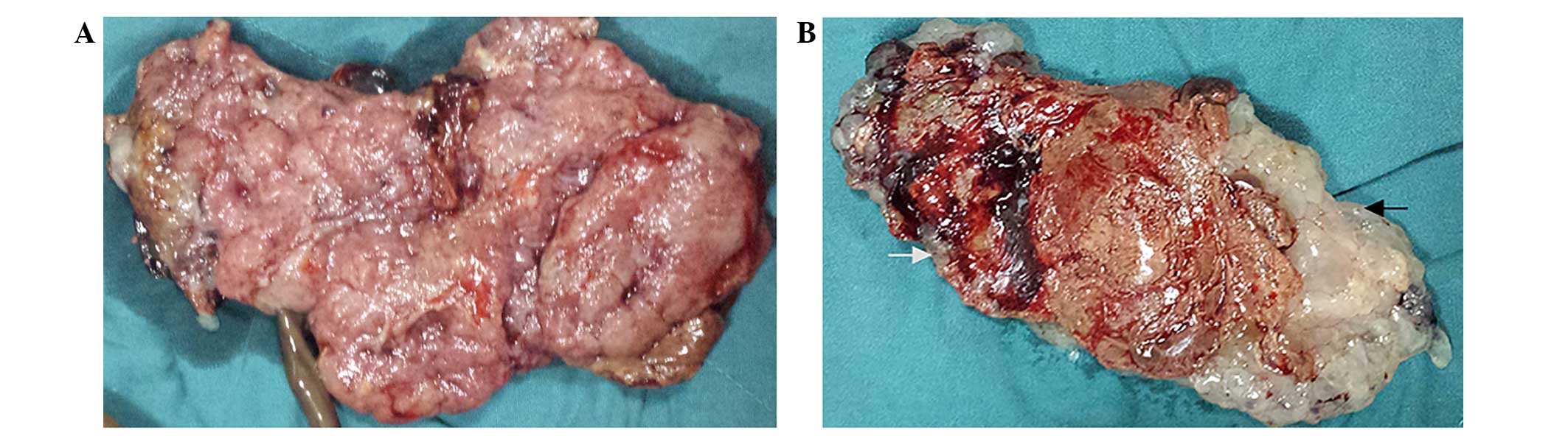
later, a fetus with a normal placenta, as well as a partially cystic placenta were delivered (Fig. 2). Suction curettage was performed due to incomplete evacuation. There were no fetal gross abnormalities and no genetic abnormalities were identified by DNA Array (Affymetrix, Inc., Santa 
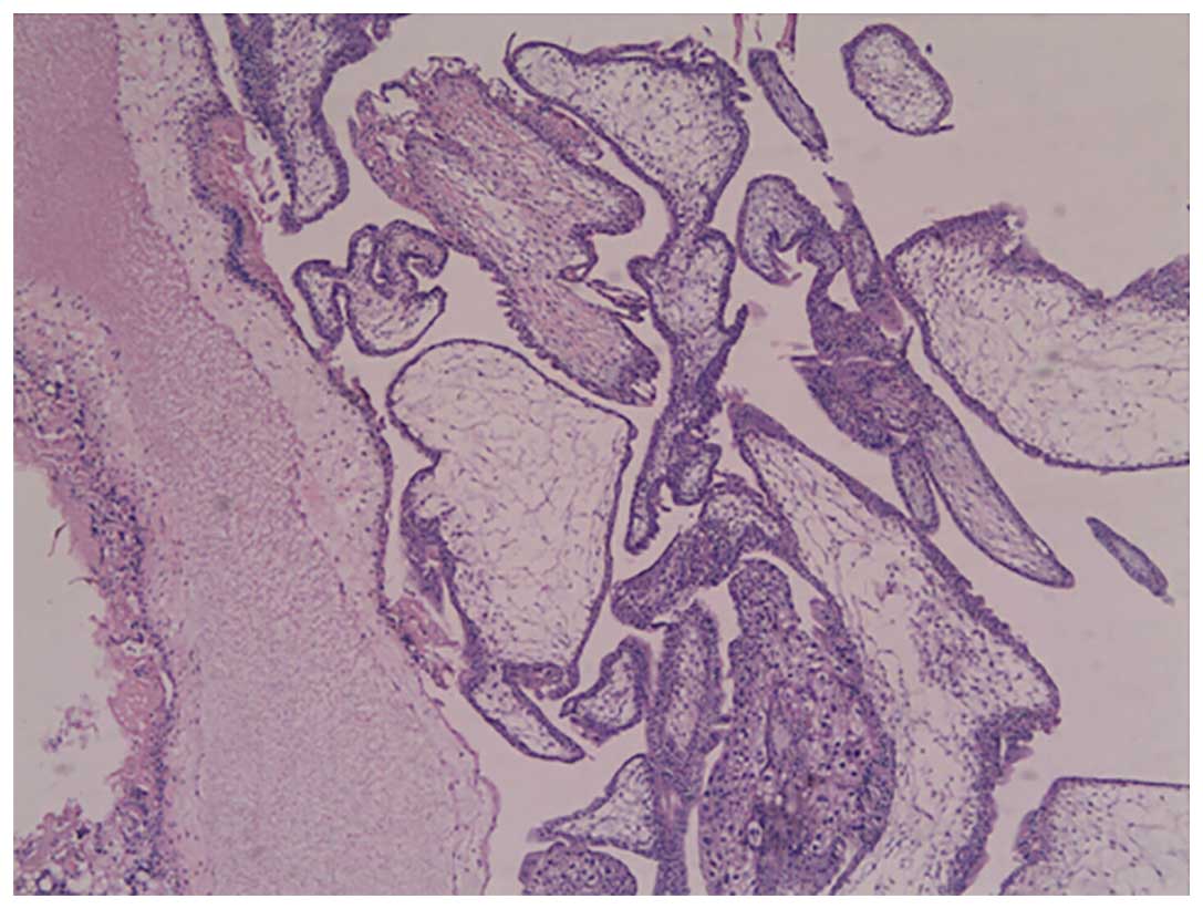
Clara, CA, USA). Histopathological analysis of the molar tissue revealed chorionic villi of varying size and shape, with focal edema and functioning villous circulation, as well as focal trophoblastic hyperplasia (Fig. 3). DNA Array (Affymetrix, Inc.) of the molar tissue revealed a triploid cell line, and thus confirmed the diagnosis of PHMCF.

Figure 1.

T1-weighted single shot fast spin echo magnetic resonance imaging scans revealing the cystic placenta with a degenerated twin in the right anterior portion of the uterus (white arrow), a normal fetus (black arrow) and placenta in the posterior of the uterus (white arrowhead). (A) Transverse scan and (B) coronal scan.

Figure 2.

Images of the gross specimen. (A) The normal placenta (specimen size, 12.0×8.0×2.0 cm), and (B) the abnormal placenta (specimen size, 11.5×6.0×1.8 cm) containing molar (black arrow) and normal (white arrow) sections.

Figure 3.

Histopathology of the hydatidiform mole (hematoxylin and eosin staining; magnification, ×40) revealed chorionic villi of varying sizes and shapes, with focal edema and functioning villous circulation, as well as focal trophoblastic hyperplasia.

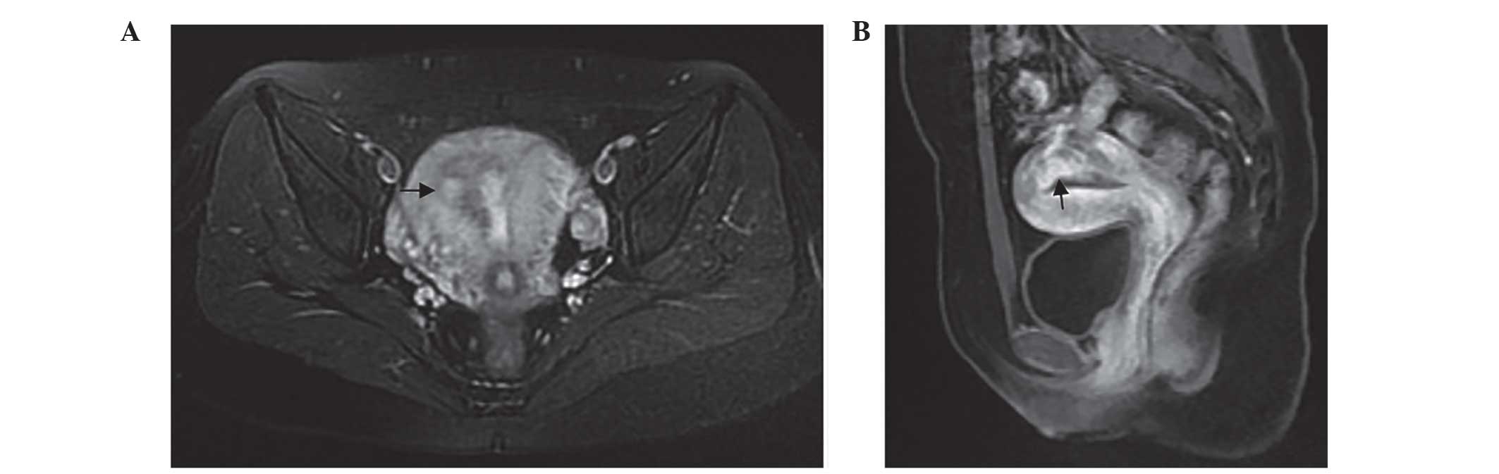
Serum β-HCG levels were monitored for three weeks following the termination of pregnancy (TOP), and initially reduced to 3,845 IU/l; however, 1 week later, β-HCG levels had increased to 3,947 IU/l. An MRI study revealed abnormal signs in the right posterior portion of the myometrium and uterine cavity, which indicated the presence of an invasive mole (Fig. 4). Serum β-HCG levels were observed to 
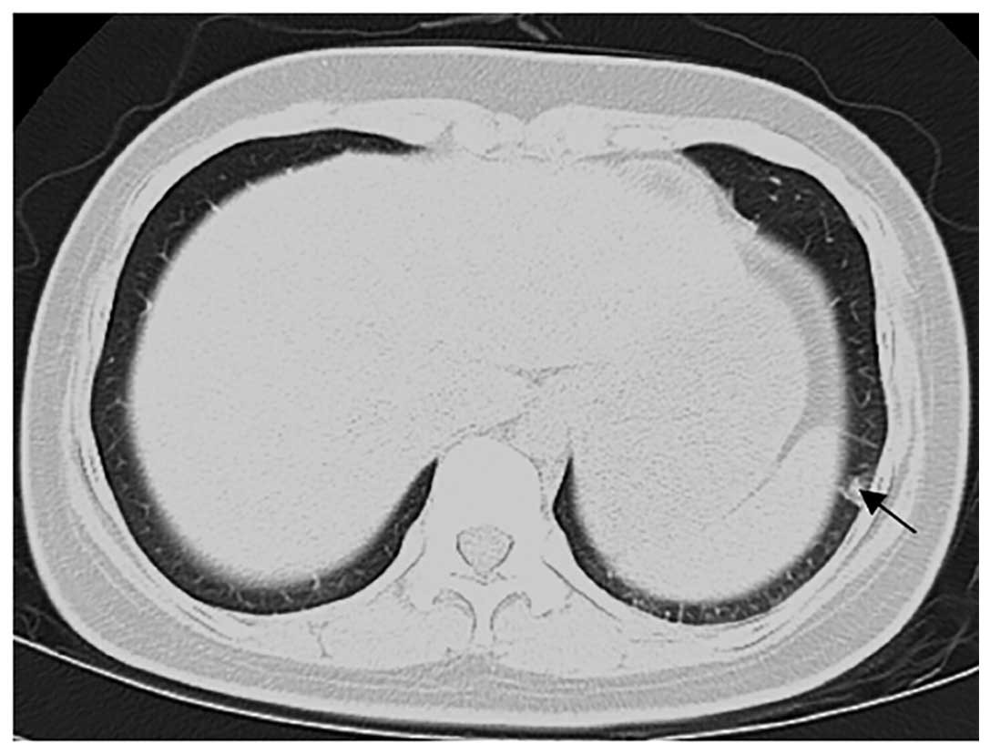
have plateaued (~1,706–2,603 IU/l) at the next two-week follow-up appointment. A follow-up chest computed tomography (CT) scan revealed a number of small, ill-defined nodules of varying sizes in the right and left lower lungs, indicative of lung metastases (Fig. 5). An MRI of the head and a CT scan of the abdomen were normal. The patient was admitted for chemotherapy, and following three courses (3-week cycles) of methotrexate (0.4 mg/kg, days 1–5), the metastases receded and β-HCG levels reduced to within the normal range. The patient demonstrated no disease recurrence during 1 year of follow-up.

Figure 4.

MRI scanning revealed abnormal signs in the right posterior portion of the myometrium and uterine cavity, indicating an invasive mole (A) Transverse T2-weighted fat-suppressed fast recovery fast spin echo MRI, and (B) sagittal LAVA-flex image with contrast enhancement. Arrows indicate the trophoblastic tumor. MRI, magnetic resonance imaging.

Figure 5.

Computed tomography chest scan revealing the lung metastasis of the trophoblast. Arrow indicates the metastatic tumor.

Discussion

Multiple pregnancy involving a partial hydatidiform mole and normal fetus is rare. The management of such pregnancies presents a dilemma for physicians and parents between expectant management and immediate intervention; particularly in pregnancies involving assisted reproductive technology (8–10). A small number of cases of PHMCF where pregnancy was allowed to continue until 28 weeks of gestation or later have previously been reported, and delivery was achieved via Caesarean section, resulting in a number of healthy babies (11,12). However, parents must also be aware of the risk of potential maternal complications associated with molar pregnancy, including early-onset preeclampsia, hyperemesis gravidarum, PTD and metastases (13).

To the best of our knowledge, following an extensive search of the relevant medical literature since the year 2000, 10 PHMCF cases have previously been reported (Table I) (8–12,14–18). Two of these cases of PHMCF were treated by termination of pregnancy during the first trimester, via dilatation and suction curettage, resulting in one case of PTD and no metastases (8,14). In 3/5 cases (60%) that were terminated during the second trimester, two by medically induced labor and the other via unspecified methodology, the patient subsequently developed PTD and lung metastases (9,10,15). Finally, three cases that were terminated by caesarean section in the third trimester did not develop PTD or metastases, and resulted in the delivery of live infants (although one infant succumbed 30 days subsequent to delivery) (11,12,18). In the present case, the PHMCF pregnancy was terminated by the induction of labor via intra-amniotic administration of Rivanol at 17 weeks of gestation. PTD and pulmonary metastases were subsequently detected, which required further treatment with chemotherapy. Certain investigators have hypothesized that the risk of PTD in HMCF pregnancies is not associated with the length of gestation at termination (5). The literature cited in the present study also supports this conclusion, as none of the three cases where pregnancy was terminated later than 28 weeks of gestation developed PTD.

Table I.

Previously reported cases of partial hydatidiform mole and coexisting fetus in the literature.

Table I.

Previously reported cases of partial hydatidiform mole and coexisting fetus in the literature.

StudyGestational age at delivery or abortion, weeksMethod of terminationLive neonatePTDMetastasesRef
Ingec et al10D&ENoYesNo(11)
Tay11D&ENoNoNo(12)
Kim et al14MedicalNoYesYesa(13)
Zhou et al16MedicalNoYesYesa(14)
Shiina et al20UnknownNoYesYesa(15)
Sánchez-Ferrer et al21Spontaneous abortionNoNoNo(16)
Chu et al24Caesarean sectionYesNoNo(17)
Copeland and Stanek28Caesarean sectionYesNoNo(8)
Navarro Amezcua et al29Caesarean sectionNoNoNo(18)
Sun et al35Caesarean sectionYesNoNo(9)
Present case17MedicalNoYesYes

a metastases located in the lung. PTD, persistent trophoblastic disease; D&E, dilatation and evacuation.

Although advances in transvaginal ultrasound and MRI may assist with the diagnosis of HMCF in the first trimester (8,19), HMCF is typically diagnosed at ~15–20 weeks of gestation or later (20). The empirical management of pregnancy in the second trimester is more difficult (10). Suction curettage is the typical treatment for females exhibiting molar pregnancy, however normal fetal structures may preclude the use of this method (7). Medical induction of labor is an effective and safe way for the termination of normal pregnancies during the second trimester (21). However, medical termination of molar pregnancy is associated with an increased risk of a subsequent requirement for chemotherapeutic treatment, due to the development of increased intrauterine pressure in the first and second trimesters (7). Performance of a caesarean section is not recommended during the second trimester of pregnancy, due to the increased risk of maternal morbidity and the fact that the reduced gestational period means that the neonate is too small to survive (7).

Evidence-based practice guidelines of the Royal College of Obstetricians and Gynecologists recommend dilatation and evacuation (D&E) as a safe and effective termination option for pregnancies of >15 weeks gestation, when undertaken by specialist practitioners with sufficient experience and skills (22). A number of studies have demonstrated that D&E is a rapid and well-tolerated method of termination, demonstrating low complication rates when performed by skilled physicians (23,24). However, to the best of our knowledge, there have been no reports concerning the risk of PTD following PHMCF termination by D&E. In a study conducted by Niemann et al (5), 5/8 HMCF patients' pregnancies were terminated by curettage during the second trimester. One of these patients developed PTD, indicating a risk factor of 20%.

Mid-trimester TOP due to PHMCF is challenging (9,10). Due to the rarity of this condition, minimal relevant data has been amassed, meaning that it is difficult to formulate valid predictions for treatment and outcomes of patients presenting with PHMCF during the second trimester. The present study concluded that medical termination may not be a safe method for patients with PHMCF, due to the high risk of PTD and metastasis associated. D&E may be an effective alternative method for termination of PHMCF in the second trimester. Pregnancy may be allowed to continue empirically, provided that maternal morbidity does not appear to be associated with the gestational age at termination, and that the acquisition of healthy neonates is a feasible outcome.

Acknowledgements

The authors would like to thank Professor Jian-Ming Zhu of the University of North Carolina School of Medicine (Chapel Hill, NC, USA) for his contribution in preparing this manuscript and for his discussion.

References

1 

Jones WB and Lauersen NH: Hydatidiform mole with coexistent fetus. Am J Obstet Gynecol. 122:267–272. 1975.PubMed/NCBI

2 

Steller MA, Genest DR, Bernstein MR, et al: Natural history of twin pregnancy with complete hydatidiform mole and coexisting fetus. Obstet Gynecol. 83:35–42. 1994.PubMed/NCBI

3 

Sebire NJ, Foskett M, Paradinas FJ, et al: Outcome of twin pregnancies with complete hydatidiform mole and healthy co-twin. Lancet. 359:2165–2166. 2002. View Article : Google Scholar : PubMed/NCBI

4 

Peng M, Li L, Zheng J, Ding Y, Yu L and Huang J: Termination of twin pregnancies with hydatidiform moles: A case series of four patients. Iran J Public Health. 43:1000–1006. 2014.PubMed/NCBI

5 

Niemann I, Sunde L and Petersen LK: Evaluation of the risk of persistent trophoblastic disease after twin pregnancy with diploid hydatidiform mole and coexisting normal fetus. Am J Obstet Gynecol. 197:45.e1–45.e5. 2007. View Article : Google Scholar

6 

Worley MJ Jr, Joseph NT, Berkowitz RS and Goldstein DP: Women with a partial mole during their first pregnancy and diagnosed earlier in gestation are at increased risk of developing gestational trophoblastic neoplasia. Int J Gynecol Cancer. 24:941–945. 2014. View Article : Google Scholar : PubMed/NCBI

7 

Tidy JA, Gillespie AM, Bright N, Radstone CR, Coleman RE and Hancock BW: Gestational trophoblastic disease: A study of mode of evacuation and subsequent need for treatment with chemotherapy. Gynecol Oncol. 78:309–312. 2000. View Article : Google Scholar : PubMed/NCBI

8 

Tay ET: Partial hydatidiform mole and coexisting viable twin pregnancy. Pediatr Emerg Care. 29:1298–1300. 2013.PubMed/NCBI

9 

Kim CH, Kim YH, Kim JW, Kim KM, Cho MK, Kim SM, Nam JH and Song TB: Triplet pregnancy with partial hydatidiform mole coexisting with two fetuses: A case report. J Obstet Gynaecol Res. 34:641–644. 2008. View Article : Google Scholar : PubMed/NCBI

10 

Zhou X, Chen Y, Li Y and Duan Z: Partial hydatidiform mole progression into invasive mole with lung metastasis following in vitro fertilization. Oncol Lett. 3:659–661. 2012.PubMed/NCBI

11 

Copeland JW and Stanek J: Dizygotic twin pregnancy with a normal fetus and a nodular embryo associated with a partial hydatidiform mole. Pediatr Dev Pathol. 13:476–480. 2010. View Article : Google Scholar : PubMed/NCBI

12 

Sun CJ, Zhao YP, Yu S, Fan L, Wu QQ, Li GH and Zhang WY: Twin pregnancy and partial hydatidiform mole following in vitro fertilization and embryos transfer: A novel case of placental mosaicism. Chin Med J (Engl). 125:4517–4519. 2012.PubMed/NCBI

13 

Vimercati A, de Gennaro AC, Cobuzzi I, Grasso S, Abruzzese M, Fascilla FD, Cormio G and Selvaggi L: Two cases of complete hydatidiform mole and coexistent live fetus. J Prenat Med. 7:1–4. 2013.PubMed/NCBI

14 

Ingec M, Borekci B, Altas S and Kadanali S: Twin pregnancy with partial hydatidiform mole and coexistent normal fetus. J Obstet Gynaecol. 26:379–380. 2006. View Article : Google Scholar : PubMed/NCBI

15 

Shiina H, Oka K, Okane M, et al: Coexisting true hermaphroditism and partial hydatidiform mole developing metastatic gestational trophoblastic tumors. A case report. Virchows Arch. 441:514–518. 2002. View Article : Google Scholar : PubMed/NCBI

16 

Sánchez-Ferrer ML, Ferri B, Almansa MT, et al: Partial mole with a diploid fetus: Case study and literature review. Fetal Diagn Ther. 25:354–358. 2009. View Article : Google Scholar : PubMed/NCBI

17 

Chu W, Chapman J, Persons DL and Fan F: Twin pregnancy with partial hydatidiform mole and coexistent fetus. Arch Pathol Lab Med. 128:1305–1306. 2004.PubMed/NCBI

18 

Navarro Amezcua ME, Castellanos Reyes J, Cardona González O and Torres Gómez LG: Twin pregnancy with partial hydatidiform mole and alive fetus: Case report. Ginecol Obstet Mex. 76:275–279. 2008.(In Spanish). PubMed/NCBI

19 

Herek D and Karabulut N: The role of magnetic resonance imaging in the diagnosis of complete hydatidiform mole in a twin pregnancy. Int J Gynaecol Obstet. 123:772013. View Article : Google Scholar : PubMed/NCBI

20 

Matsui H, Sekiya S, Hando T, Wake N and Tomoda Y: Hydatidiform mole coexistent with a twin live fetus: A national collaborative study in Japan. Hum Reprod. 15:608–611. 2000. View Article : Google Scholar : PubMed/NCBI

21 

Mentula MJ, Niinimäki M, Suhonen S, Hemminki E, Gissler M and Heikinheimo O: Immediate adverse events after second trimester medical termination of pregnancy: Results of a nationwide registry study. Hum Reprod. 26:927–932. 2011. View Article : Google Scholar : PubMed/NCBI

22 

Morrison J: Audit of the care of women requesting induced abortion. J Obstet Gynaecol. 23:521–524. 2003. View Article : Google Scholar : PubMed/NCBI

23 

Panoskaltsis TA: Mid-trimester termination of pregnancy by dilatation and evacuation. J Obstet Gynaecol. 21:280–284. 2001. View Article : Google Scholar : PubMed/NCBI

24 

Kent A: Second trimester termination of pregnancy. Rev Obstet Gynecol. 4:952011.PubMed/NCBI

Related Articles

  • Abstract
  • View
  • Download
  • Twitter
Copy and paste a formatted citation
Spandidos Publications style
Wang Y, Qian H and Wang J: Medical termination of a partial hydatidiform mole and coexisting fetus during the second trimester: A case report. Oncol Lett 10: 3625-3628, 2015.
APA
Wang, Y., Qian, H., & Wang, J. (2015). Medical termination of a partial hydatidiform mole and coexisting fetus during the second trimester: A case report. Oncology Letters, 10, 3625-3628. https://doi.org/10.3892/ol.2015.3743
MLA
Wang, Y., Qian, H., Wang, J."Medical termination of a partial hydatidiform mole and coexisting fetus during the second trimester: A case report". Oncology Letters 10.6 (2015): 3625-3628.
Chicago
Wang, Y., Qian, H., Wang, J."Medical termination of a partial hydatidiform mole and coexisting fetus during the second trimester: A case report". Oncology Letters 10, no. 6 (2015): 3625-3628. https://doi.org/10.3892/ol.2015.3743
Copy and paste a formatted citation
x
Spandidos Publications style
Wang Y, Qian H and Wang J: Medical termination of a partial hydatidiform mole and coexisting fetus during the second trimester: A case report. Oncol Lett 10: 3625-3628, 2015.
APA
Wang, Y., Qian, H., & Wang, J. (2015). Medical termination of a partial hydatidiform mole and coexisting fetus during the second trimester: A case report. Oncology Letters, 10, 3625-3628. https://doi.org/10.3892/ol.2015.3743
MLA
Wang, Y., Qian, H., Wang, J."Medical termination of a partial hydatidiform mole and coexisting fetus during the second trimester: A case report". Oncology Letters 10.6 (2015): 3625-3628.
Chicago
Wang, Y., Qian, H., Wang, J."Medical termination of a partial hydatidiform mole and coexisting fetus during the second trimester: A case report". Oncology Letters 10, no. 6 (2015): 3625-3628. https://doi.org/10.3892/ol.2015.3743
Follow us
  • Twitter
  • LinkedIn
  • Facebook
About
  • Spandidos Publications
  • Careers
  • Cookie Policy
  • Privacy Policy
How can we help?
  • Help
  • Live Chat
  • Contact
  • Email to our Support Team