Introduction
As the primary cause of cancer-associated mortality
globally, lung cancer is an important public health problem. In
men, lung cancer is ranked first among the commonly observed
malignant tumors, while in females, the disease is ranked second or
third in the majority of developed countries. The 5-year survival
rate is ~15% and the cure rate remains low (1). Therefore, the elucidation of the
tumorigenesis of lung carcinoma is of paramount importance for the
improvement of public health.
Thyroid transcription factor-1 (TTF-1), also known
as NKX2-1 or TITF-1, is mapped to chromosome 14q13 and the protein
a 38-kDa transcription factor that is normally expressed in the
thyroid gland, lungs and brain. This type of exhibits significant
functions in the development, cell growth and differentiation
processes. TTF-1 reportedly possesses oncogenic and suppressive
functions within the same tumor type and may play a dual function
in the progression of lung cancer (2). TTF-1 is also used in the differentiation
between primary and metastatic lung tumors (3,4).
Immunohistochemistry (IHC) has been used (5–7) to detect
TTF-1 expression in lung carcinomas. TTF-1 expression differs among
the subtypes of lung carcinoma, with strong expression in lung
adenocarcinoma and small cell lung carcinoma (SCLC), and weak or no
expression in squamous cell lung carcinoma and large cell lung
carcinoma. In our previous studies, mRNA in situ hybridization
(ISH) was used to detect TTF-1 mRNA expression in different
subtypes of lung cancer. The TTF-1 protein expression level
detected by IHC is consistent with that detected by mRNA ISH
(8). However, no comparison of the
statistical significance of the two methods has been performed.
In the present study, TTF-1 expression was
investigated using IHC and mRNA ISH, and the concordance rate of
the two methods was compared for detecting TTF-1 expression in
non-SCLC (NSCLC).
Materials and methods
Patients
A total of 196 patients with NSCLC who were treated
in Nanfang Hospital (Southern Medical University and Guangdong
Provincial Institute of Nephrology, Guangzhou, Guangdong, China)
between January 2000 and December 2010 were selected for this
study. The diagnosis of NSCLC mainly included adenocarcinoma and
squamous cell carcinoma based on the pathological classification of
the International Association for the Study of Lung Cancer/American
Thoracic Society/European Respiratory Society (9). The inclusion criteria for this study
were a diagnosis of NSCLC, no radiotherapy or chemotherapy, and the
availability of an adequate paraffin block for the analysis.
Specimens from these patients were obtained from the Departments of
Pathology and Thoracic Surgery of Nanfang Hospital. The specimens
included tumor tissues from 92 primary lung adenocarcinomas and 104
primary squamous cell lung carcinomas.
Written informed consent was obtained from all
patients according to the protocols of the Southern Medical
University Ethics Committee, and the study was approved by the
ethics committee of South Hospital of Southern Medical
University.
IHC and ISH in the tissue microarray
(TMA)
A TMA was constructed from the 196 paraffin-embedded
blocks according to a previously described procedure (10–12). Four
tissue cores selected from each sample were used to construct a
30×30 matrix microarray. In the final row of the microarray, 16
cores were used as sample location indicators. Sections (4-µm
thick) were cut from the TMA blocks and used for the IHC and mRNA
ISH labeling.
For IHC, TMA sections (4-µm thick) were stained with
mouse anti-human TTF-1 monoclonal antibody (clone 8G7G3/1; Dako,
Carpinteria, CA, USA) using the standard streptavidin-biotin
complex method according to the manufacturer's instructions.
For mRNA ISH, a TTF-1 mRNA–specific oligonucleotide
probe designed by Bioasia Co., Ltd., (Shanghai, China) was used to
detect TTF-1 mRNA expression in the TMA samples. The sequence of
the probe was 5′-GCCGACAGGTACTTCTGTTGCTTGAAGCGT-3′. The probe was
labeled with digoxin on the 3′ and 5′ ends. TMA sections (4-µm
thick) were deparaffinized, rehydrated, rinsed in
phosphate-buffered saline (PBS) and digested with pepsin at 37°C
for 20 min. The slides were hybridized with the riboprobe at 38°C
for 16 h. The hybridization samples were then washed sequentially
in 2X sodium chloride and sodium citrate (SSC) at 37°C for 5 min
and in 0.5X SSC at 37°C for 30 min. The slides were treated with 3%
hydrogen peroxide to inactivate the endogenous peroxidase. Pepsin
diluted with 3% citric acid was subsequently used to expose mRNA
fragments. The sections were hybridized with a probe diluted with
hybridization solution for 16 h at 38°C. The hybridization samples
were sequentially washed three times in 2X SSC for 5 min at 37°C,
twice in 0.5X SSC for 5 min at 37°C and once in 0.2X SSC for 15 min
at 37°C.
The hybridized probe was detected using an in
situ hybridization detection kit (Wuhan Boster Biological
Technology, Ltd., Wuhan, China) according to the manufacturer's
instructions (13). Briefly, the
slides were incubated with blocking reagent for 30 min at 37°C, and
then with biotin-labeled sheep antidigoxigenin for 1 h at 37°C. The
slides were sequentially washed with 0.5 M PBS at 37°C for 5 min,
and then incubated with streptavidin-biotin complex for 30 min and
washed four times in 0.5 M PBS for 5 min. Finally, the slides were
incubated with biotin peroxidase for 30 min at 37°C and washed four
times in 0.5 M PBS for 5 min. The peroxidase reaction was enhanced
using 3,3′-diaminobenzidine. The IHC and ISH slides were
counterstained with hematoxylin and cover-slipped.
Two cases of normal lung tissue were used as
internal positive controls for IHC and ISH. PBS or hybridization
solution was used to replace the primary antibody or the probe in
order to serve as negative controls.
Analysis of IHC and mRNA ISH
slides
The samples were analyzed under a 40X objective lens
using a BX51 light microscope (Olympus, Tokyo, Japan). A total of
20 fields were randomly selected from four randomly chosen
representative tissue cores of a specimen on the TMA. In total, 200
tumor cells were observed. Those with dark brown particles in the
nuclei or cytoplasm were considered to demonstrate positive protein
or mRNA expression. Two independent analysts assessed IHC and ISH
staining effects.
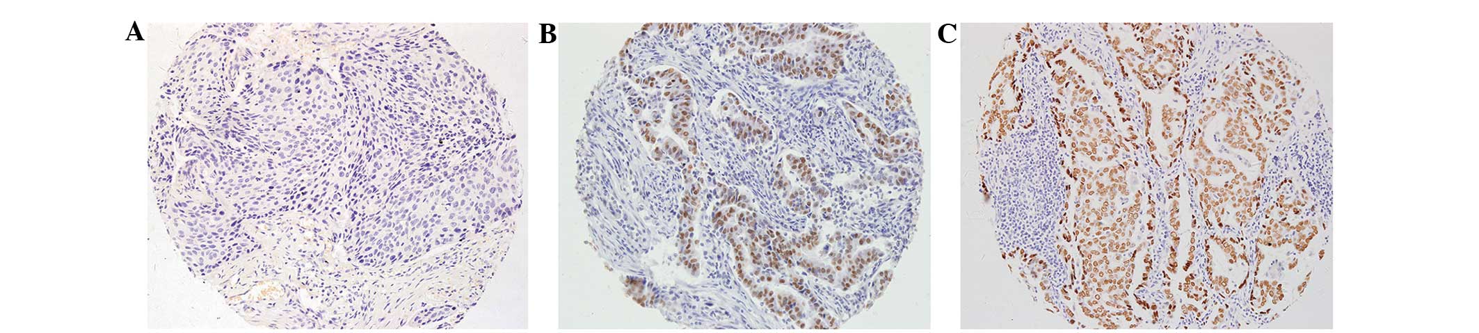
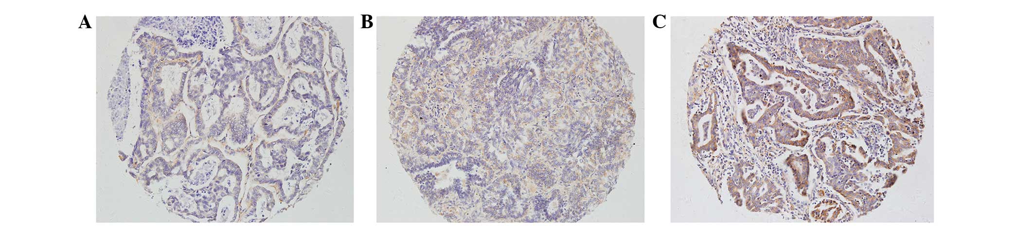
TTF-1 IHC and ISH staining was recorded as no
expression (absent staining, −), low expression (≥30% of tumor
cells with weak staining intensity, +) or high expression (≥30% of
tumor cells with strong staining intensity, ++). No expression was
considered as negative, while low or high expression was considered
as positive.
Statistical analysis
Comparisons between IHC and mRNA ISH methods were
analyzed using the McNemar-Bowker test. Concordance data obtained
from IHC and mRNA ISH were determined. The κ statistic method was
used to measure the agreement of positive ratios between the two
assays. The κ statistic evaluates the level of agreement following
adjustment for agreement expected to occur by chance alone, with a
κ-coefficient of >0.80 indicating near-perfect agreement,
0.61–0.80 indicating substantial agreement, 0.41–0.60 indicating
moderate agreement, 0.21–0.40 indicating fair agreement, >0–0.20
indicating slight agreement and 0 indicating no agreement or a
random association (14). SPSS 13.0
(SPSS Inc., Chicago, IL, USA) was used for all statistical
analyses. P<0.05 was considered to indicate a statistically
significant difference.
Results
In the 196 cases of NSCLC detected by IHC and mRNA
ISH using TMAs, the two techniques agreed that 109 were
TTF-1-negative (−), 27 exhibited low expression (+) and 43
exhibited high expression (++). Representative IHC images are shown
in Fig. 1 and mRNA ISH images are
shown in Fig. 2. In the 46 cases
detected with ++ TTF-1 protein expression on IHC, 43 were ++ and 3
were +, as detected by mRNA ISH. No cases were negative when
detected by mRNA ISH. Of the 50 cases detected with ++ TTF-1 mRNA
expression on ISH, 43 were ++, 4 were + and 3 were negative, as
detected by IHC. The agreement between IHC and mRNA ISH was
near-perfect at 91.3% (179/196), with a κ-coefficient of 0.848
(Table I). There was no significant
difference between the two methods on the McNemar-Bowker test
(P=0.219) (Table I).
 | Table I.Comparison of IHC vs. mRNA ISH for
detecting TTF-1 expression in non-small cell lung cancer. |
Table I.
Comparison of IHC vs. mRNA ISH for
detecting TTF-1 expression in non-small cell lung cancer.
|
| mRNA ISH |
|
|---|
|
|
|
|
|---|
| IHC | − | + | ++ | Total |
|---|
| − | 109 |
5 |
3 | 117 |
| + |
2 | 27 |
4 | 33 |
| ++ |
0 |
3 | 43 | 46 |
| Total | 111 | 35 | 50 | 196 |
Discussion
TMAs, also known as tissue chips, are a novel
technology that were invented in 1998 by Konenen et al
(15) based on cDNA microarrays. This
technique has a high throughput and is considered to be a
resource-conserving technology in which tens of thousands of
typical minute cylindrical tissue samples or cells from numerous
tumor types are transferred to a new paraffin block. TMA can be
used to detect DNA, RNA or proteins in a range of clinical or basic
studies (16–18).
In the present study, IHC and mRNA ISH were used to
detect TTF-1 gene expression in NSCLC. The McNemar-Bowker test
demonstrated that there was no significant difference between the
two methods (P=0.219), and the κ-test revealed near-perfect
agreement between them (κ-coefficient, 0.848). This finding shows
that the highly conserved transcription factor TTF-1 is crucial at
the protein and mRNA levels in lung cancer development and
progression.
With the development and progression of scientific
technology of targeted agents in NSCLC, the majority of patients
are not considered suitable for molecularly targeted therapy, as
they do not express any presently known genetic alterations that
make them suitable candidates for such treatment (19).
According to the central dogma of genetics, genes
are first transcribed into mRNA and then translated into proteins.
In the present study, the TTF-1 gene status was detected and
analyzed at the transcriptional level using mRNA ISH and at the
translational level using IHC. The concordance rate of the two
methods was near-perfect (91.3%; κ-coefficient, 0.848; P=0.219).
However, of the 50 cases with ++ TTF-1 mRNA expression, 3 cases
were negative on IHC. Of the 46 cases with ++ TTF-1 protein
expression, no cases were negative on mRNA ISH. This finding
indicates that the transcriptional step is the crucial process
prior to translation. Similar to TTF-1 protein expression, positive
TTF-1 mRNA expression in NSCLC indicates a good prognosis in lung
cancer patients. The expression patterns of TTF-1 protein and mRNA
in the present study are consistent with those of our previous
studies (10,11). TTF-1 and p63 immunostaining has been
utilized successfully in previous studies (20,21) to
facilitate pathological differentation between small cell carcinoma
and poorly differentiated pulmonary SCC using cytological samples.
Kapila et al (20) were able
to catergorize NSCLC samples using a restricted panel of
antibodies. The authors were also able to differentiate between
adenocarcinoma and SCC samples. Furthermore, Kalhor et al
(21) employed TTF-1 and p63
immunostaining to identify primary lung tumors, which could be
further catergorized as SCCs.
During the construction of the TMAs, representative
tumor areas were carefully chosen from the donor blocks. The four
cores biopsied in each donor block highly represented the
characteristics of each lung cancer. All 196 samples were detected
simultaneously on a single glass slide to ensure a comparable and
consistent data analysis.
Furthermore, additional studies are required to
authenticate the possible diagnostic value of the current findings.
For example, future studies should include intraobserver and
interobserver variability in their evaluation; sensitivity,
specificity, predictive values for the presence of TTF-1
positivity; and possibly a correlation between the different growth
patterns, TTF-1 gene expression and TTF-1 amplification.
In conclusion, TTF-1 expression, as detected by IHC
and mRNA ISH using TMA technology, could reveal the biological
features of samples and detect gene expression.
Acknowledgements
This study was supported by a grant (no. 81100496)
from the National Natural Science Foundation of China.
References
|
1
|
Torre LA, Bray F, Siegel RL, Ferlay J,
Lortet-Tieulent J and Jemal A: Global cancer statistics, 2012. CA
Cancer J Clin. 65:87–108. 2015. View Article : Google Scholar : PubMed/NCBI
|
|
2
|
Winslow MM, Dayton TL, Verhaak RG,
Kim-Kiselak C, Snyder EL, Feldser DM, Hubbard DD, DuPage MJ,
Whittaker CA, Hoersch S, et al: Suppression of lung adenocarcinoma
progression by Nkx2-1. Nature. 473:101–104. 2011. View Article : Google Scholar : PubMed/NCBI
|
|
3
|
Mishra M, Morgan V, Hamati AK and
Al-Abbadi M: Carcinoma of unknown primary: Check the liver… thanks
to TTF-1. Tenn Med. 105:35–36. 2012.PubMed/NCBI
|
|
4
|
Ye J, Findeis-Hosey JJ, Yang Q, McMahon
LA, Yao JL, Li F and Xu H: Combination of napsin A and TTF-1
immunohistochemistry helps in differentiating primary lung
adenocarcinoma from metastatic carcinoma in the lung. Appl
Immunohistochem Mol Morphol. 19:313–317. 2011. View Article : Google Scholar : PubMed/NCBI
|
|
5
|
Nakamura N, Miyagi E, Murata S, Kawaoi A
and Katoh R: Expression of thyroid transcription factor-1 in normal
and neoplastic lung tissues. Mod Pathol. 15:1058–1067. 2002.
View Article : Google Scholar : PubMed/NCBI
|
|
6
|
Tang X, Kadara H, Behrens C, Liu DD, Xiao
Y, Rice D, Gazdar AF, Fujimoto J, Moran C, Varella-Garcia M, et al:
Abnormalities of the TITF-1 lineage-specific oncogene in NSCLC:
Implications in lung cancer pathogenesis and prognosis. Clin Cancer
Res. 17:2434–2443. 2011. View Article : Google Scholar : PubMed/NCBI
|
|
7
|
Tanaka H, Yanagisawa K, Shinjo K, Taguchi
A, Maeno K, Tomida S, Shimada Y, Osada H, Kosaka T, Matsubara H, et
al: Lineage-specific dependency of lung adenocarcinomas on the lung
development regulator TTF-1. Cancer Res. 67:6007–6011. 2007.
View Article : Google Scholar : PubMed/NCBI
|
|
8
|
Bai XY and Shen H: Quantitative analysis
of thyroid transcription factor-1 mRNA expressions in primary lung
cancer and its metastatic foci. Nan Fang Yi Ke Da Xue Xue Bao.
28:20–25. 2008.(In Chinese). PubMed/NCBI
|
|
9
|
Feng RE: IASLC/ATS/ERS international
multidisciplinary new classification of lung adenocarcinoma and its
clinical significance. Zhonghua Jie He He Hu Xi Za Zhi. 35:95–96.
2012.(In Chinese). PubMed/NCBI
|
|
10
|
Li X, Wan L, Shen H, Geng J, Nie J, Wang
G, Jia N, Dai M and Bai X: Thyroid transcription factor-1
amplification and expressions in lung adenocarcinoma tissues and
pleural effusions predict patient survival and prognosis. J Thorac
Oncol. 7:76–84. 2012. View Article : Google Scholar : PubMed/NCBI
|
|
11
|
Li X, Wan L, Geng J, Wu CL and Bai X:
Aldehyde dehydrogenase 1A1 possesses stem-like properties and
predicts lung cancer patient outcome. J Thorac Oncol. 7:1235–1245.
2012. View Article : Google Scholar : PubMed/NCBI
|
|
12
|
Jiang H, Bai X, Zhang C and Zhang X:
Evaluation of Her2 gene amplification in breast cancer using nuclei
microarray in situ hybridization. Int J Mol Sci. 13:5519–5527.
2012. View Article : Google Scholar : PubMed/NCBI
|
|
13
|
Ma H, Zhang X, Zhang X, Yang D, Meng L,
Zhang Y and Zhou S: The effect of esculentoside A on lupus
nephritis-prone BXSB mice. Arch Med Sci. 9:354–360. 2013.
View Article : Google Scholar : PubMed/NCBI
|
|
14
|
Landis JR and Koch GG: A one-way
components of variance model for categorical data. Biometric.
33:671–679. 1977. View
Article : Google Scholar
|
|
15
|
Kononen J, Bubendorf L, Kallioniemi A,
Bärlund M, Schraml P, Leighton S, Torhorst J, Mihatsch MJ, Sauter G
and Kallioniemi OP: Tissue microarrays for high-throughput
molecular profiling of tumor specimens. Nat Med. 4:844–847. 1998.
View Article : Google Scholar : PubMed/NCBI
|
|
16
|
Rimm DL, Camp RL, Charette LA, Olsen DA
and Provost E: Amplification of tissue by construction of tissue
microarrays. Exp Mol Pathol. 70:255–264. 2001. View Article : Google Scholar : PubMed/NCBI
|
|
17
|
von Wasielewski R, Mengel M, Wiese B,
Rüdiger T, Müller-Hermelink HK and Kreipe H: Tissue array
technology for testing interlaboratory and interobserver
reproducibility of immunohistochemical estrogen receptor analysis
in a large multicenter trial. Am J Clin Pathol. 118:675–682. 2002.
View Article : Google Scholar : PubMed/NCBI
|
|
18
|
Braunschweig T, Chung JY and Hewitt SM:
Tissue microarrays: Bridging the gap between research and the
clinic. Expert Rev Proteomics. 2:325–336. 2005. View Article : Google Scholar : PubMed/NCBI
|
|
19
|
Fennell DA, Myrand SP, Nguyen TS, Ferry D,
Kerr KM, Maxwell P, Moore SD, Visseren-Grul C, Das M and Nicolson
MC: Association between gene expression profiles and clinical
outcome of pemetrexed-based treatment in patients with advanced
non-squamous non-small cell lung cancer: exploratory results from a
phase II study. PLoS One. 9:e1074552014. View Article : Google Scholar : PubMed/NCBI
|
|
20
|
Kapila K, Al-Ayadhy B, Francis IM, George
SS and Al-Jassar A: Subclassification of pulmonary non-small cell
lung carcinoma in fine needle aspirates using a limited
immunohistochemistry panel. J Cytol. 30:223–225. 2013. View Article : Google Scholar : PubMed/NCBI
|
|
21
|
Kalhor N, Zander DS and Liu J: TTF-1 and
p63 for distinguishing pulmonary small-cell carcinoma from poorly
differentiated squamous cell carcinoma in previously pap-stained
cytologic material. Mod Pathol. 19:1117–1123. 2006.PubMed/NCBI
|