Spandidos Publications Logo
  • About
    • About Spandidos
    • Aims and Scopes
    • Abstracting and Indexing
    • Editorial Policies
    • Reprints and Permissions
    • Job Opportunities
    • Terms and Conditions
    • Contact
  • Journals
    • All Journals
    • Oncology Letters
      • Oncology Letters
      • Information for Authors
      • Editorial Policies
      • Editorial Board
      • Aims and Scope
      • Abstracting and Indexing
      • Bibliographic Information
      • Archive
    • International Journal of Oncology
      • International Journal of Oncology
      • Information for Authors
      • Editorial Policies
      • Editorial Board
      • Aims and Scope
      • Abstracting and Indexing
      • Bibliographic Information
      • Archive
    • Molecular and Clinical Oncology
      • Molecular and Clinical Oncology
      • Information for Authors
      • Editorial Policies
      • Editorial Board
      • Aims and Scope
      • Abstracting and Indexing
      • Bibliographic Information
      • Archive
    • Experimental and Therapeutic Medicine
      • Experimental and Therapeutic Medicine
      • Information for Authors
      • Editorial Policies
      • Editorial Board
      • Aims and Scope
      • Abstracting and Indexing
      • Bibliographic Information
      • Archive
    • International Journal of Molecular Medicine
      • International Journal of Molecular Medicine
      • Information for Authors
      • Editorial Policies
      • Editorial Board
      • Aims and Scope
      • Abstracting and Indexing
      • Bibliographic Information
      • Archive
    • Biomedical Reports
      • Biomedical Reports
      • Information for Authors
      • Editorial Policies
      • Editorial Board
      • Aims and Scope
      • Abstracting and Indexing
      • Bibliographic Information
      • Archive
    • Oncology Reports
      • Oncology Reports
      • Information for Authors
      • Editorial Policies
      • Editorial Board
      • Aims and Scope
      • Abstracting and Indexing
      • Bibliographic Information
      • Archive
    • Molecular Medicine Reports
      • Molecular Medicine Reports
      • Information for Authors
      • Editorial Policies
      • Editorial Board
      • Aims and Scope
      • Abstracting and Indexing
      • Bibliographic Information
      • Archive
    • World Academy of Sciences Journal
      • World Academy of Sciences Journal
      • Information for Authors
      • Editorial Policies
      • Editorial Board
      • Aims and Scope
      • Abstracting and Indexing
      • Bibliographic Information
      • Archive
    • International Journal of Functional Nutrition
      • International Journal of Functional Nutrition
      • Information for Authors
      • Editorial Policies
      • Editorial Board
      • Aims and Scope
      • Abstracting and Indexing
      • Bibliographic Information
      • Archive
    • International Journal of Epigenetics
      • International Journal of Epigenetics
      • Information for Authors
      • Editorial Policies
      • Editorial Board
      • Aims and Scope
      • Abstracting and Indexing
      • Bibliographic Information
      • Archive
    • Medicine International
      • Medicine International
      • Information for Authors
      • Editorial Policies
      • Editorial Board
      • Aims and Scope
      • Abstracting and Indexing
      • Bibliographic Information
      • Archive
  • Articles
  • Information
    • Information for Authors
    • Information for Reviewers
    • Information for Librarians
    • Information for Advertisers
    • Conferences
  • Language Editing
Spandidos Publications Logo
  • About
    • About Spandidos
    • Aims and Scopes
    • Abstracting and Indexing
    • Editorial Policies
    • Reprints and Permissions
    • Job Opportunities
    • Terms and Conditions
    • Contact
  • Journals
    • All Journals
    • Biomedical Reports
      • Information for Authors
      • Editorial Policies
      • Editorial Board
      • Aims and Scope
      • Abstracting and Indexing
      • Bibliographic Information
      • Archive
    • Experimental and Therapeutic Medicine
      • Information for Authors
      • Editorial Policies
      • Editorial Board
      • Aims and Scope
      • Abstracting and Indexing
      • Bibliographic Information
      • Archive
    • International Journal of Epigenetics
      • Information for Authors
      • Editorial Policies
      • Editorial Board
      • Aims and Scope
      • Abstracting and Indexing
      • Bibliographic Information
      • Archive
    • International Journal of Functional Nutrition
      • Information for Authors
      • Editorial Policies
      • Editorial Board
      • Aims and Scope
      • Abstracting and Indexing
      • Bibliographic Information
      • Archive
    • International Journal of Molecular Medicine
      • Information for Authors
      • Editorial Policies
      • Editorial Board
      • Aims and Scope
      • Abstracting and Indexing
      • Bibliographic Information
      • Archive
    • International Journal of Oncology
      • Information for Authors
      • Editorial Policies
      • Editorial Board
      • Aims and Scope
      • Abstracting and Indexing
      • Bibliographic Information
      • Archive
    • Medicine International
      • Information for Authors
      • Editorial Policies
      • Editorial Board
      • Aims and Scope
      • Abstracting and Indexing
      • Bibliographic Information
      • Archive
    • Molecular and Clinical Oncology
      • Information for Authors
      • Editorial Policies
      • Editorial Board
      • Aims and Scope
      • Abstracting and Indexing
      • Bibliographic Information
      • Archive
    • Molecular Medicine Reports
      • Information for Authors
      • Editorial Policies
      • Editorial Board
      • Aims and Scope
      • Abstracting and Indexing
      • Bibliographic Information
      • Archive
    • Oncology Letters
      • Information for Authors
      • Editorial Policies
      • Editorial Board
      • Aims and Scope
      • Abstracting and Indexing
      • Bibliographic Information
      • Archive
    • Oncology Reports
      • Information for Authors
      • Editorial Policies
      • Editorial Board
      • Aims and Scope
      • Abstracting and Indexing
      • Bibliographic Information
      • Archive
    • World Academy of Sciences Journal
      • Information for Authors
      • Editorial Policies
      • Editorial Board
      • Aims and Scope
      • Abstracting and Indexing
      • Bibliographic Information
      • Archive
  • Articles
  • Information
    • For Authors
    • For Reviewers
    • For Librarians
    • For Advertisers
    • Conferences
  • Language Editing
Login Register Submit
  • This site uses cookies
  • You can change your cookie settings at any time by following the instructions in our Cookie Policy. To find out more, you may read our Privacy Policy.

    I agree
Search articles by DOI, keyword, author or affiliation
Search
Advanced Search
presentation
Oncology Letters
Join Editorial Board Propose a Special Issue
Print ISSN: 1792-1074 Online ISSN: 1792-1082
Journal Cover
December-2015 Volume 10 Issue 6

Full Size Image

Sign up for eToc alerts
Recommend to Library

Journals

International Journal of Molecular Medicine

International Journal of Molecular Medicine

International Journal of Molecular Medicine is an international journal devoted to molecular mechanisms of human disease.

International Journal of Oncology

International Journal of Oncology

International Journal of Oncology is an international journal devoted to oncology research and cancer treatment.

Molecular Medicine Reports

Molecular Medicine Reports

Covers molecular medicine topics such as pharmacology, pathology, genetics, neuroscience, infectious diseases, molecular cardiology, and molecular surgery.

Oncology Reports

Oncology Reports

Oncology Reports is an international journal devoted to fundamental and applied research in Oncology.

Experimental and Therapeutic Medicine

Experimental and Therapeutic Medicine

Experimental and Therapeutic Medicine is an international journal devoted to laboratory and clinical medicine.

Oncology Letters

Oncology Letters

Oncology Letters is an international journal devoted to Experimental and Clinical Oncology.

Biomedical Reports

Biomedical Reports

Explores a wide range of biological and medical fields, including pharmacology, genetics, microbiology, neuroscience, and molecular cardiology.

Molecular and Clinical Oncology

Molecular and Clinical Oncology

International journal addressing all aspects of oncology research, from tumorigenesis and oncogenes to chemotherapy and metastasis.

World Academy of Sciences Journal

World Academy of Sciences Journal

Multidisciplinary open-access journal spanning biochemistry, genetics, neuroscience, environmental health, and synthetic biology.

International Journal of Functional Nutrition

International Journal of Functional Nutrition

Open-access journal combining biochemistry, pharmacology, immunology, and genetics to advance health through functional nutrition.

International Journal of Epigenetics

International Journal of Epigenetics

Publishes open-access research on using epigenetics to advance understanding and treatment of human disease.

Medicine International

Medicine International

An International Open Access Journal Devoted to General Medicine.

Journal Cover
December-2015 Volume 10 Issue 6

Full Size Image

Sign up for eToc alerts
Recommend to Library

  • Article
  • Citations
    • Cite This Article
    • Download Citation
    • Create Citation Alert
    • Remove Citation Alert
    • Cited By
  • Similar Articles
    • Related Articles (in Spandidos Publications)
    • Similar Articles (Google Scholar)
    • Similar Articles (PubMed)
  • Download PDF
  • Download XML
  • View XML
Case Report Open Access

Artificial finger joint replacement due to a giant cell tumor of the tendon sheath with bone destruction: A case report

  • Authors:
    • Hui Lu
    • Hui Shen
    • Qiang Chen
    • Xiang‑Qian Shen
    • Shou‑Cheng Wu
  • View Affiliations / Copyright

    Affiliations: Department of Hand Surgery, The First Affiliated Hospital, College of Medicine, Zhejiang University, Hangzhou, Zhejiang 310003, P.R. China
    Copyright: © Lu et al. This is an open access article distributed under the terms of Creative Commons Attribution License.
  • Pages: 3502-3504
    |
    Published online on: October 19, 2015
       https://doi.org/10.3892/ol.2015.3813
  • Expand metrics +
Metrics: Total Views: 0 (Spandidos Publications: | PMC Statistics: )
Metrics: Total PDF Downloads: 0 (Spandidos Publications: | PMC Statistics: )
Cited By (CrossRef): 0 citations Loading Articles...

This article is mentioned in:


Abstract

The current study presents the case of a 25-year‑old male who developed tumor recurrence of the proximal phalange of the ring finger on the right hand 4 years after partial tumor resection surgery. An X‑ray of the right hand showed that the distal bone of the proximal phalange on the ring finger was destroyed. An artificial finger joint replacement was performed using a silicone joint for this unusual tumor recurrence. The pathological findings were indicative of a giant cell tumor of the tendon sheath. As a result of surgery, the patient's proximal interphalangeal point motion recovered to the pre‑operative level. The pre‑operative and post‑operative disabilities of the arm, at shoulder and hand and total activity measurement values were 1.67 and 3.33, and 255 and 243˚, respectively. Complications such as tumor recurrence, joint dislocation and the requirement for prosthetic training were not observed during the 5-year follow-up period.

Introduction

Giant cell tumor of the tendon sheath (GCTTS) is also known as giant-cell synovioma and localized nodular tenosynovitis. GCTTS is most commonly attached to the tendons of the fingers, hands, and wrists, and affects both males and females between the ages of 20 and 50 (1). It is a common treatment to remove the tumor by the surgery, after which it has a high recurrence (2). GCTTS with bone destruction occurs rarely and is hard to remove completely (3). Previous case studies have shown that traditional surgical methods impact joint function and that the tumors easily recur (3–5). The present study reports the case of a young patient with a GCTTS who underwent an artificial finger joint replacement; the tumor was excised completely and the function of the joints was recovered.

Case report

A 25-year-old male was admitted to The First Affiliated Hospital, College of Meicine, ZheJiang University (Hangzhou, Zhejiang, China) with tumor recurrence of the proximal phalange of the ring finger on the right hand 4 years after partial tumor resection surgery. The patient had no history of trauma or infection and was previously healthy. A physical examination revealed swelling of the proximal phalange of the finger without clear cause, multiple nodules in the local areas of the proximal interphalangeal point (PIP) without marked pain, and a lack of phalangeal paresthesia, a blood circulation disorder or a finger 
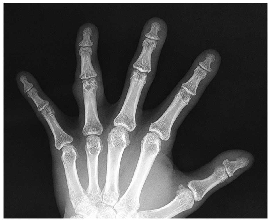
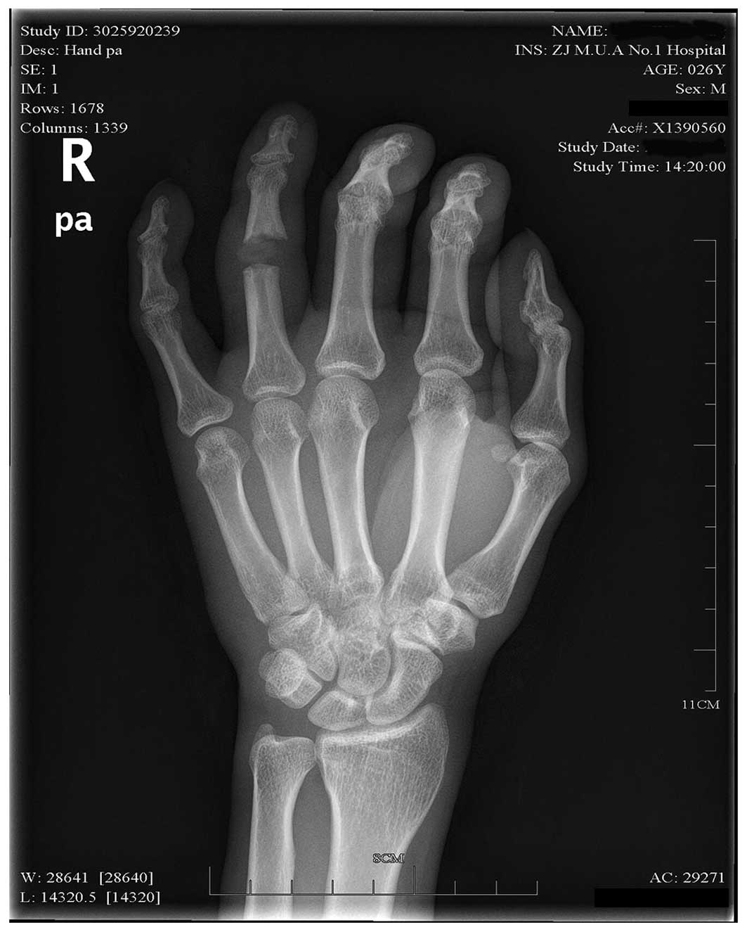
movement disorder. An X-ray of the right hand showed that the distal bone of the proximal phalange of the ring finger on the right hand had been destroyed. Although the diagnosis was unfavorable, a local resection of the extra-articular joint was performed to remove the tumor mass. The surgical pathology report confirmed GCTSS. The tumor recurred after 1 year, with multiple nodules found in the proximal phalange. According to the physical examination, the soft tissue in the PIP of the finger was swollen, but no skin redness was found. Multiple nodules could be locally palpated with mild tenderness. The blood supply to the fingertip was good, and the range of motion of the PIP was satisfactory. An X-ray of the right hand showed distal bone destruction of the proximal phalange of the ring finger on the right hand and swelling 
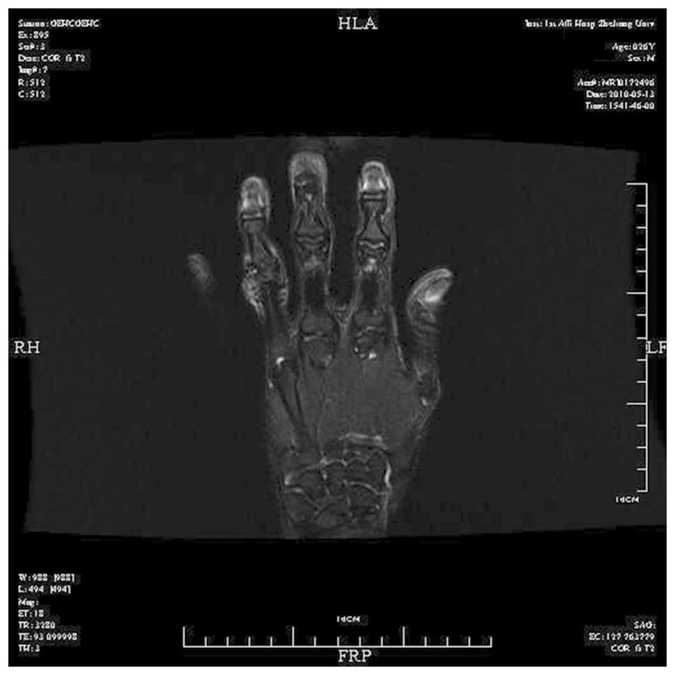
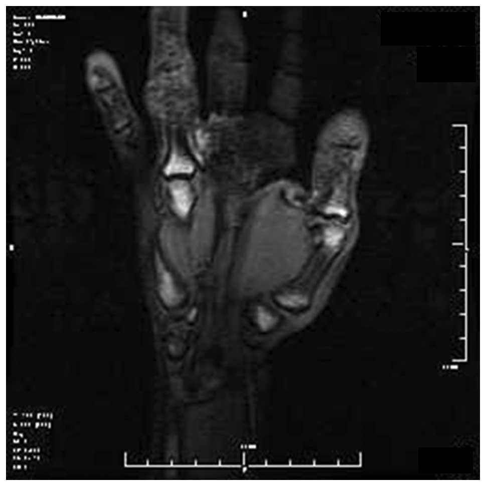
of the soft tissue. Magnetic resonance image of the finger (Fig. 1) showed that the distal bone of the proximal phalange of the finger was structurally destroyed, and the adjacent PIP capsule and periarticular soft tissue were markedly swollen. Slightly elongated T1- and T2-weighted signals were abnormal. The joint space remained. A giant cell tumor of the bone was subsequently diagnosed (Figs. 2 and 3).

Figure 1.

An X-ray of the right hand showing distal bone destruction of the proximal phalange of the ring finger and swelling of the soft tissue.

Figure 2.

Slightly elongated T1-weighted signal on magnetic resonance imaging.

Figure 3.

Abnormal T2-weighted signals on magnetic resonance imaging. The joint space remained. A giant cell tumor of the bone was diagnosed.

Resection of the tumor of the distal phalange was performed under general anesthesia. Local swelling was found around the PIP of the ring finger. Following incision of the skin and subcutaneous tissue of the dorsal digit, the bulk of the tumor was observed to be derived from the extensor tendon, with infiltration into the PIP capsule and ligament. The local soft tissue was severely damaged. The tumor mass was yellow-brown in color. Subsequent to removal of the tumor and involved soft tissue, and opening of the joint capsule, it was found that the tumor had infiltrated into the joint and destroyed the bone. Part of the tumor reached the joint, causing severe local damage of the bone. No indication of a simple tumor resection was found. Subsequently, an osteotomy was performed in the segment 0.5 cm from the proximal and distal regions of the joint to completely remove the tumor tissue. No significant tumor infiltration was found in the palmar tendon or soft tissue. A drill was used to expand the space to encompass nearly the entire bone marrow cavity of the intermediate phalange, and a Swanson #2 artificial silicone joint (Wright Medical Technology, Inc., Memphis, TN, USA) was placed inside the joint space. The residual dorsal capsule and ligaments on either side were sutured to strengthen and protect the joint. The wound was repeatedly washed. The local soft tissue and skin were sutured and wrapped with aseptic dressing. The treated finger was fixed in a dorsiflexion position with an external plaster support.

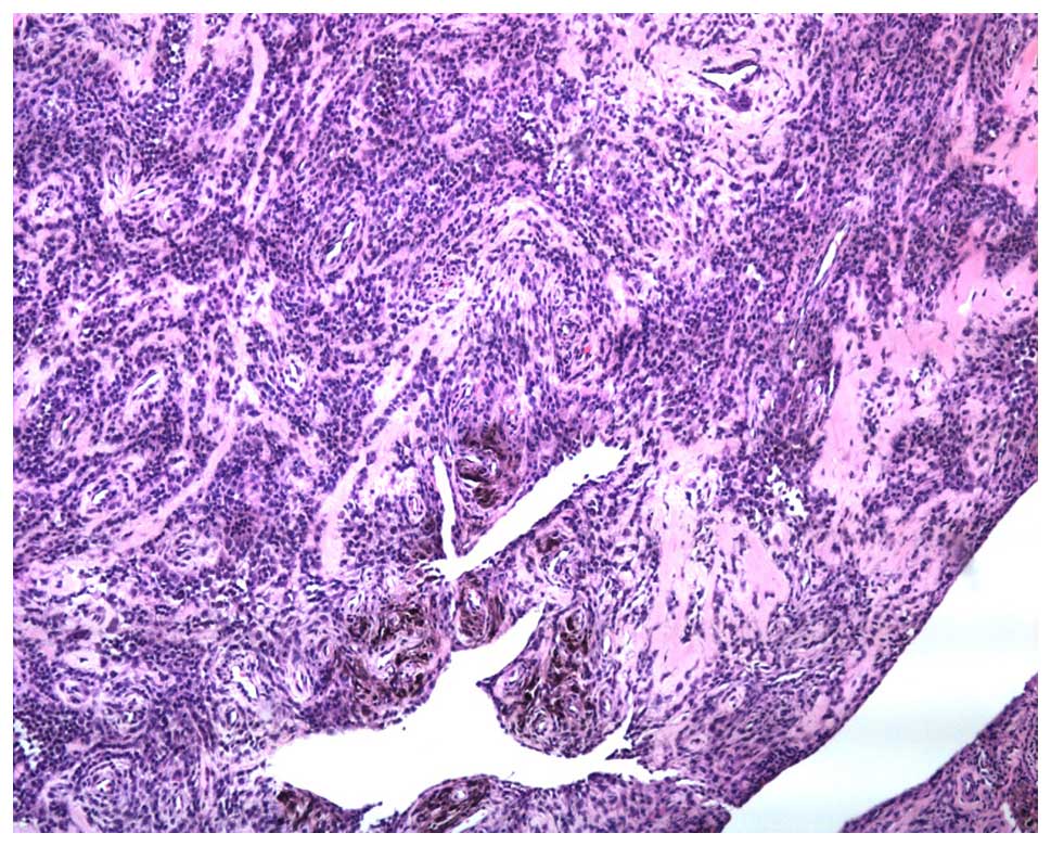
Irregular tissue measuring 2×1.2×0.2 cm was submitted for examination. Upon magnification, the tumor was found to be composed of a large number of round and synovial cells and a few multinucleated giant cells. The tumor was lamellar in arrangement, and a large amount of hemosiderin deposition was found. These findings were indicative of a GCTTS (Fig. 4).

Figure 4.

Pathological examination indicative of a giant cell tumor of the tendon sheath. The tumor consists of large rounded synoviocytes and a small number of multinuclear giant cells, arranged in diffuse sheets. Extensive hemosiderin deposition is also visible (stain, hematoxylin and eosin; magnification, ×50).

At 3–5 days post-surgery, the external plaster was removed and functional exercises were begun. Orderly and gradual exercise of the passive joint was conducted early during recovery, with three groups of exercises being performed every day, and each group being performed 10 times, while avoiding joint dislocation and tendon rupture. Once the patient's pain and local inflammatory edema subsided, active motion of the joint was gradually conducted. A quantitative exercise program was begun based on the use of a hand continuous passive motion machine. At discharge, the patient's PIP motion had recovered to the pre-operative level (Fig. 5), with pre-operative and post-operative disabilities of the arm, at shoulder and hand, and total activity measurement values of 1.67 and 3.33, and 255 and 243°, respectively. Complications such as tumor recurrence, joint dislocation and the requirement for prosthetic training were not observed during a 5-year follow-up period.

Figure 5.

X-ray at 24 months post-surgery.

Discussion

GCTTS is a common benign tumor arising in the tendon sheath GCTTS occurs most often in the hands and feet (6). Pigmented villonodular synovitis (PVNS) is a rare, idiopathic proliferative disorder of the synovium (7). It usually affects the hip or keen, but also occurs in hand or foot and the joint becomes swollen. Therefore, PVNS is often confused with GCTTS. The use of MRI may show a combination of synovial proliferation and soft tissue masses, together with deposits of hemosiderin and bone erosion, is highly diagnostic for PVNS, may also adequately displays the location and extent of the lesion and its internal structure (8).

Studies on GCTTS with bone destruction are inconsistent. In a report by Moore et al, no bone destruction was found among 115 cases of GCTTS (9). Bone destruction was detected in only 1 out of 51 cases of GCTTS reported by Fyfe and MacFarlane (10). Jones et al (11) studied 91 cases of GCTTS in the hand, among which 11 cases showed evidence of cortical bone erosion without bone destruction. However, certain studies have suggested that GCTTS with bone destruction is not uncommon. According to a study by Pan et al (12), among 98 cases, the bone was normal in 55 cases, the tumor infiltrated into the bone surface in 16 cases and the bone was destroyed in 27 cases. In a study by Uriburu and Levy (13), 23.3% of 133 total cases showed tumor intruding onto the bone surface and 11.3% exhibited bone destruction. Additionally, in another study, X-ray analysis showed lucent areas in single locular or multilocular cysts (14). On magnetic resonance imaging a further study showed that the tumor grew along the sheath, revealing a low signal intensity on T1-weighted images, but a high signal intensity on T2-weighted images. After joint intrusion by GCTTS with bone destruction, the occurrence rate of degenerative arthritis is 45% (11).

During treatment, elimination of the tumor lesions, including the soft tissue and bone, is favored. Recurrence is associated with the use of a surgical resection. In the present study, the previous tumor resection was performed at the expense of PIP function and appearance. Taking into account the patient's age, amputation was not an acceptable treatment. Complete resection of the tumor is critical to curing the disease. Due to the invasive properties of the tumor, joint stability must be considered if the joint is to be retained. The tumor cannot be completely resected, and thus the recurrence rate is high. The use of an artificial joint as a treatment for PVNS can achieve complete tumor resection, good functional recovery after surgery and reduce the likelihood of relapse (15).

Cases of GCTTS on the hand that infringes the joints, the tumor requires complete excision including the affected articular surface and joint capsule in order to have a lower rate of recurrence. The function of joints following reconstruction is a problem after the surgery. Artificial joint replacement may excise the tumor completely and also reconstruct the function of the finger joint and it is therefore a good choice for the clinic.

Artificial joint replacement surgery of the GCTTS that intruded into the distal region of the proximal phalange was successfully performed in the present case. Lesions were removed and PIP function was reconstructed.

References

1 

Scott SJ and Jenkinson MD: Giant-cell tumour of the tendon sheath. J Bone Joint Surg Br. 82:12062000.PubMed/NCBI

2 

Garg B and Kotwal PP: Giant cell tumour of the tendon sheath of the hand. J Orthop Surg (Hong Kong). 19:218–220. 2011.PubMed/NCBI

3 

Athanasou NA, Quinn J, Ferguson DJ and McGee JO: Bone resorption by macrophage polykaryons of giant cell tumour of tendon sheath. Br J Cancer. 63:527–533. 1991. View Article : Google Scholar : PubMed/NCBI

4 

Relwani J, Factor D, Khan F and Dutta A: Giant cell tumour of the patellar tendon sheath - an unusual cause of anterior knee pain: A case report. Knee. 10:145–148. 2003. View Article : Google Scholar : PubMed/NCBI

5 

Kitagawa Y, Ito H, Yokoyama M, Sawaizumi T and Maeda S: The effect of cellular proliferative activity on recurrence and local tumour extent of localized giant cell tumour of tendon sheath. J Hand Surg Br. 29:604–607. 2004. View Article : Google Scholar : PubMed/NCBI

6 

Demouy EH, Kaneko K, Bear HM and Rodriguez RP: Giant cell tumor of the plantar tendon sheath: Role of MR imaging in diagnosis. Case report. Clin Imaging. 17:153–155. 1993. View Article : Google Scholar : PubMed/NCBI

7 

Kanagawa H, Niki Y, Matsumoto H, Kosaki N, Enomoto H, Morioka H, Toyama Y and Suda Y: Localized pigmented villonodular synovitis presenting as a loose body following minor trauma in the knee: A case report. Knee. 14:395–397. 2007. View Article : Google Scholar : PubMed/NCBI

8 

Cheng XG, You YH, Liu W, Zhao T and Qu H: MRI features of pigmented villonodular synovitis (PVNS). Clinical rheumatology. 23:31–34. 2004. View Article : Google Scholar : PubMed/NCBI

9 

Moore JR, Weiland AJ and Curtis RM: Localized nodular tenosynovitis: Experience with 115 cases. J hand surg Am. 9:412–417. 1984. View Article : Google Scholar : PubMed/NCBI

10 

Fyfe IS and MacFarlane AU: Pigmented villonodular synovitis of the hand. Hand. 12:179–188. 1980. View Article : Google Scholar : PubMed/NCBI

11 

Jones FE, Soule EH and Conventry MB: Fibrous xanthoma of synovium (giant cell tumor of tendon sheath, pigmented nodular synovitis). A study of one hundred and eighteen cases. J Bone Joint Surg Am. 51:76–86. 1969.PubMed/NCBI

12 

Pan YW, Tian GL, Rong GW, Li C, Wang ZZ and Tian W: Giant cell tumor of tendon sheath in hand combined with bone invasion. Zhonghua Shou Wai Ke Za Zhi. 20:152–154. 2004.(In Chinese).

13 

Uriburu IJ and Levy VD: Intraosseous growth of giant cell tumors of the tendon sheath (localized nodular tenosynovitis) of the digits: Report of 15 cases. J hand surg Am. 23:732–736. 1998. View Article : Google Scholar : PubMed/NCBI

14 

Midletton WD, Patel V, Teefey SA and Boyer MI: Giant cell tumor of the tendon sheath: Analysis of scanographic finding. AJR Am J Roentgenol. 183:337–339. 2004. View Article : Google Scholar : PubMed/NCBI

15 

Lang L and Guo W: Treatment of diffused giant cell tumors of tendon sheath by rotation hinge prosthesis. Zhongguo Gu Yu Yuan Jie Za Zhi. 5:83–86. 2006.(In Chinese).

Related Articles

  • Abstract
  • View
  • Download
  • Twitter
Copy and paste a formatted citation
Spandidos Publications style
Lu H, Shen H, Chen Q, Shen XQ and Wu SC: Artificial finger joint replacement due to a giant cell tumor of the tendon sheath with bone destruction: A case report. Oncol Lett 10: 3502-3504, 2015.
APA
Lu, H., Shen, H., Chen, Q., Shen, X., & Wu, S. (2015). Artificial finger joint replacement due to a giant cell tumor of the tendon sheath with bone destruction: A case report. Oncology Letters, 10, 3502-3504. https://doi.org/10.3892/ol.2015.3813
MLA
Lu, H., Shen, H., Chen, Q., Shen, X., Wu, S."Artificial finger joint replacement due to a giant cell tumor of the tendon sheath with bone destruction: A case report". Oncology Letters 10.6 (2015): 3502-3504.
Chicago
Lu, H., Shen, H., Chen, Q., Shen, X., Wu, S."Artificial finger joint replacement due to a giant cell tumor of the tendon sheath with bone destruction: A case report". Oncology Letters 10, no. 6 (2015): 3502-3504. https://doi.org/10.3892/ol.2015.3813
Copy and paste a formatted citation
x
Spandidos Publications style
Lu H, Shen H, Chen Q, Shen XQ and Wu SC: Artificial finger joint replacement due to a giant cell tumor of the tendon sheath with bone destruction: A case report. Oncol Lett 10: 3502-3504, 2015.
APA
Lu, H., Shen, H., Chen, Q., Shen, X., & Wu, S. (2015). Artificial finger joint replacement due to a giant cell tumor of the tendon sheath with bone destruction: A case report. Oncology Letters, 10, 3502-3504. https://doi.org/10.3892/ol.2015.3813
MLA
Lu, H., Shen, H., Chen, Q., Shen, X., Wu, S."Artificial finger joint replacement due to a giant cell tumor of the tendon sheath with bone destruction: A case report". Oncology Letters 10.6 (2015): 3502-3504.
Chicago
Lu, H., Shen, H., Chen, Q., Shen, X., Wu, S."Artificial finger joint replacement due to a giant cell tumor of the tendon sheath with bone destruction: A case report". Oncology Letters 10, no. 6 (2015): 3502-3504. https://doi.org/10.3892/ol.2015.3813
Follow us
  • Twitter
  • LinkedIn
  • Facebook
About
  • Spandidos Publications
  • Careers
  • Cookie Policy
  • Privacy Policy
How can we help?
  • Help
  • Live Chat
  • Contact
  • Email to our Support Team