Introduction
Although breast cancer is the most common malignancy
among female individuals, the breast is an uncommon site of
metastasis from extramammary malignant neoplasms. In addition,
metastases to the breast represent only 0.3–2.7% of all malignant
mammary tumours (1). Excluding
contralateral mammary tumours, the most common sources of primary
tumour metastasis to the breast are, in decreasing order of
frequency, melanoma, lymphoma, lung and ovarian cancer (2). Metastases from the head and neck cancer
are rare, and those from nasopharyngeal carcinoma (NPC) are
extremely rare (2).
Diagnosis of breast metastasis from NPC is
difficult, even when the patient has a medical history of another
primary cancer. Experience of dealing with this clinical setting is
lacking; in fact, only four documented cases have been reported in
the literature thus far (3–5), and comprehensive descriptions of the
biological behavior and optimal treatment of this entity are
lacking. To reveal important aspects of breast metastasis from NPC,
the present study reports two cases from the Sun Yat-Sen University
Cancer Center (SYSUCC; Guangzhou, China) and reviews previous case
reports of breast metastasis from NPC, emphasizing the
clinicopathological and radiological features, diagnosis, treatment
strategies, and outcomes.
To the best of our knowledge, the current case study
describes the only case of a male patient in this clinical setting
exhibiting long-term survival after undergoing salvage treatment.
In addition, this is the first study to review all available cases
of breast metastasis from NPC.
Case report
Patients
From the pathology files of the SYSUCC (Guangzhou,
China), two cases of breast metastasis from NPC were identified.
Clinical data, including patient information, tumour
characteristics, treatment and outcomes, were collected. Informed
consent was obtained from the patients at their initial visit for
the collection of clinical information. The review, analysis and
publication of the current data were approved by the Research
Ethics Board of SYSUCC. Tumour response to treatment was assessed
according to the Response Evaluation Criteria in Solid Tumours
(6), and survival was traced through
medical records and phone interviews.
Pathological analysis
Immunohistochemistry (IHC) was performed on
paraffin-embedded sections using the avidin-biotin-peroxidase
complex method (7), with antibodies
for human estrogen receptor (ER), progesterone receptor (PR) and
human epidermal growth factor receptor 2 (HER2; Santa Cruz
Biotechnology, Inc., Santa Cruz, CA, USA). In situ
hybridization (ISH) for Epstein-Barr virus (EBV)-encoded small RNAs
(EBER) was performed on paraffin-embedded sections, according to
the manufacturer's instructions (Dako, Glostrup, Denmark). The
samples were obtained from lumpectomy for case 1 and halsted
mastectomy in case 2. Briefly, 4-µm paraffin embedded sections were
deparaffinized, rehydrated, and predigested with proteinase K. A
fluorescein-conjugated EBER probe was applied and the sections were
incubated at 37°C for 2 h. Subsequently, alkaline
phosphatase-conjugated antibody to fluorescein was applied,
followed by chromogen. Sections were then counterstained with
hematoxylin (Sigma-Aldrich, St. Louis, MO, USA). Dark brown
staining of the cell nucleus was regarded as positive
expression.
Case 1
A 41-year-old female patient presented in July 2008
to Sun Yat-Sen University Cancer Center (Shenzen, China) with a
1-month history of left upper neck swelling, tinnitus and
epistaxis. Physical examination revealed a mass in the nasopharynx
and bilateral enlargement of the jugular-carotid lymph nodes. The
size of the lymph node at the left upper neck was 7×6 cm and the
diameter of the lymph node in the right neck was 1.5 cm. Analysis
of biopsies obtained from the growth in the nasopharynx were used
to determine a diagnosis of poorly differentiated squamous cell
carcinoma. Magnetic resonance imaging scans of the head and neck
revealed tumour infiltration of the roof and lateral walls, and the
skull base. No distant metastasis was identified upon initial
diagnosis and the patient was classified with stage T3N3M0 NPC
(8). The patient was treated with one
cycle of cisplatin (80 mg/m2 on day 1) and
5-fluorouracil (5-FU; 500 mg/m2/day on days 1–5) regimen
followed by curative two-dimensional radiotherapy. The patient
received doses of 70 Gy in 35 fractions to the primary tumour, 66
Gy in 33 fractions to the right neck and 62 Gy in 31 fractions to
the left neck. Chemotherapy was performed between 25 July 2008 to 1
August 2008. Radiotherapy was performed between 12 August 2008 and
30 September 2008. Complete remission was achieved after
radiotherapy was finished. Upon termination of treatment, complete
remission was achieved.
In January 2009, the patient presented with a mass
in the right breast. Physical examination revealed a firm mass in
the lateral upper quadrant of the right breast measuring 1.5 cm in
diameter. No mass was identified in the left breast or bilateral
axillary fossa.
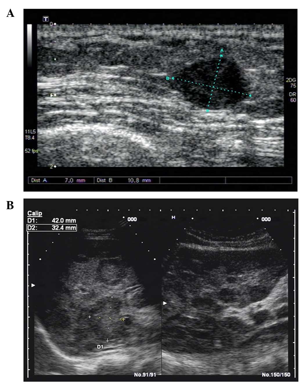
Ultrasonography identified a hypoechoic lesion with
a smooth margin in the lateral upper quadrant of the right breast,
and no retrotumour acoustic shadowing, microcalcification, skin
change or obvious blood signal, as indicated in Fig. 1A. Furthermore, no obvious lymph node
enlargement was detected in the two auxiliary fossa. Concurrently,
multiple diffuse hypoechoic lesions with smooth margins were
identified in the liver, as demonstrated in Fig. 1B. Chemical profiling revealed anemia
(88 g/l Hb; normal range, 130–160 g/l Hb) and enzyme elevation,
including alanine aminotransferase (115.1 U/l; normal range,
0.0–40.0 U/l), alkaline phosphatase (253.3 U/l; normal range,
0.0–45.0 U/l), γ-glutamyl transferase (226.4 U/l; normal range,
11.0–50.0 U/l) and lactate dehydrogenase (1,328.2 U/l; normal
range, 109.0–245.0 U/l). In addition, carcinoembryonic antigen
levels (CEA; 74.4 ng/ml; normal range, 0–5 ng/ml) were notably
higher than the normal range. Prior to characterisation of the
breast mass, a diagnosis of liver metastasis was considered. Thus,
a segmental resection (lumpectomy) was performed on the breast on
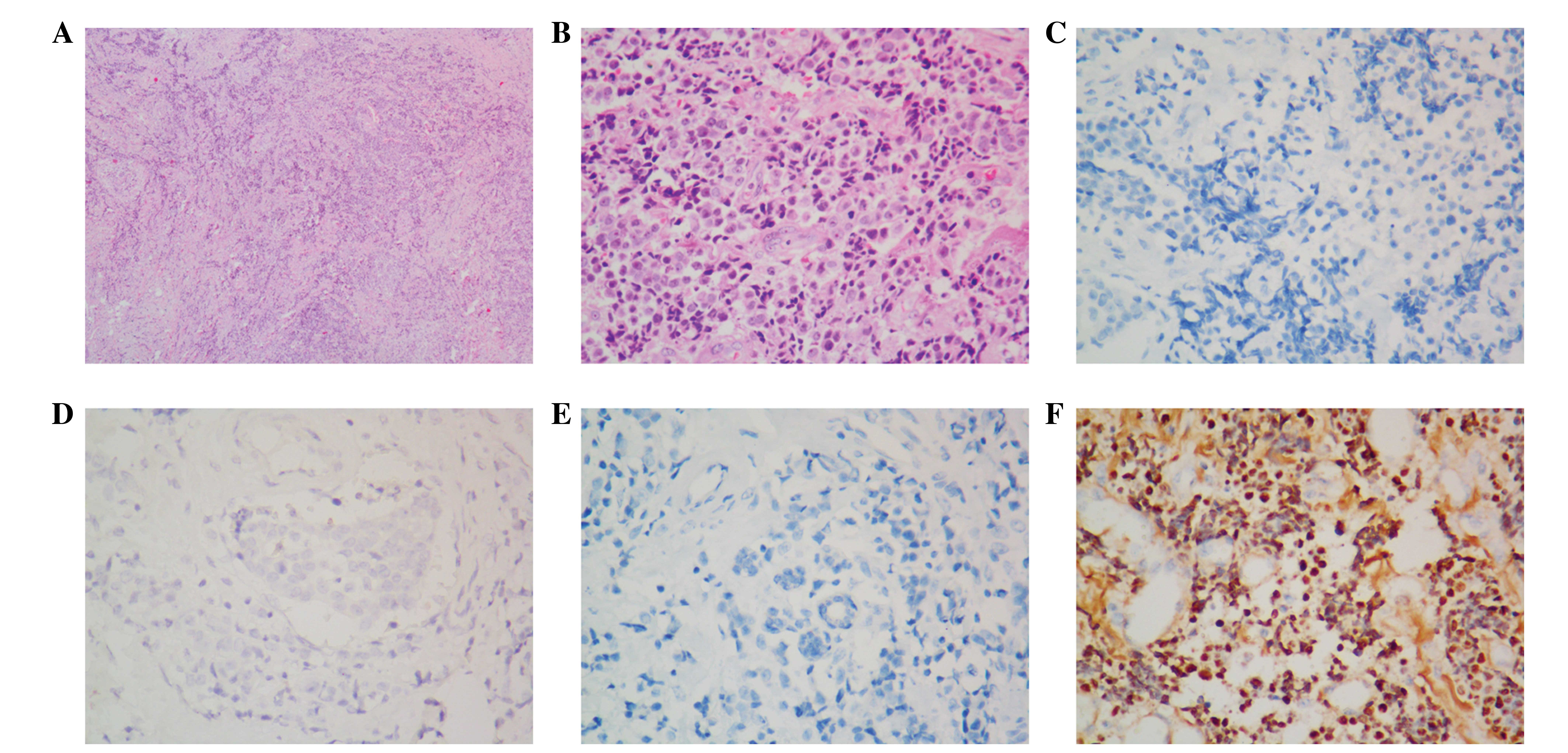
January 20, 2009. Histological analysis performed on the metastatic
breast tissue demonstrated malignant anaplastic carcinoma
proliferating in a nested and trabecular pattern (Fig. 2A), and neoplastic cells exhibited a
short spindle or round appearance with little cytoplasm (Fig. 2B). Immunostaining results were as
follows: ER (−), PR (−) and HER2 (−) (Fig. 2C–E). ISH determined that the
neoplastic cells were positively labeled with the EBER probe
(Fig. 2F).
Palliative care with chemotherapy was planned,
however, the patient refused further treatment, was discharged from
hospital on 25 January 2009 and succumbed to cachexia 5 months
after leaving hospital.
Case 2
A 46-year-old male patient presented in October 2000
to Sun Yat-Sen University Cancer with a 6-month history of nasal
and 1-month history of right neck swelling. Physical examination
identified a mass in the roof of the nasopharynx and a lump in the
right neck. The size of lymph node at the right side was 8×4 cm.
Computed tomography (CT) scan of the nasopharynx indicated tumour
infiltration of the lateral wall and roof of the nasopharyngeal
cavity, bilateral walls of the oropharynx, and the two posterior
portions of the nasal cavity, as well as invasion of the skull
base, right maxillary sinus and sphenoid sinus. No distant
metastasis was identified on chest X-ray, abdominal ultrasonography
or emission CT examination. Thus, the patient was diagnosed with
T4N3M0 stage IV NPC. Prior to radiotherapy, one cycle of PF regimen
chemotherapy (30 mg cisplatin for 5 days plus 750 mg floxuridine
for 5 days) was administered to the patient. Curative radiotherapy
was performed between November 2000 and January 2001. The dose to
the primary tumour was 74 Gy in 37 fractions, to the right neck was
70 Gy in 35 fractions and to the left neck was 60 Gy in 30
fractions. At the termination of treatment the size of the residual
lymph node at the right side had reduced to 1.5×1.5 cm and, 3
months after treatment, complete remission was achieved.
In July 2001, the patient identified a palpable mass
in the left breast and returned to Sun Yat-Sen University Cancer
for a consultation. Physical examination revealed a mass measuring
3×2 cm in the lateral upper quadrant of the left breast.
Fine-needle biopsy identified a small number of atypical cells,
with an appearance of bare and obvious nucleus. Metastasis from the
nasopharynx was suspected. Blood profiling identified elevation of
EBV-DNase antibodies from 49% at diagnosis to 71%. However, CEA
(0.1 ng/ml; normal range, 0–5 ng/ml) and squamous cell carcinoma
antigen (0.3 ng/ml; normal range, 0–1.5 ng/ml) levels were normal,
and there was no evidence of distant metastasis in the lung or
abdomen.
A solitary metastatic lesion was identified in the
breast. Such patients have potentially good prognoses, therefore, a
Halsted mastectomy was performed on the left breast on 10 August
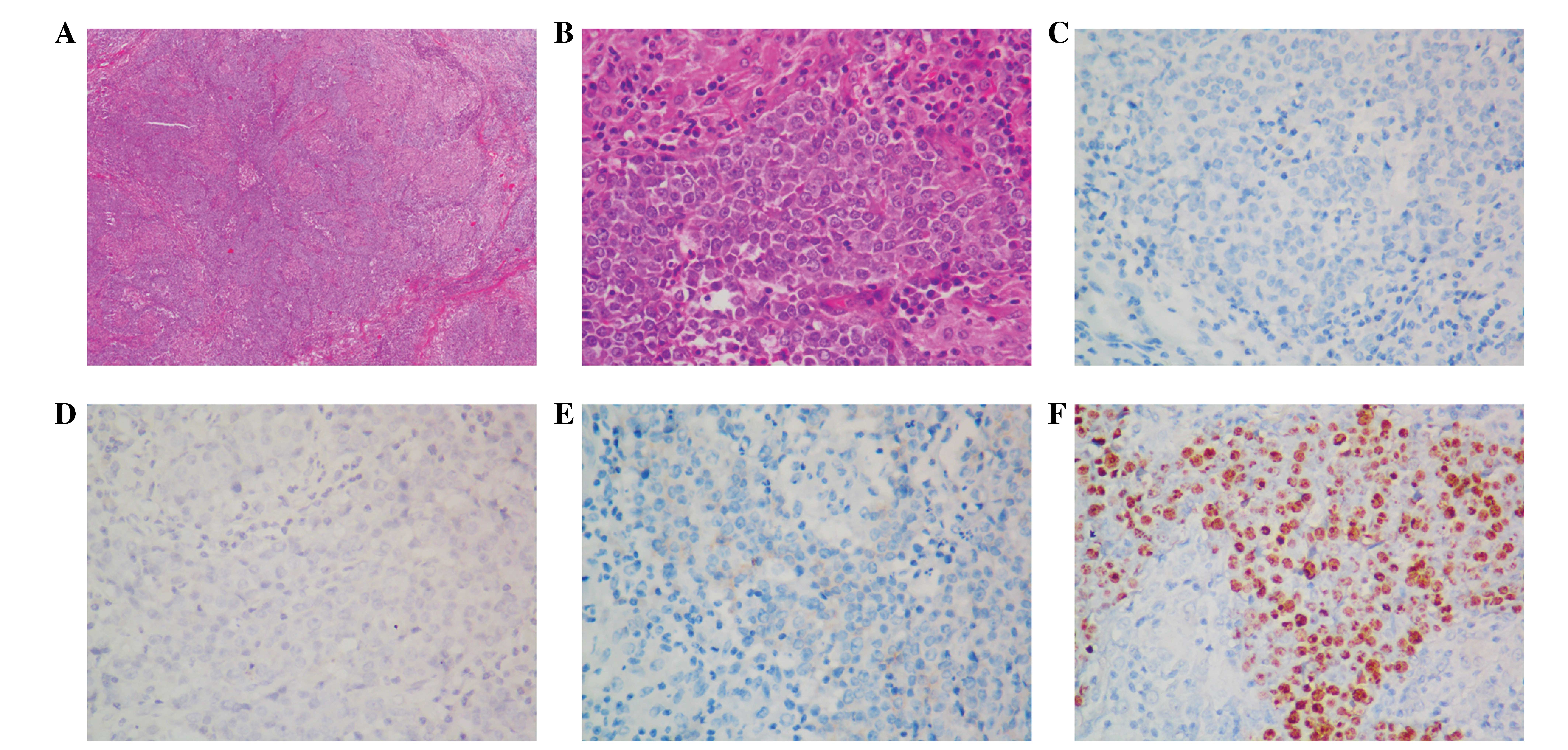
2001. Histological analysis revealed poorly and moderately
differentiated squamous cell carcinoma diffusely infiltrated in the
left breast and pectoralis muscles (Fig.
3A and B). Immunostaining results were as follows: ER (−), PR
(−) and HER2 (−) (Fig. 3C–E). ISH
revealed that the neoplastic cells were positively labeled with an
EBER probe (Fig. 3F). No tumour cells
were observed in the axillary lymph nodes.
The patient was disease-free and without discomfort
at the most recent follow-up on May 8, 2010, but was then lost to
follow-up.
Literature review
Search strategy
The PubMed database was searched for all English
literature publications using the following keywords: ‘Breast
metastasis’ ‘breast metastases’ ‘metastases to the breast’
‘metastasis to the breast’ ‘metastasis in the breast’ ‘metastases
in the breast’ and ‘nasopharyngeal carcinoma’ and ‘nasopharyngeal
cancer’. Four cases of breast metastasis from NPC were identified
and are included in the current analysis, termed cases 3–6
(2–4).
The six cases, including cases 1 and 2 of the present study, are
summarized in Table I and discussed
hereafter.
 | Table I.Demographic data of patients
exhibiting nasopharyngeal carcinoma with breast metastasis. |
Table I.
Demographic data of patients
exhibiting nasopharyngeal carcinoma with breast metastasis.
| Case no. | Age,
years/gender | Histology of NP
tumor | Stage | Primary Tx | Latency of
metastasis, months | Site of BM | Size, cma | Axillary LN | DM | Local recurrence | Treatment | Survival |
|---|
| 1 | 41/F | Poorly differentiated
squamous cell carcinoma | T3N3M0 | CT/RT | 6 | R/LUQ | 1.2 | N | Liver | N | Refused
treatment | 5 months |
| 2 | 46/M | Poorly/moderately
differentiated squamous cell carcinoma | T4N3M0 | CT/RT | 9 | L/LUQ |
3×3.5 | N | No | N | Halsted
mastectomy | ≥10 years |
| 3b | 39/F | Anaplastic carcinoma
of the nasopharynx | T2N2M0e | RT | 28 | Central | 6.0 | Y | Bone, left
supraclavicular LN | Y | Two cycles of DDP +
5-FU, RT to the breast mass (32 Gy in 8 daily fractions), bilateral
nephrostomy and pelvic radiotherapy | 3 weeks |
| 4b | 51/F | Anaplastic carcinoma
of the nasopharynx | T1N2M0e | RT | 27 | R/MLQ | 2.0 | Yf | Lung | N | DDP + 5-FU | / |
| 5c | 46/F | Poorly differentiated
carcinoma | / | CT/RT | / | R/LLQ | 2.8×1.4 | N | / | / | / | / |
| 6d | 25/F | Anaplastic
carcinoma | T4N3M0 | CT | 42 | L/MUQ R/upper | 5.0 (L) 8.0 (R) | Y | Left supraclavicular
LN, liver, bone | N | Palliative care | / |
Clinical manifestation
Five of the six patients were female while case 2
from the current series was male. The age at initial diagnosis of
NPC ranged between 25 and 51 years, with a median age of 43.5
years. The primary staging classifications of five of the
documented patients were all loco-regionally advanced without
distant metastasis. Radiotherapy with or without chemotherapy was
administered in cases 1–5; however, case 6 received chemotherapy
alone. Following a latency period of 6–42 months, patients were
diagnosed with metastasis in the breast (median latency period, 27
months). The predominant symptoms at relapse were the presence of a
breast mass and axillary lymph node enlargement. Three of the six
patients (cases 3, 4 and 6) exhibited enlarged axillary lymph
nodes. Local recurrence at the nasopharynx was only observed in
case 3.
Distant metastasis pattern
All the patients exhibited locally and regionally
confined disease without synchronous metastasis upon initial
diagnosis. All breast metastasis occurred subsequent to treatment
as metachronous metastasis. Multiple metastases were the
predominant pattern, with distant metastases occurring in other
sites in cases 1, 3, 4 and 6, including in the liver, bone and lung
and supraclavicular lymph node. However, the breast was the only
site of metastasis in case 2.
Pathological analysis
The histology of all of the primary nasopharyngeal
tumours was poorly differentiated squamous or anaplastic carcinoma.
The five documented patients also exhibited anaplastic carcinoma or
poorly differentiated squamous cell carcinoma in the breast
metastasis, mimicking the primary tumour. IHC and EBV analyses were
performed in a number of cases. IHC of the malignant cells revealed
negativity for ER and PR in cases 1, 2 and 6, and negativity for
HER2 in cases 1 and 2. Positive EBER staining was identified in
cases 1, 2 and 6.
Ultrasonographic findings
Ultrasonography was performed for cases 1 and 5.
Similar features were observed in the two cases, including a
hypoechoic lesion with a heterogeneous internal echo pattern, and
no retrotumour acoustic shadowing, microcalcification or skin
change. No obvious lymph node enlargement was detected in the two
axillary fossa. However, a smooth margin was observed in case 1
while case 5 exhibited an irregular margin.
Treatment strategies and outcomes
Chemotherapy is the first choice treatment strategy
if multiple metastases are diagnosed, such as in cases 1, 3 and 4.
A combination of cisplatin and 5-FU was the standard regimen if the
patient was able to tolerate it. However, palliative care was
selected if the general situation of the patient was poor, such as
for case 6. Radiotherapy was used to reduce the size of the breast
metastasis and control the pain associated bone metastasis, as in
case 3. By contrast, a Halsted mastectomy was performed with
curative intent in case 2 of the current series.
Survival
Median follow-up could not be calculated due to
missing data in certain cases. Cases 1 and 3 had short survival
times following the diagnosis of metastasis, possibly due to
cachexia. The survival times were not reported for cases 4–6;
however, their prognosis was considered to be poor due to the
presence of multiple metastases. Case 2 from the current series
survived without any discomfort for 10 years and was then lost
during follow-up.
Discussion
NPC is a malignant tumour of the nasopharyngeal
epithelium with unique epidemiological features, clinical
manifestations, treatment strategies and survival outcome (9). It is an endemic disease in Southern
China and the incidence of NPC is higher in male individuals, with
a male:female ratio of 2–3:1 (9).
Furthermore, NPC predominantly occurs between the ages of 50–60
years. It is characterised by a tendency to spread regionally to
cervical lymph nodes and diffusely to distant organs. Radiotherapy
and chemotherapy are the predominant treatment strategies for
patients with NPC (2).
Breast metastases from NPC are rare, with 6 cases
summarized in the present study (3–5). In
contrast to primary NPC, the disease is more frequent among female
individuals; however, the median age is similar to that of primary
NPC. To the best of our knowledge, case 2 from the current study is
the first and only male patient in this clinical setting.
The five female patients (cases 1, 3–6) shared the
following characteristics: Female, loco-regionally advanced disease
at initial diagnosis, metachronous breast metastasis following
curative treatment and concurrent disseminated disease at other
sites, similar to breast metastasis from other extramammary
malignancies (10). The latency
period from the initial diagnosis of NPC to the diagnosis of breast
metastasis ranged between 6 and 42 months, with a median latency
period of 27 months. Breast metastasis may occur in the short term
or 2 years after the first course treatment. Case 2 in the current
report is unique, as the patient was male. A male patient with
breast metastasis from NPC has not previously been reported,
highlighting that this disease entity can also occur in males.
The major problem in establishing such a diagnosis
is differentiating between primary breast cancer and breast
metastasis. Misdiagnosis as a primary breast cancer may lead to an
unnecessary mastectomy. Thus, differential diagnosis is vital for
ensuring that appropriate chemoradiotherapy is administered for
patients with breast metastasis from NPC.
Clinically, breast metastases typically present as a
firm mass, may be located at any quadrant and are often
superficial. Skin involvement may also exist, while nipple
discharge is absent (2). Half of the
patients analyzed in the present study demonstrated enlarged
axillary lymph nodes. Concomitant local recurrence at the
nasopharynx is not frequent; however, distant metastases at other
sites are common, including in the lung, liver and bone. These
sites are similar to the frequent metastatic sites of primary
breast cancer (11). Breast
metastases from NPC are rare and typically occur as part of
disseminated disease; thus, a previous clinical diagnosis of NPC
alone may indicate the metastatic nature of the breast tumour
(12,13). The identification of a solitary breast
metastasis may also occur, as in case 2. However, ultrasonographic
features of breast metastasis are not particularly informative or
unique; observations may include regular or irregular margins,
hypoechoic lesions, heterogeneous internal echo patterns, an
absence of retrotumour acoustic shadowing and no skin changes.
The most important aspect of this differential
diagnosis is the pathological morphology and molecular staining of
the metastatic lesion. Morphologically, metastatic carcinoma has
specific features, characterized by multinodular architecture and a
lack of an intraductal carcinoma or lobular neoplasia components.
However, Driss et al (5)
reported that breast metastasis and primary breast cancer may still
exhibit a number of similarities, such as mimicking certain
histological types of primary carcinoma, including
lymphoepithelioma-like carcinoma, medullary carcinoma or a variety
of infiltrating ductal or lobular carcinoma with inflammatory
stroma. Lymphoepithelioma-like carcinoma of the breast may also
display multinodular growth. However, Dadmanesh et al
(14) identified that
lymphoepithelioma-like carcinoma of the breast was not associated
with EBV infection, while EBER positivity was identified in the
breast metastatic lesions of the three patients tested (cases 1, 2
and 6). Thus, ISH is recommended for differential diagnosis.
Furthermore, IHC indicated negativity for ER, PR and HER2, a
phenotype unique to patients with triple-negative breast cancer.
Therefore, pathological, IHC and radiological findings, in
conjunction with the patient's clinical history, should be
considered in differentiating a secondary mass from a primary
breast cancer.
No consensus has yet been reached for the optimum
treatment of breast metastasis from NPC. In principle, appropriate
chemoradiotherapy or chemotherapy should be administered to
patients with multiple metastases, while mastectomy appears
unnecessary due to poor prognosis. Radiotherapy may be effective
for reducing tumour size as observed in case 3 and may relieve pain
in bone metastasis. Supportive treatment should be used for
patients with cachexia. The prognoses for these patients is poor,
with a survival time of <6 months in cases 1 and 3. The final
treatment result was not documented for cases 4–6, however, it is
reasonable to predict poor prognosis of these three patients due to
the presence of multiple distant metastases.
Breast metastasis was only identified in case 2 and,
after receiving radical mastectomy, the patient survived for ≥10
years with no recurrence of disease. In such cases, overall
examination and an accurate diagnosis are vital for oncologists to
determine the appropriate treatment regimen and predict the
patients' survival.
In a patient with a medical history of
loco-regionally advanced NPC, metastatic disease should be
considered when a breast mass is identified, particularly when the
patient is female, with multiple site of malignancies and
positivity upon EBER staining. Breast metastasis may also occur in
male individuals. Palliative treatment, including chemotherapy and
radiotherapy, is the primary treatment strategy for such patients.
However, it is considered that patients exhibiting NPC with
solitary breast metastasis may still gain long-term survival,
therefore, radical mastectomy should be considered.
Acknowledgements
The present study was funded by the National Natural
Science Foundation (grant no. 81372814).
References
|
1
|
Canda AE, Sevinc AI, Kocdor MA, Canda T,
Balci P, Saydam S and Harmancioglu O: Metastatic tumours in the
breast: A report of 5 cases and review of the literature. Clin
Breast Cancer. 7:638–643. 2007. View Article : Google Scholar : PubMed/NCBI
|
|
2
|
Yeh CN, Lin CH and Chen MF:
Characteristics of metastasis in the breast from extramammary
malignancies. J Surg Oncol. 101:137–140. 2010. View Article : Google Scholar : PubMed/NCBI
|
|
3
|
Sham JS and Choy D: Breast metastasis from
nasopharyngeal carcinoma. Eur J Surg Oncol. 17:91–93.
1991.PubMed/NCBI
|
|
4
|
Yeh CN, Lin CH and Chen MF: Clinical and
ultrasonographic characteristics of breast metastases from
extramammary malignancies. Am Surg. 70:287–290. 2004.PubMed/NCBI
|
|
5
|
Driss M, Abid L, Mrad K, Dhouib R, Charfi
L, Bouzaein A and Ben Romdhane K: Breast metastases from
undifferentiated nasopharyngeal carcinoma. Pathologica. 99:428–430.
2007.PubMed/NCBI
|
|
6
|
Therasse P, Arbuck SG, Eisenhauer EA,
Wanders J, Kaplan RS, Rubinstein L, Verweij J, Van Glabbeke M, van
Oosterom AT, Christian MC, et al: New guidelines to evaluate the
response to treatment in solid tumours. European organization for
research and treatment of cancer, National Cancer Institute of the
United States, National Cancer Institute of Canada. J Natl Cancer
Inst. 92:205–216. 2000. View Article : Google Scholar : PubMed/NCBI
|
|
7
|
Li H, Xiao W, Ma J, Zhang Y, Li R, Ye J,
Wang X, Zhong X and Wang S: Dual high expression of STAT3 and
cyclinD1 is associated with poor prognosis after curative resection
of esophageal squamous cell carcinoma. Int J Clin Exp Pathol.
7:7989–7998. 2014.PubMed/NCBI
|
|
8
|
Hong MH, Mai HQ, Min HQ, Ma J, Zhang EP
and Cui NJ: A comparison of the Chinese 1992 and fifth-edition
International Union Against Cancer staging systems for staging
nasopharyngeal carcinoma. Cancer. 89:242–247. 2000. View Article : Google Scholar : PubMed/NCBI
|
|
9
|
Chua ML, Wee JT, Hui EP and Chan AT:
Nasopharyngeal carcinoma. Lancet. 2015.[Epub ahead of print].
View Article : Google Scholar : PubMed/NCBI
|
|
10
|
Lee SK, Kim WW, Kim SH, Hur SM, Kim S,
Choi JH, Cho EY, Han SY, Hahn BK and Choe JH: Characteristics of
metastasis in the breast from extramammary malignancies. J Surg
Oncol. 101:137–140. 2010. View Article : Google Scholar : PubMed/NCBI
|
|
11
|
Marino N, Woditschka S, Reed LT, Nakayama
J, Mayer M, Wetzel M and Steeg PS: Breast cancer metastasis: Issues
for the personalization of its prevention and treatment. Am J
Pathol. 183:1084–1095. 2013. View Article : Google Scholar : PubMed/NCBI
|
|
12
|
Silverman JF, Feldman PS, Covell JL and
Frable WJ: Fine needle aspiration cytology of neoplasms metastatic
to the breast. Acta Cytol. 31:291–300. 1987.PubMed/NCBI
|
|
13
|
Domanski HA: Metastases to the breast from
extramammary neoplasms. A report of six cases with diagnosis by
fine needle aspiration cytology. Acta Cytol. 40:1293–1300. 1996.
View Article : Google Scholar : PubMed/NCBI
|
|
14
|
Dadmanesh F, Peterse JL, Sapino A, Fonelli
A and Eusebi V: Lymphoepithelioma-like carcinoma of the breast:
Lack of evidence of Epstein-Barr virus infection. Histopathology.
38:54–61. 2001. View Article : Google Scholar : PubMed/NCBI
|


