Introduction
Non-small-lung cancer (NSCLC) is a common malignant
tumor, and a leading cause of mortality (1). Invasion and metastasis of NSCLC are the
most dominant reasons for cancer mortality and treatment failure.
Findings of previous studies have suggested that tumor pathogenesis
was induced by tumor stem cells (2).
Tumor stem cells were identified in many solid tumors, such as
glioma, as well as breast, prostate, pancreatic and colon cancer
(2). These stem cells have been
associated with tumor pathogenesis and development, as well as
invasion and metastasis of tumor cells (2). Octamer-binding transcription factor 4
(Oct-4) is a marker of tumor stem cells. Epithelial-mesenchymal
transition (EMT) indicates the phenomenon that epithelial cells
transit to fibroblast or mesenchymal cells and acquire migration
ability (3). The most important
marker of EMT is the downregulation or silencing of E-cadherin
(E-cad) expression. The occurrence of EMT causes a loose cellular
arrangement and increased cell mobility, leading to further
invasion and metastasis of tumor cells (3).
The present study analyzed the expression of Oct-4
and E-cad in NSCLC and normal lung tissue, and examined the
association between tumor stem cells in NSCLC and EMT.
Materials and methods
Specimen source
Specimens were obtained from 65 NSCLC patients at
the Department of Thoracic Surgery, Hebei People's Hospital
(Shijiazhuang, China) between January 2013 and June 2014. The
patients included 38 males and 27 females, with an age range of
26–78 years and a median age of 53 years. The pathogenic grade of
the patients was: high differentiation, 22 cases; medium
differentiation, 30 cases; and low differentiation, 13 cases. The
clinical stage of the patients was: stage 0-I, 9 cases; stage
II–III, 31 cases; and stage IV, 25 cases. The control group
included 15 cases of normal lung tissue following lobectomy for
bronchiectasis and pulmonary bullae resection, which were confirmed
pathologically. The age range for the control group was 18–48
years, with a median age of 35 years. These specimens were fixed in
10% formaldehyde, embedded with paraffin, and cut into serial
tissue sections (5 µm).
Reagents
Mouse anti-human E-cad mAb (Beckman Coulter, Brea,
CA, USA), and rabbit anti-Oct-4 polyclonal antibodies (Abcam,
Cambridge, UK) were used. The dilutions for the antibodies was
1:500. Diaminobenzidine (DAB) chromogenic agent and ELISA kit were
purchased from Beijing Zhongshan Jinqiao Biotechnology Co., Ltd.
(Beijing, China). The protocols followed were as specified in the
kit instructions.
Interpretation of results
Brown granules in the cell nucleus indicated a
positive expression of Oct-4. The score was calculated by combining
the staining intensity and the percentage of positive cells in the
tumor cells. Values for the staining intensity were: colorless, 0;
light yellow, 1; tan, 2; and brown, 3. The score for positive cell
percentage was: <5%, 0; 5–25%, 1; 26–50%, 2; and >50%, 3. The
final score was calculated using the score of staining intensity
multiplied by the score of positive cell percentage, as follows:
0–1, negative (−); 2–6, weak positive (+); and 7–12, strongly
positive (++). Both (+) and (++) were classified as a positive
expression (4). Light-yellow to brown
granules were observed in the cells with a positive expression of
E-cad, and the staining was located on the cell membrane with a few
granules in the cytoplasm. The scoring criteria used were as
previously described by Mahler-Araujo et al (5): <10% positive cells, 0: 10–25%
positive cells, 1; 25–50% positive cells, 2; 50–75% positive cells,
3; ≥75% positive cells, 4. Scores of 0–3 indicated a negative
expression, i.e., abnormal expression; and ≥3 indicated a positive
expression, i.e., normal expression.
Statistical analysis
SPSS 17.0 software (SPSS, Inc., Chicago, IL, USA)
was used for statistical analysis. The χ2 test was used
for enumeration data, with an inspection level of α=0.05. P<0.05
was considered to indicate a statistically significant
difference.
Results
Relationship between grade, stage of
Oct-4 and E-cad
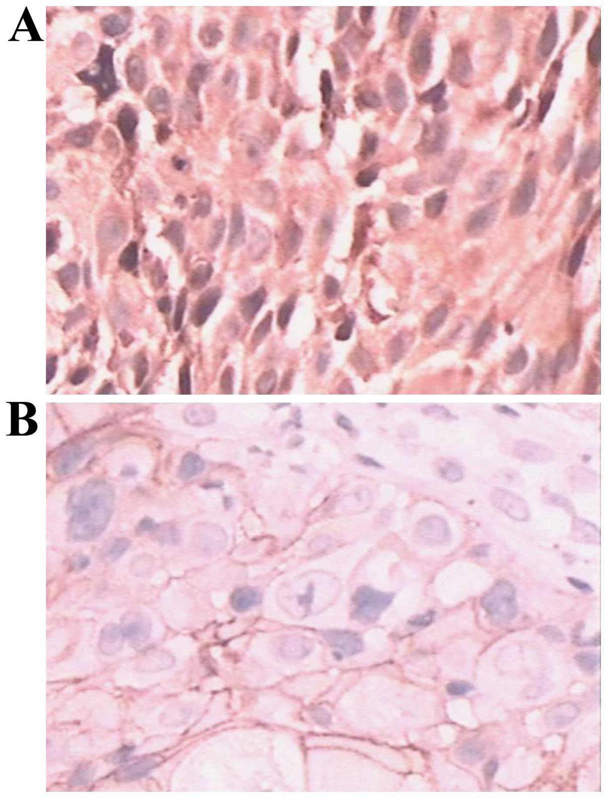
Abnormal expression of E-cad was observed in NSCLC
tissue and the staining demonstrated light-yellow to brown
granules. E-cad expression was located on the cell membrane, with a
few granules in the cytoplasm (Fig.
1A). A positive expression of Oct-4 was observed in the NSCLC
tissue, and the staining demonstrated brown granules. Oct-4
expression was located on the cell membrane (Fig. 1B). The abnormal expression of E-cad
and positive expression of Oct-4 in the NSCLC specimens was higher
than that of the normal lung tissue, and statistically significant
(P<0.05). The expression of Oct-4 and E-cad were associated with
the pathological grade and clinical stage of the patient, and were
increased as the NSCLC malignancy increased. The differences in
each grade and each stage were statistically significant
(P<0.05) (Table I).
 | Table I.Correlation between the expression of
Oct-4 and E-cad in NSCLC tissue and between pathological grade and
clinical stage. |
Table I.
Correlation between the expression of
Oct-4 and E-cad in NSCLC tissue and between pathological grade and
clinical stage.
|
|
| Oct-4 |
| E-cad |
|
|---|
|
|
|
|
|
|
|
|---|
| Group | No. of cases | No. of positive cases
(%) | P-value | No. of abnormal
expression cases (%) | P-value |
|---|
| Normal lung
tissue | 15 | 0
(0.00) |
| 2
(13.3) |
|
| Pathological
grade |
|
|
|
|
|
| High
differentiation | 13 |
4 (30.77) |
|
3 (23.08) |
|
| Medium
differentiation | 30 | 16
(53.33) | 0.009a | 19
(63.33) | 0.002a |
| Low
differentiation | 22 | 18
(81.82) |
| 18
(81.82) |
|
| Clinical stage |
|
|
|
|
|
| 0-I | 9 |
2 (22.22) |
|
1 (11.11) |
|
|
II–III | 31 | 18
(58.06) | 0.034a | 18
(58.06) | 0.001a |
| IV | 25 | 18
(72.00) |
| 21
(84.00) |
|
Correlation between Oct-4 and E-cad
expression
Of the 65 NSCLC specimens, the abnormal expression
rate of E-cad was 61.54% and the positive expression rate of Oct-4
was 58.46% (Table II). The
difference between E-cad and Oct-4 was statistically significant
(P=0.000, coefficient of contingency=0.439), and demonstrated that
E-cad expression was correlated with Oct-4 expression.
| Table II.Correlation between Oct-4 and E-cad
expression. |
Table II.
Correlation between Oct-4 and E-cad
expression.
|
| E-cad | Oct-4 |
|
|
|---|
|
|
|
|
|
|
|---|
| Characteristics | Negative | Positive | Total | P-value |
|---|
| Normal
expression | 18 | 7 | 25 |
|
| Abnormal
expression | 9 | 31 | 40 | 0.000a |
| In total | 27 | 38 | 65 |
|
Discussion
Invasion and metastasis are basic characteristics of
malignant tumor. Tumor invasion and metastasis includes three key
features: decreased adhesion, degraded matrix and enhanced
migration, and EMT attributes to these features. E-cad is
distributed in various epithelial cells and may mediate cell
adhesion. The most important EMT tumor marker is the downregulation
or silencing of E-cad, which is considered as the prerequisite for
the ability of invasion and metastasis of epithelial cells
(6). The findings of the present
study have demonstrated that the increase in abnormal expression of
E-cad in the NSCLC specimens was higher than that of the normal
lung tissue, and the result was statistically significant. The
decreased NSCLC differentiation level and increased clinical stage
led to the abnormal expression of E-cad being increased, and the
result was statistically significant.
Tumor stem cells are the cells that are
characteristic of self-renewal with differentiation potential, and
contribute to tumor relapse, metastasis and drug resistance
(7). The expression of Oct-4 is a
marker of tumor stem cells. Tumor cells with a positive Oct-4
expression have increased tumorigenic capacity in vitro and
the characteristics of tumor stem cells. Akunuru et al
(8) reported that although tumor stem
cells have various phenotypes, they can express the genes of
pluripotent stem cells, particularly Oct-4. The overexpression of
Oct-4 protein may enhance tumor malignancy and promote tumor growth
(9). The results of the present study
have demonstrated that Oct-4 was not expressed in normal lung
tissue. This indicates that the normal lung tissue achieved
developmental maturation and lost the differentiation ability. The
expression of Oct-4 was higher in NSCLC tissue compared to the
normal lung tissue. This finding suggested that the expression of
Oct-4 was associated with NSCLC pathogenesis, as well as
pathological grade and clinical stage. Thus, the positive
expression of Oct-4 is associated with NSCLC pathogenesis.
Tumor stem cells have the characteristics of
mesenchymal cells. The tumor microenvironment theory suggests that
the tumor epithelial cells may have the characteristics of
mesenchymal cells after EMT. This finding indicates that the
formation of tumor stem cells is associated with the EMT of tumor
cells. It was previously reported that the EMT promoted mammary
epithelial cells and breast cancer cells to acquire the properties
of stem cells, leading to limitless proliferation and cell growth
(3). The tumorigenicity of tumor
cells was significantly increased after EMT, indicating that EMT is
associated with the formation of tumor stem cells (10). Our results have demonstrated that the
positive expression of Oct-4 was associated with the abnormal
expression of E-cad, i.e., the expression of Oct-4 may be
associated with the EMT of NSCLC.
In summary, Oct-4 is highly expressed in NSCLC
tissue, and is a potential new biomarker of NSCLC pathogenesis,
development and differentiation. Currently, E-cadherin is
considered as a tumor suppressor. The downregulation or deficiency
of E-cadherin can induce EMT, leading to invasion and metastasis of
tumor cells. Specific therapy against tumor stem cells may block
EMT, which is a promising target of NSCLC therapy, and can assist
in the clinical treatment of NSCLC.
References
|
1
|
Siegel R, DeSantis C, Virgo K, Stein K,
Mariotto A, Smith T, Cooper D, Gansler T, Lerro C, Fedewa S, et al:
Cancer treatment and survivorship statistics, 2012. CA Cancer J
Clin. 62:220–241. 2012. View Article : Google Scholar : PubMed/NCBI
|
|
2
|
Kellner S and Kikyo N: Transcriptional
regulation of the Oct4 gene, a master gene for pluripotency. Histol
Histopathol. 25:405–412. 2010.PubMed/NCBI
|
|
3
|
Guarino M: Epithelial-mesenchymal
transition and tumour invasion. Int J Biochem Cell Biol.
39:2153–2160. 2007. View Article : Google Scholar : PubMed/NCBI
|
|
4
|
Dong C, Tu J, Tao L, et al: The expression
of OCT4 and miRNA 155 in non-small lung cancer tissue and the
relationship to clinical pathogenic features. Res Cancer Prev
Treat. 40:776–780. 2013.
|
|
5
|
Mahler-Araujo B, Savage K, Parry S and
Reis-Filho JS: Reduction of E-cadherin expression is associated
with non-lobular breast carcinomas of basal-like and triple
negative phenotype. J Clin Pathol. 61:615–620. 2008. View Article : Google Scholar : PubMed/NCBI
|
|
6
|
Berx G and van Roy F: Involvement of
members of the cadherin superfamily in cancer. Cold Spring Harb
Perspect Biol. 1:a0031292009. View Article : Google Scholar : PubMed/NCBI
|
|
7
|
Millane RC, Kanska J, Duffy DJ, Seoighe C,
Cunningham S, Plickert G and Frank U: Induced stem cell neoplasia
in a cnidarian by ectopic expression of a POU domain transcription
factor. Development. 138:2429–2439. 2011. View Article : Google Scholar : PubMed/NCBI
|
|
8
|
Akunuru S, James Zhai Q and Zheng Y:
Non-small cell lung cancer stem/progenitor cells are enriched in
multiple distinct phenotypic subpopulations and exhibit plasticity.
Cell Death Dis. 3:e3522012. View Article : Google Scholar : PubMed/NCBI
|
|
9
|
Hu J, Qin K, Zhang Y, Gong J, Li N, Lv D,
Xiang R and Tan X: Downregulation of transcription factor Oct4
induces an epithelial-to-mesenchymal transition via enhancement of
Ca2+ influx in breast cancer cells. Biochem Biophys Res
Commun. 411:786–791. 2011. View Article : Google Scholar : PubMed/NCBI
|
|
10
|
Mani SA, Guo W, Liao MJ, Eaton EN, Ayyanan
A, Zhou AY, Brooks M, Reinhard F, Zhang CC, Shipitsin M, et al: The
epithelial-mesenchymal transition generates cells with properties
of stem cells. Cell. 133:704–715. 2008. View Article : Google Scholar : PubMed/NCBI
|