Introduction
Colon cancer is a type of malignancy with high
levels of incidence; worldwide, it is the third most commonly
occurring malignancy in males, and the second most common in
females (1). According to estimates
from the International Agency for Research on Cancer, ~1.2 million
novel cases of colon cancer were diagnosed in 2008, of which ~8%
led to mortality (2). Colon cancer is
more common in developed countries and regions, however due to
economic development and the acceleration of urbanization, the diet
and lifestyle of the population in developing countries have been
altered (3). In recent years, the
incidence and mortality of colon cancer has demonstrated an
increasing trend in developing countries, which is higher than the
world average (4).
Nuclear factor-κB (NF-κB) is a ubiquitous
transcription factor that is able to mediate inflammation and the
immune response (5). Previous studies
have revealed that NF-κB is associated with the occurrence and
development of tumors, the inhibition of apoptosis and the
induction of drug resistance (6,7).
Identifying a natural inhibitor of NF-κB activity, and blocking the
NF-κB signaling pathway which inhibits apoptosis, may be able to
provide promising drug candidates for the treatment of malignant
tumors (8). Yu et al (9) demonstrated that berberine was able to
enhance the chemosensitivity of colon cancer cells to irinotecan
via the suppression of NF-κB. Tanwar et al (10) concluded that etoricoxib reduced colon
cancer development by inhibition of NF-κB.
The levels and interaction of B-cell lymphoma-2
(Bcl-2) family gene products are important for the regulation of
apoptosis, during which the ratio of Bcl-2/Bcl-2-associated X
protein (Bax) is critical (11).
Investigating the expression of Bcl-2/Bax may be significant in the
improvement of the study, diagnosis, treatment efficacy and
prognosis assessment of tumors (12,13). Ko
et al (14) demonstrated that
soy soluble polysaccharide induced Bcl-2/Bax-mediated apoptosis of
HCT-116 human colon cancer cells. Mao et al (15) reported that gastrin accelerated the
action of the cell apoptosis regulation complex Bcl-2/Bax in large
intestine carcinoma. Zhao et al (16) revealed that β-sitosterol inhibited
cell growth and induced apoptosis of human stomach cancer cells via
a reduction of the Bcl-2/Bax ratio.
Current research has revealed that psoralidin
contains a number of compounds, including coumarin, flavonoids and
monoterpene phenols, which possess immunomodulatory,
anti-inflammatory, antioxidant and anti-tumor effects (17). Furthermore, Hao et al (18) reported that psoralidin inhibited the
proliferation of A549 human lung cancer cells through the
generation of reactive oxygen species (ROS). Additionally,
psoralidin has been observed to inhibit cell proliferation and
induce apoptosis of androgen-independent prostate cancer cells
through phosphatidylinositol 3-kinase-mediated Akt signaling
(17). However, to the best of our
knowledge, the mechanisms underlying the anticancer effects of
psoralidin on colon cancer cells have not previously been studied.
Therefore, in the present study, the mechanism of action of
psoralidin was investigated in human colon cancer cells.
Materials and methods
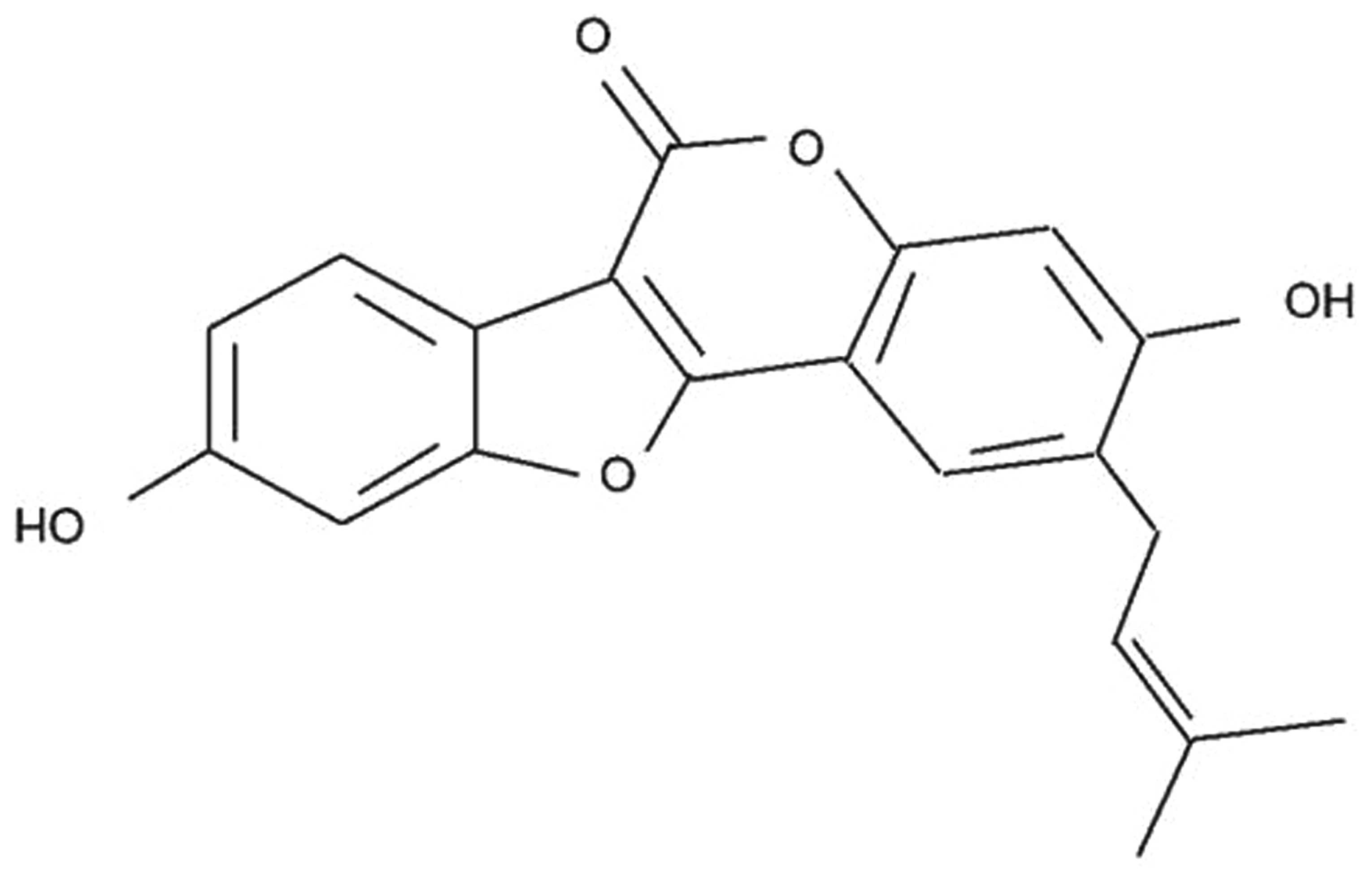
Reagents and chemicals
The chemical structure of psoralidin (with purity
≥98%) is presented in Fig. 1.
Psoralidin and DAPI reagents were obtained from Sigma-Aldrich (St.
Louis, MO, USA). RPMI-1640 medium was obtained from KeyGen
Biotechnology Co., Ltd. (Nanjing, China). Fetal bovine serum (FBS)
was obtained from HyClone (GE Healthcare Life Sciences, Logan, UT,
USA). MTT was purchased from Sangon Biotech Co., Ltd. (Shanghai,
China). Annexin V-fluorescein isothiocyanate (FITC)/propidium
iodide (PI) apoptosis detection kit, caspase-3 colorimetric assay
and NF-κB ELISA assay kits were purchased from Beyotime Institute
of Biotechnology (Nanjing, China). ABT-737 was purchased from EMD
Millipore (#HY-50907; Billerica, MA, USA).
Cell line and culture
The SW480 human colon cancer cell line was obtained
from the Department of Oncology (Central Hospital of Jingzhou,
Jingzhou, China). Cells were grown in RPMI-1640 medium supplemented
with 10% FBS, 100 U/ml penicillin and 100 mg/ml streptomycin
(Invitrogen Life Technologies, Carlsbad, CA, USA) at 37°C in a
humidified atmosphere containing 5% CO2. The culture
medium was replaced every 2–3 days with fresh complete medium.
MTT assay
SW480 cells (2.0×10⁴ cells/well) were
cultured with psoralidin (0, 5, 10 and 20 µM) at 37°C in a
humidified atmosphere containing 5% CO2 for 0, 24, 48
and 72 h in 96-well plates. SW480 cells were washed twice with
phosphate-buffered saline (PBS; Sangon Biotech Co., Ltd.), prior to
the addition of 10 µl MTT to each well. SW480 cells were incubated
at 37°C for 4 h. Subsequently, the culture medium was removed and
150 µl dimethyl sulfoxide (Invitrogen Life Technologies) was added
to each well. SW480 cells were incubated for 20 min at room
temperature with agitation. Cell viability of SW480 cells was
determined by the MTT assay as described previously (19). Briefly, absorbance was measured at a
wavelength of 570 nm using a microplate reader (2104-0010;
PerkinElmer, Inc., Waltham, MA, USA). Cell viability was calculated
as follows: Cell viability (%) = (mean absorbance of psoralidin
treated groups/mean absorance of 0 µM psoralidin group) × 100.
Flow cytometric analysis of cellular
apoptosis
SW480 cells (2×10⁶ cells/well) were
cultured with psoralidin (0, 5, 10 and 20 µM) (17) at 37°C in a humidified atmosphere
containing 5% CO2 for 48 h in 6-well plates. SW480 cells
were harvested and washed twice with cold PBS. Subsequently, SW480
cells (1×10⁶ cells/ml) were resuspended in Annexin-V
binding buffer. Annexin V-FITC (10 µl) was added and cells were
incubated for 30 min in the dark at 4°C. Subsequently, 5 µl PI was
added to the cells and incubated for 10 min in the dark at room
temperature. Cellular apoptosis of SW480 cells was immediately
detected using flow cytometry (EPICS® ALTRA™; Beckman Coulter,
Inc., Brea, CA, USA).
DAPI staining assay
SW480 cells (2×10⁶ cells/well) were
cultured with psoralidin (0, 5, 10 and 20 µM) at 37°C in a
humidified atmosphere containing 5% CO2 for 0, 24, 48
and 72 h in 6-well plates. SW480 cells were washed twice with PBS.
Subsequently, 0.5 ml 4% paraformaldehyde (Beijing DingGuo
ChangSheng Biotechnology Co., Ltd., Beijing, China) was added to
each well and fixed for 30 min at 4°C. SW480 cells were washed
twice with PBS, prior to the addition of sodium citrate (0.1%)
containing 0.1% Triton X-100 and incubated for 5 min at 4°C.
Subsequently, DAPI was added to each well and incubated for 10–15
min at 4°C in the dark. Apoptotic cells were excited by ultraviolet
light. SW480 cells were observed and images were captured under a
fluorescent microscope (Zeiss Axio Observer A1; Ziess AG,
Oberkochen, Germany) at 340 nm.
Detection of caspase-3 activity
SW480 cells (2×10⁶ cells/well) were
cultured with psoralidin (0, 5, 10 and 20 µM) at 37°C in a
humidified atmosphere containing 5% CO2 for 48 h in
6-well plates. Caspase-3 activity was detected by measuring
fluorescence at a wavelength of 405 nm using a microplate reader
(SN11693; Bio-Rad Laboratories, Inc., Hercules, CA, USA) with
caspase-3 colorimetric assay kits.
Measurement of NF-κB activity
SW480 cells (1×10⁶ cells/well) were
cultured with psoralidin (0, 5, 10 and 20 µM) at 37°C in a
humidified atmosphere containing 5% CO2 for 0, 24, 48
and 72 h in 6-well plates. Working solution (100 µl; 1% biotin
labeling, 99% antibody) was added into every well and incubated at
37°C for 1 h. Following incubation, the supernatant was dried and
discarded, and 350 µl wash buffer was added to each well and washed
for 1 min. Next, 3.90 µl tetramethylbenzidine substrate was added
to each well, followed by incubation at 37°C for 30 min in
darkness. Then, 50 µl stop solution was added into every well.
NF-κB activity was analyzed using ELISA assay kits (DRE20177;
Shanghai Bioleaf Biotech Co., Ltd., Shanghai, China) using a
microplate reader (SN11693; Bio-Rad Laboratories, Inc.) at 450 nm,
as described previously (20).
Western blot analysis
SW480 cells (1×10⁶ cells/well) were
cultured with psoralidin (0, 5, 10 and 20 µM) at 37°C in a
humidified atmosphere containing 5% CO2 for 0, 24, 48
and 72 h in 6-well plates. SW480 cells were subsequently incubated
with ice-cold lysis buffer (Beyotime Institute of Biotechnology)
for 30 min on ice and then centrifuged at 12,000 × g for 10 min at
4°C. The total protein concentration of soluble materials was
determined using a bicinchoninic acid protein assay (Thermo Fisher
Scientific, Inc., Waltham, MA, USA). Equal quantities of protein
were separated using 12% SDS-PAGE (Invitrogen Life Technologies)
and then transferred onto a polyvinylidene difluoride membrane
(0.22 mm; GE Healthcare Life Sciences, Piscataway, NJ, USA).
Following blocking with Tris-buffered saline (Beyotime Institute of
Biotechnology) containing 5% non-fat milk for 2 h, the membranes
were incubated with monoclonal mouse anti-human Bcl-2 (1:1,500;
cat. no. sc-7382; Santa Cruz Biotechnology, Inc., Dallas, TX, USA),
monoclonal mouse anti-human Bax (1:2,000; cat. no. sc-8432; Santa
Cruz Biotechnology, Inc.) and monoclonal mouse anti-human β-actin
(1:500; cat. no. sc-8432; Tiangen Biotech Co., Ltd., Beijing,
China) overnight at 4°C. The membrane was washed twice with
Tris-buffered saline with Tween-20 (Sigma-Aldrich) for 2 h and then
incubated with horseradish peroxidase-conjugated sheep anti-mouse
immunoglobulin G (1:1,000; cat. no. sc-358922; Santa Cruz
Biotechnology, Inc.) at room temperature for 2 h. Proteins were
subsequently detected using enhanced chemiluminescence (DAB
Horseradish Peroxidase Color Development Kit; Beyotime Institute of
Biotechnology).
Effects of Bcl-2 inhibition
To further investigate the potential association
between Bcl-2 inhibition and the effect of psoralidin on SW480
cells, SW480 cells were incubated with 1 µM Bcl-2 inhibitor
(ABT-737; 99% purity; Sigma-Aldrich) and 10 µM psoralidin for 48 h
at 37°C in a humidified atmosphere containing 5% CO2.
The aforementioned MTT assay and western blotting protocols were
then performed to determine cell viability and Bcl-2 protein
expression levels, respectively.
Statistical analysis
Statistical analysis was performed using SPSS 17.0
software (SPSS, Inc., Chicago, IL, USA). Data are expressed as the
mean ± standard deviation. Data were analyzed using Student's
t-test. P<0.05 was considered to indicate a statistically
significant difference.
Results
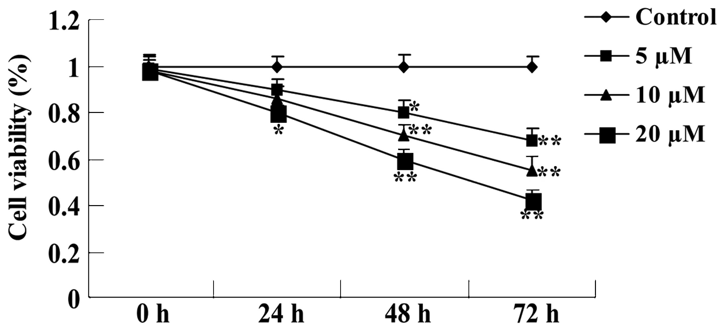
Psoralidin treatment reduces the
viability of SW480 cells
The anti-proliferative effect of psoralidin (0, 5,
10 and 20 µM) on the viability of SW480 cells was detected by MTT
assay. As demonstrated in Fig. 2,
psoralidin (5, 10 and 20 µM) treatment reduced the viability of
SW480 cells in a dose- and time-dependent manner. This effect was
particularly evident at a concentration of 20 µM, at which the
proliferation of SW480 cells was significantly inhibited at 24, 48
and 72 h (P<0.01; Fig. 2).
Additionally, treatment with 5 µM psoralidin for 72 h and 10 µM
psoralidin for 48 and 72 h also significantly reduced SW480 cell
viability (P<0.01; Fig. 2).
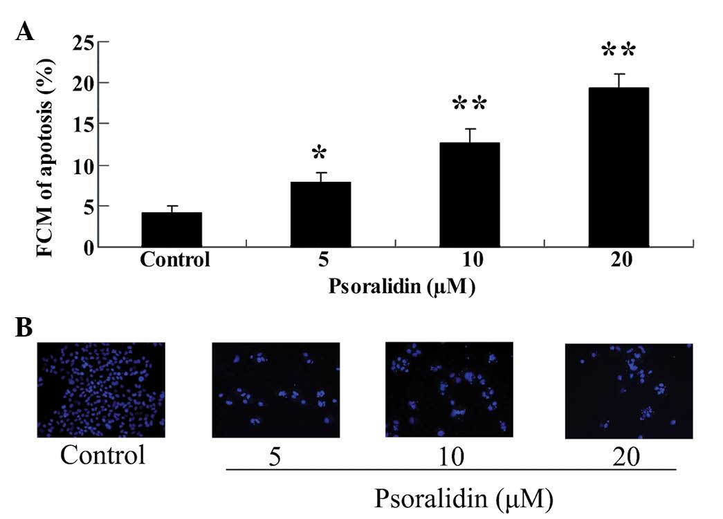
Psoralidin treatment promotes the
apoptosis of SW480 cells
To determine whether apoptosis mediated the
anti-proliferative effect of psoralidin (0, 5, 10 and 20 µM),
apoptosis of SW480 cells was analyzed using flow cytometry and a
DAPI staining assay. As demonstrated in Fig. 3A, psoralidin (0, 5, 10 and 20 µM)
treatment promoted the apoptosis of SW480 cells in a dose-dependent
manner. This effect of psoralidin was particularly evident at
concentrations of 10 and 20 µM, at which the apoptosis of SW480
cells at 48 h was significantly increased (P<0.01; Fig. 3A). In addition, treatment with 5 µM
psoralidin for 72 h significantly enhanced the apoptosis of SW480
cells at 48 h (P<0.05; Fig. 3A).
Notably, the results of the DAPI staining assay revealed that the
apoptosis of SW480 cells was augmented in cells treated with
psoralidin (5, 10 and 20 µM) at 48 h, compared with the control
group (Fig. 3B).
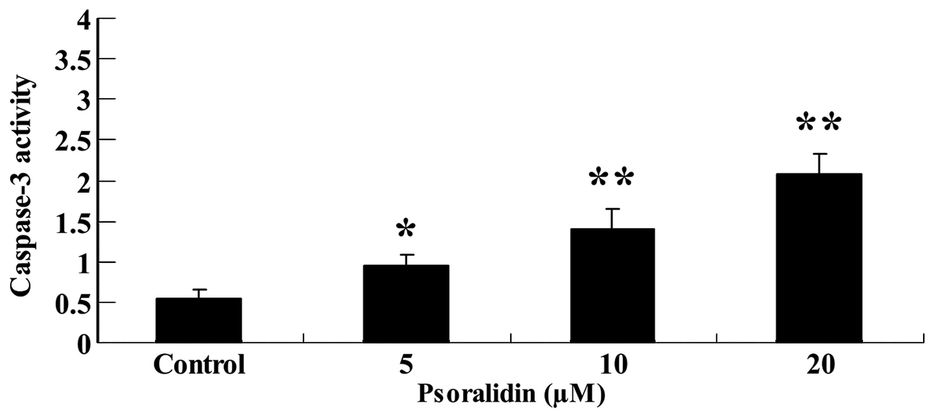
Psoralidin treatment enhances
caspase-3 activity of SW480 cells
To further investigate the anti-proliferative effect
of psoralidin (0, 5, 10 and 20 µM) on the caspase-3 activity of
SW480 cells, caspase-3 activity was measured using colorimetric
assay kits. As demonstrated in Fig.
4, psoralidin (5, 10 and 20 µM) treatment increased the
caspase-3 activity of SW480 cells in a dose-dependent manner.
Following psoralidin (10 and 20 µM) treatment for 48 h, the
caspase-3 activity of SW480 cells was significantly augmented
(P<0.01), and following psoralidin (5 µM) treatment for 48 h,
the caspase-3 activity of SW480 cells was also augmented
(P<0.05; Fig. 4A and B).
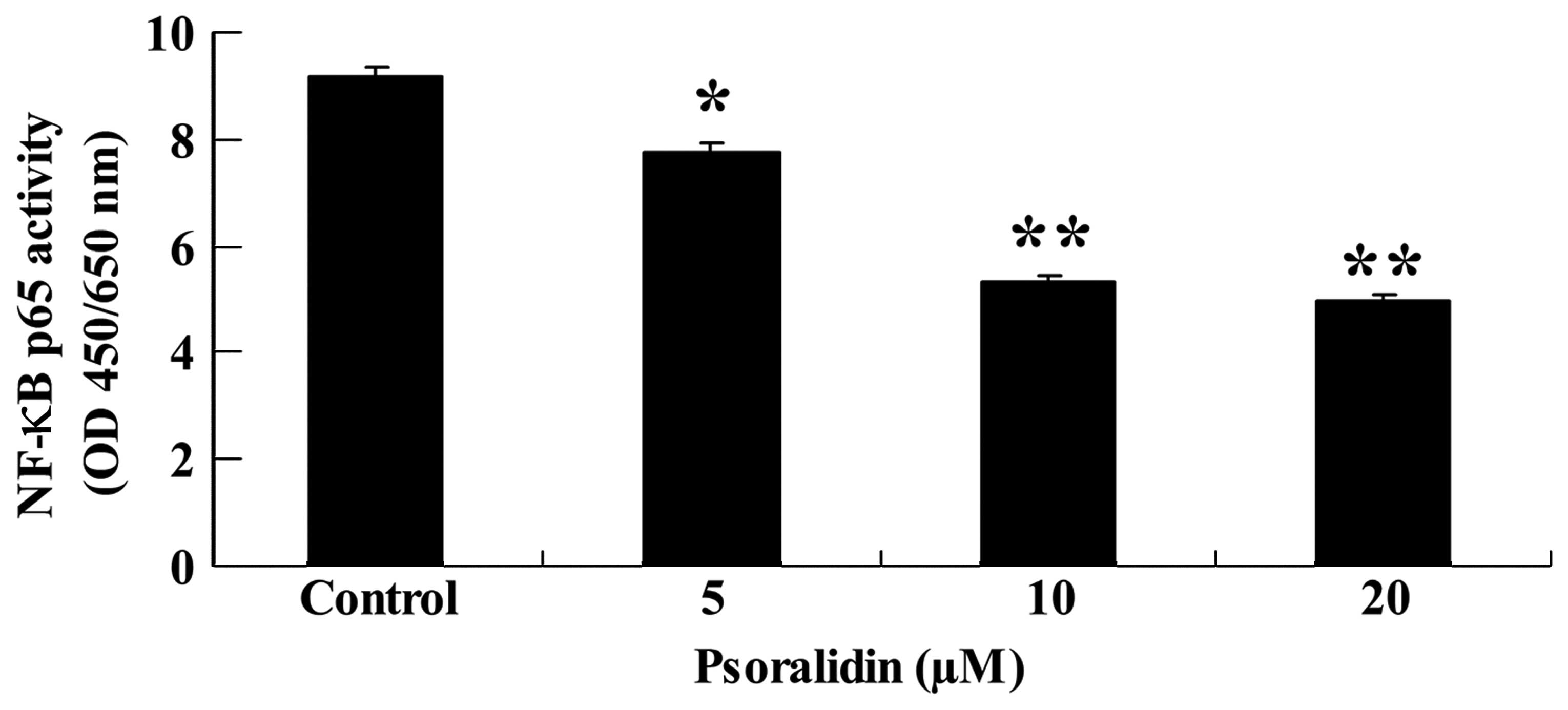
Psoralidin treatment reduces the NF-κB
activity of SW480 cells
The potential association between the
anti-proliferative effect of psoralidin (0, 5, 10 and 20 µM) and
NF-κB activity in SW480 cells was evaluated. NF-κB p65 activity was
investigated using an ELISA assay kit. As demonstrated in Fig. 5, NF-κB p65 activity was reduced by
psoralidin (5, 10 and 20 µM) treatment in a dose-dependent manner.
NF-κB p65 activity was significantly decreased following psoralidin
(10 and 20 µM) treatment for 48 h (P<0.01), and markedly
decreased following psoralidin (5 µM) treatment for 48 h
(P<0.05; Fig. 5).
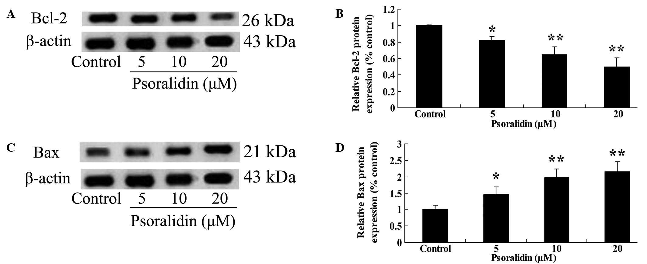
Psoralidin treatment inhibits Bcl-2
and enhances Bax protein expression in SW480 cells
To determine whether there was an association
between the anti-proliferative effect of psoralidin (0, 5, 10 and
20 µM) treatment on SW480 cells, and the Bcl-2/Bax signaling
pathway, Bcl-2 and Bax protein expression in SW480 cells was
detected by western blot analysis. As demonstrated in Fig. 6A and B, Bcl-2 protein expression was
significantly inhibited by psoralidin (10 and 20 µM) treatment for
48 h (P<0.01), and markedly decreased following psoralidin (5
µM) treatment for 48 h (P<0.05; Fig.
6A and B). By contrast, Bax protein expression in SW480 cells
was markedly promoted by psoralidin (5, 10 and 20 µM) treatment:
Bax protein expression was significantly increased following
psoralidin (10 and 20 µM) treatment for 48 h (P<0.01), and
markedly increased following psoralidin (5 µM) treatment for 48 h
(P<0.05; Fig. 6C and D).
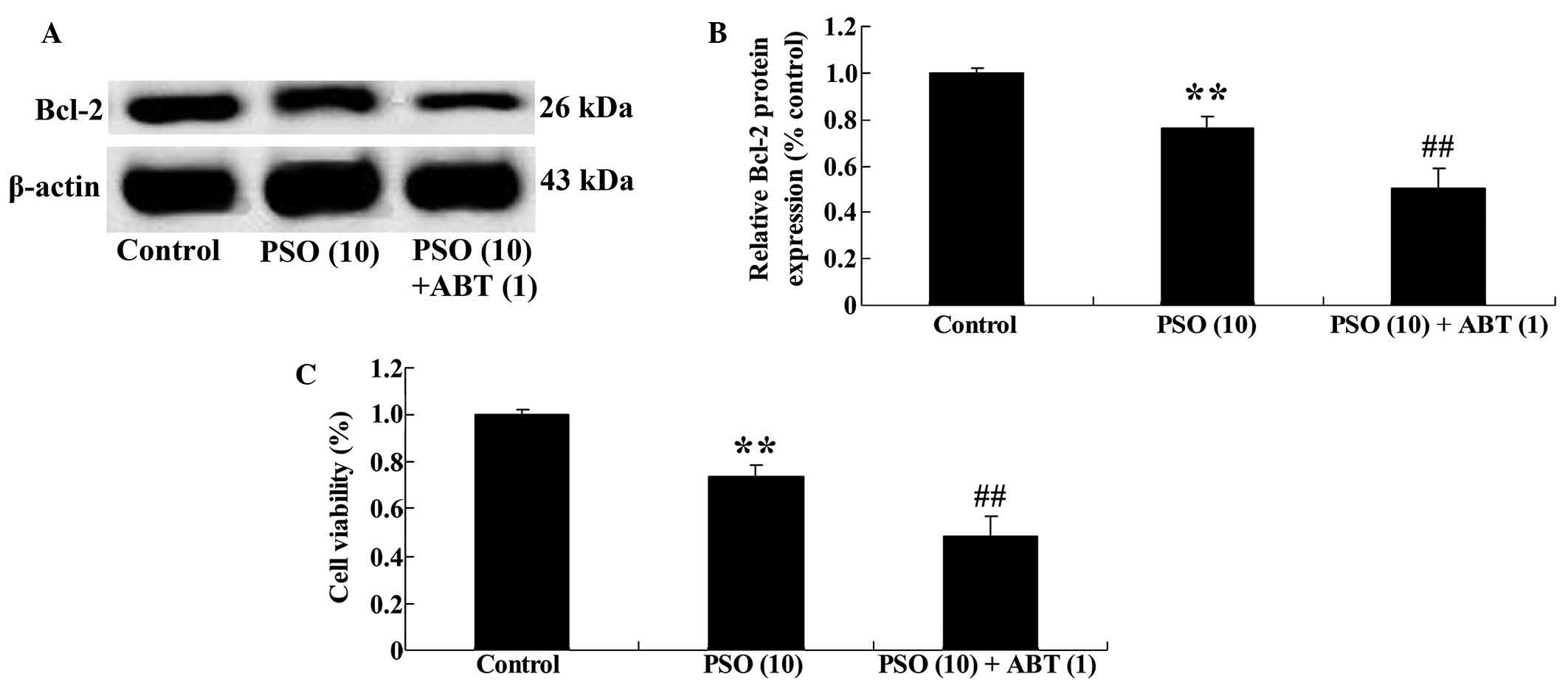
Bcl-2 inhibitor is able to enhance the
anti-proliferative effect of psoralidin on SW480 cells
To further investigate the potential association
between Bcl-2 inhibition and the effect of psoralidin on SW480
cells, psoralidin-treated SW480 cells were incubated with a Bcl-2
inhibitor (ABT-737) for 48 h. ABT-737 inhibited Bcl-2 protein
expression (Fig. 7A and B) and
decreased the viability of SW480 cells (Fig. 7C), compared with the psoralidin (10
µM) treatment control group.
Discussion
Worldwide, colon cancer is the third most common
cancer in terms of incidence, following lung and breast cancer
(21). There are differences in the
geographical distribution of colon cancer (22). In developed countries or regions,
including Australia, New Zealand, Europe and North America, there
is a higher prevalence of colon cancer, whereas in African and
South Asian countries, incidence is reduced, and may be up to ten
times lower (23). Colon cancer
mortality rates are highest in Central and Eastern Europe, and
lowest in Central Africa (24). In
the present study, psoralidin treatment reduced the viability and
promoted the apoptosis of SW480 cells in a dose-dependent manner.
Furthermore, Hao et al (18)
reported that psoralidin inhibited the proliferation of A549 cells
by inducing ROS production. Szliszka et al (25) identified that psoralidin inhibited
cell proliferation of prostate cancer cells by enhancement of tumor
necrosis factor-related apoptosis-inducing ligand-mediated
apoptosis. Additionally in the present study, caspase-3 activity of
SW480 cells was significantly enhanced following psoralidin
treatment. Das et al (26)
suggested that psoralidin may promote growth arrest in prostate
cancer cells by activation of caspase-3 and caspase-9.
A number of studies have revealed that NF-κB is
highly expressed within tumor cells, and is involved in cell
proliferation, invasion, angiogenesis and inhibition of apoptosis
(27). Activation of the NF-κB signal
transduction pathway may weaken the antitumor effects of
chemoradiation therapy (28). The
Hut-78 cell line, which highly expresses NF-κB, is resistant to the
anticancer drug Taxol; however, Jurket cells, which do not express
NF-κB, are sensitive to Taxol. This suggests that NF-κB
participates in regulation of the sensitivity of tumor cells to
anticancer therapies (29). In the
present study, it was observed that psoralidin treatment markedly
reduced the NF-κB p65 activity of SW480 cells. Chiou et al
(30) observed that psoralidin
inhibited lipopolysaccharide-induced inducible nitric oxide
synthase expression by mediating NF-κB signaling. Yang et al
(17) reported that psoralidin
regulated ionizing radiation-induced pulmonary inflammation by
regulation of the phosphoinositide 3-kinase/Akt and NF-κB
pathways.
Apoptosis is the primary mechanism for removal of
unwanted cells during embryonic development and tissue repair, and
disorders of the apoptotic mechanism are closely associated with
tumorigenesis. Apoptosis is a process of active cell death that is
controlled by genes (31). In
particular, Bcl-2 family genes are significant for apoptosis and
have been extensively studied. Bcl-2 family proteins may act on the
apoptotic pathway and exert regulatory effect on apoptosis via
various routes (32). The levels of
and interaction between Bcl-2 and Bax gene products may also be the
regulatory pathway center of Bcl-2 apoptosis, while the Bcl-2/Bax
protein ratio is a significant switch in apoptosis regulation
(33). It is known that psoralidin
may decrease Bcl-2 protein expression and increase Bax protein
expression in SW480 cells (34).
Based on these findings, the present study demonstrated that Bcl-2
protein expression was markedly decreased following psoralidin
treatment. It was previously revealed that psoralidin inhibited
androgen-independent prostate cancer cells via Bcl-2/Bax signaling
pathway inhibition (17). In the
present study, it was demonstrated that inhibition of Bcl-2 protein
expression was capable of enhancing the anticancer effects of
psoralidin on SW480 cells. Srinivasan et al (35) suggested that psoralidin inhibited cell
proliferation and induced apoptosis of prostate cancer cells by
mediation of NF-κB and Bcl-2 signaling pathways.
In conclusion, psoralidin decreased the expression
levels of NF-κB and Bcl-2, increased the expression levels of Bax,
promoted caspase-3 activity and subsequently induced apoptosis in
human colon cancer SW480 cells. The results of the present study
demonstrate the potential benefits of psoralidin in clinical
practice.
References
|
1
|
Isik A, Peker K, Firat D, Yilmaz B, Sayar
I, Idiz O, Cakir C, Demiryilmaz I and Yilmaz I: Importance of
metastatic lymph node ratio in non-metastatic, lymph node-invaded
colon cancer: A clinical trial. Med Sci Monit. 20:1369–1375. 2014.
View Article : Google Scholar : PubMed/NCBI
|
|
2
|
Lu ZJ, Lu LG, Tao KZ, Chen DF, Xia Q, Weng
JJ, Zhu F, Wang XP and Zheng P: MicroRNA-185 suppresses growth and
invasion of colon cancer cells through inhibition of the
hypoxia-inducible factor-2α pathway in vitro and in
vivo. Mol Med Rep. 10:2401–2408. 2014.PubMed/NCBI
|
|
3
|
Su CC: Tanshinone IIA potentiates the
efficacy of 5-FU in Colo205 colon cancer cells in vivo
through downregulation of P-gp and LC3-II. Exp Ther Med. 3:555–559.
2012.PubMed/NCBI
|
|
4
|
Grijelmo C, Rodrigue C, Svrcek M, Bruyneel
E, Hendrix A, de Wever O and Gespach C: Proinvasive activity of
BMP-7 through SMAD4/src-independent and ERK/Rac/JNK-dependent
signaling pathways in colon cancer cells. Cell Signal.
19:1722–1732. 2007. View Article : Google Scholar : PubMed/NCBI
|
|
5
|
Miskolci V, Rollins J, Vu HY, Ghosh CC,
Davidson D and Vancurova I: NFkappaB is persistently activated in
continuously stimulated human neutrophils. Mol Med. 13:134–142.
2007. View Article : Google Scholar : PubMed/NCBI
|
|
6
|
Luo W, Liu Y, Zhang J, Luo X, Lin C and
Guo J: Andrographolide inhibits the activation of NF-κB and MMP-9
activity in H3255 lung cancer cells. Exp Ther Med. 6:743–746.
2013.PubMed/NCBI
|
|
7
|
Chelvarajan RL, Liu Y, Popa D, Getchell
ML, Getchell TV, Stromberg AJ and Bondada S: Molecular basis of
age-associated cytokine dysregulation in LPS-stimulated
macrophages. J Leukoc Biol. 79:1314–1327. 2006. View Article : Google Scholar : PubMed/NCBI
|
|
8
|
Nakanishi C and Toi M: Nuclear
factor-kappaB inhibitors as sensitizers to anticancer drugs. Nat
Rev Cancer. 5:297–309. 2005. View
Article : Google Scholar : PubMed/NCBI
|
|
9
|
Yu M, Tong X, Qi B, Qu H, Dong S, Yu B,
Zhang N, Tang N, Wang L and Zhang C: Berberine enhances
chemosensitivity to irinotecan in colon cancer via inhibition of
NF-κB. Mol Med Rep. 9:249–254. 2014.PubMed/NCBI
|
|
10
|
Tanwar L, Vaish V and Sanyal SN:
Chemoprevention of 1,2-dimethylhydrazine-induced colon
carcinogenesis by a non-steroidal anti-inflammatory drug,
etoricoxib, in rats: Inhibition of nuclear factor kappaB. Asian Pac
J Cancer Prev. 10:1141–1146. 2009.PubMed/NCBI
|
|
11
|
Sato T, Hanada M, Bodrug S, Irie S, Iwama
N, Boise LH, Thompson CB, Golemis E, Fong L, Wang HG, et al:
Interactions among members of the Bcl-2 protein family analyzed
with a yeast two-hybrid system. Proc Natl Acad Sci USA.
91:9238–9242. 1994. View Article : Google Scholar : PubMed/NCBI
|
|
12
|
Zhao L, Yu N, Guo T, Hou Y, Zeng Z, Yang
X, Hu P, Tang X, Wang J and Liu M: Tissue biomarkers for prognosis
of prostate cancer: A systematic review and meta-analysis. Cancer
Epidemiol Biomarkers Prev. 23:1047–1054. 2014. View Article : Google Scholar : PubMed/NCBI
|
|
13
|
Giatromanolaki A, Stathopoulos GP,
Koukourakis MI, Rigatos S, Vrettou E, Kittas C, Fountzilas G and
Sivridis E: Angiogenesis and apoptosis-related protein (p53, bcl-2,
and bax) expression versus response of gastric adenocarcinomas to
paclitaxel and carboplatin chemotherapy. Am J Clin Oncol.
24:222–226. 2001. View Article : Google Scholar : PubMed/NCBI
|
|
14
|
Mirjolet JF, Barberi-Heyob M, Didelot C,
Peyrat JP, Abecassis J, Millon R and Merlin JL: Bcl-2/Bax protein
ratio predicts 5-fluorouracil sensitivity independently of p53
status. Br J Cancer. 83:1380–1386. 2000. View Article : Google Scholar : PubMed/NCBI
|
|
15
|
Ko YJ, Jeong JW, Choi YH and Ryu CH: Soy
soluble polysaccharide induces apoptosis in HCT-116 human colon
cancer cells via reactive oxygen species generation. Mol Med Rep.
8:1767–1772. 2013.PubMed/NCBI
|
|
16
|
Mao JD, Wu P, Xia XH, Hu JQ, Huang WB and
Xu GQ: Correlation between expression of gastrin, somatostatin and
cell apoptosis regulation gene bcl-2/bax in large intestine
carcinoma. World J Gastroenterol. 11:721–725. 2005. View Article : Google Scholar : PubMed/NCBI
|
|
17
|
Yang HJ, Youn H, Seong KM, Yun YJ, Kim W,
Kim YH, Lee JY, Kim CS, Jin YW and Youn B: Psoralidin, a dual
inhibitor of COX-2 and 5-LOX, regulates ionizing radiation
(IR)-induced pulmonary inflammation. Biochem Pharmacol. 82:524–534.
2011. View Article : Google Scholar : PubMed/NCBI
|
|
18
|
Hao W, Zhang X, Zhao W and Chen X:
Psoralidin induces autophagy through ROS generation which inhibits
the proliferation of human lung cancer A549 cells. PeerJ.
2:e5552014. View Article : Google Scholar : PubMed/NCBI
|
|
19
|
Pu Z, Zhang X, Chen Q, Yuan X and Xie H:
Establishment of an expression platform of OATP1B1 388GG and 521CC
genetic polymorphism and the therapeutic effect of tamoxifen in
MCF-7 cells. Oncol Rep. 33:2420–2428. 2015.PubMed/NCBI
|
|
20
|
Wang S, Liu K, Seneviratne CJ, Li X,
Cheung GS, Jin L, Chu CH and Zhang C: Lipoteichoic acid from an
Enterococcus faecalis clinical strain promotes TNF-α expression
through the NF-κB and p38 MAPK signaling pathways in differentiated
THP-1 macrophages. Biomed Rep. 3:697–702. 2015.PubMed/NCBI
|
|
21
|
Han SA, Lee WY, Park CM, Yun SH and Chun
HK: Comparison of immunologic outcomes of laparoscopic vs open
approaches in clinical stage III colorectal cancer. Int J
Colorectal Dis. 25:631–638. 2010. View Article : Google Scholar : PubMed/NCBI
|
|
22
|
Zhang L, Cai Q, Lin J, Fang Y, Zhan Y,
Shen A, Wei L, Wang L and Peng J: Chloroform fraction of
Scutellaria barbata D. Don promotes apoptosis and suppresses
proliferation in human colon cancer cells. Mol Med Rep. 9:701–706.
2014.PubMed/NCBI
|
|
23
|
Touil Y, Igoudjil W, Corvaisier M, Dessein
AF, Vandomme J, Monté D, Stechly L, Skrypek N, Langlois C, Grard G,
et al: Colon cancer cells escape 5FU chemotherapy-induced cell
death by entering stemness and quiescence associated with the
c-Yes/YAP axis. Clin Cancer Res. 20:837–846. 2014. View Article : Google Scholar : PubMed/NCBI
|
|
24
|
Lo AC, Soliman AS, Khaled HM, Aboelyazid A
and Greenson JK: Lifestyle, occupational, and reproductive factors
and risk of colorectal cancer. Dis Colon Rectum. 53:830–837. 2010.
View Article : Google Scholar : PubMed/NCBI
|
|
25
|
Szliszka E, Czuba ZP, Sędek Ł, Paradysz A
and Król W: Enhanced TRAIL-mediated apoptosis in prostate cancer
cells by the bioactive compounds neobavaisoflavone and psoralidin
isolated from Psoralea corylifolia. Pharmacol Rep.
63:139–148. 2011. View Article : Google Scholar : PubMed/NCBI
|
|
26
|
Das TP, Suman S and Damodaran C: Induction
of reactive oxygen species generation inhibits
epithelial-mesenchymal transition and promotes growth arrest in
prostate cancer cells. Mol Carcinog. 53:537–547. 2014. View Article : Google Scholar : PubMed/NCBI
|
|
27
|
Cohen M, Meisser A, Haenggeli L and
Bischof P: Involvement of MAPK pathway in TNF-alpha-induced MMP-9
expression in human trophoblastic cells. Mol Hum Reprod.
12:225–232. 2006. View Article : Google Scholar : PubMed/NCBI
|
|
28
|
Davoudi Z, Akbarzadeh A, Rahmatiyamchi M,
Movassaghpour AA, Alipour M, Nejati-Koshki K, Sadeghi Z,
Dariushnejad H and Zarghami N: Molecular target therapy of AKT and
NF-kB signaling pathways and multidrug resistance by specific cell
penetrating inhibitor peptides in HL-60 cells. Asian Pac J Cancer
Prev. 15:4353–4358. 2014. View Article : Google Scholar : PubMed/NCBI
|
|
29
|
Ali S, El-Rayes BF, Sarkar FH and Philip
PA: Simultaneous targeting of the epidermal growth factor receptor
and cyclooxygenase-2 pathways for pancreatic cancer therapy. Mol
Cancer Ther. 4:1943–1951. 2005. View Article : Google Scholar : PubMed/NCBI
|
|
30
|
Chiou WF, Don MJ, Liao JF and Wei BL:
Psoralidin inhibits LPS-induced iNOS expression via repressing
Syk-mediated activation of PI3K-IKK-IκB signaling pathways. Eur J
Pharmacol. 650:102–109. 2011. View Article : Google Scholar : PubMed/NCBI
|
|
31
|
Edwards MJ: Apoptosis, the heat shock
response, hyperthermia, birth defects, disease and cancer. Where
are the common links? Cell Stress Chaperones. 3:213–220. 1998.
View Article : Google Scholar : PubMed/NCBI
|
|
32
|
Xiong Y, Ma XY, Zhang Z, Shao ZJ, Zhang YY
and Zhou LM: Apoptosis induced by β,β-dimethylacrylshikonin is
associated with Bcl-2 and NF-κB in human breast carcinoma MCF-7
cells. Oncol Lett. 6:1789–1793. 2013.PubMed/NCBI
|
|
33
|
Gu Y, Liu SL, Ju WZ, Li CY and Cao P:
Analgesic-antitumor peptide induces apoptosis and inhibits the
proliferation of SW480 human colon cancer cells. Oncol Lett.
5:483–488. 2013.PubMed/NCBI
|
|
34
|
Nam H and Kim MM: Ursolic acid induces
apoptosis of SW480 cells via p53 activation. Food Chem Toxicol.
62:579–583. 2013. View Article : Google Scholar : PubMed/NCBI
|
|
35
|
Srinivasan S, Kumar R, Koduru S,
Chandramouli A and Damodaran C: Inhibiting TNF-mediated signaling:
A novel therapeutic paradigm for androgen independent prostate
cancer. Apoptosis. 15:153–161. 2010. View Article : Google Scholar : PubMed/NCBI
|