Introduction
Breast cancer is the most frequently diagnosed type
of cancer, and the leading cause of cancer-associated mortality
among females, accounting for 23% of the total cases of cancer and
14% of the cancer-associated mortalities (1). Metastasis, characterized by its complex
multistep processes and mechanism, is the most common aspect and
malignant phenotype of breast cancer, and is responsible for ~90%
of the lethality associated with this type of cancer (2). Despite early detection and opportune
treatments, metastasis remains hardly inevitable, and predicts a
poor prognosis in patients with breast cancer (3,4). The
current treatments for breast cancer, including surgery,
chemotherapy and radiation therapy, are not efficient in targeting
metastasis. Therefore, the development of novel strategies for the
treatment of breast cancer is required, particularly those
targeting metastasis.
Metastasis via the lymphatic system is the primary
step in the progression of breast cancer. The majority of patients
with breast cancer present with lymph node metastasis when
initially diagnosed or following surgery, which usually results in
a poor prognosis (5,6). Vascular endothelial growth factor
(VEGF)-C, a member of the VEGF family, has been defined as a
lymphangiogenic growth factor, which participates in tumor
lymphangiogenesis and spreading of tumor cells to lymph nodes in
breast cancer (7–9). It has been previously observed that the
overexpression of VEGF-C in tumor tissues of patients with breast
cancer is negatively correlated with the survival rate and
prognosis of these patients (10,11).
Previous studies have demonstrated that knocking down the
expression of VEGF-C with a functional blocking antibody or with
siRNA prevents lymphogenesis and enhances chemosensitivity in human
breast cancer cells (12). Therefore,
VEGF-C can be considered a representative marker to estimate
lymphogenesis and lymphatic metastasis in breast cancer. Previous
studies have reported that the presence of lymphatic metastases and
the expression levels of VEGF-C are associated with the levels of
survivin in breast cancer (13).
Survivin, the smallest but most efficient member of
the inhibitory apoptotic protein family, is involved in cell
proliferation and inhibition of apoptosis (14,15). It
has been previously reported that the downregulation of the
expression or function of survivin is able to inhibit tumor growth
and increase spontaneous apoptosis in different tumor models
(16,17). Furthermore, previous studies have
implicated survivin in cell motility, which may suggest a role for
this protein in promoting tumor metastasis (18). In addition, survivin was observed to
be positively correlated with tumor metastasis in laryngeal
carcinoma, small adenocarcinoma of the lung and breast cancer
(19–21). The overexpression of survivin in
patients with cancer usually indicates a poor prognosis and high
risk of mortality of these patients, since survivin is commonly
expressed in neoplasms and embryonic tissue, but rarely detected in
normal differentiated adult tissues (22). Therefore, survivin may be a promising
target for the diagnosis and therapy of cancer.
Survivin and VEGF-C are highly expressed in breast
cancer tissues, and a positive correlation exists between the
expression levels of these proteins in cancer (23). However, to the best of our knowledge,
the complex association between survivin and lymphatic metastasis
remains poorly characterized. In the present study, a murine breast
cancer cell line termed 4T1, which exhibits metastatic
characteristics similar to those of human breast cancer, was
employed to investigate whether knocking down survivin by siRNA
provided antitumor effects in these cells, and to study the
association between survivin and lymphatic metastasis in mouse
breast cancer.
Materials and methods
Cell culture
Murine mammary carcinoma 4T1 cells were acquired
from the Shanghai Laboratory Animal Center of the Chinese Academy
of Sciences (Shanghai, China). The cells were maintained in high
glucose Dulbeccos modified Eagle medium (DMEM) (Hyclone, GE
Healthcare Life Sciences, Logan, UT, USA), containing 10% fetal
bovine serum (Gibco™; Thermo Fisher Scientific, Inc., Carlsbad, CA,
USA), 1 mM mixed non-essential amino acids, 2 mM L-glutamine, 100
U/ml penicillin and 100 µg/ml streptomycin, and incubated at 37°C
in a humidified atmosphere with 5% CO2. Cells in
exponential growth phase were used throughout the experiments.
siRNA design and cell
transfection
The sequences of the siRNA oligonucleotides were
designed as follows: Survivin, 5′-GGAAUUGGAAGGCUGGGAATT-3 (sense)
and 5′-UUCCCAGCCUUCCAAUUCCTT-3 (antisense); and VEGF-C,
5′-GCAAGACGUUGUUUGAAAUTT-3 (sense) and 5′-AUUUCAAACAACGUCUUGCTT-3
(antisense). In order to enhance the stability of the siRNAs, the
sense strands were methylated, while the antisense strands were not
modified. Scrambled siRNA (siRNAscr), 5′-UUCUCCGAACGUGUCACGUTT-3
(sense) and 5′-ACGUGACACGUUCGGAGAATT-3 (antisense), was used as
negative control, since the selected sequences did not share any
homology with any of the known mRNA databases. The 3′-end of the
siRNAscr was labeled with fluorescein isothiocyanate to evaluate
the efficiency of the transfection. All the siRNA sequences were
chemically synthesized by Shanghai GenePharma Co., Ltd. (Shanghai,
China).
The siRNAs were transfected into cultured cells at
50% confluence, which had been seeded on dishes 24 h earlier. For
transfection, Invitrogen™ Lipofectamine 2000 reagent (Thermo Fisher
Scientific, Inc.) was used, according to the manufacturers
protocol. Three groups of cells were established, as follows: i) An
untransfected (UT) group, serving as blank control; ii) a group
transfected with 100 nmol/l siRNAscr; and iii) a group transfected
with 100 nmol/l survivin-siRNA. The cells in the UT group were
treated with Opti-MEM (Thermo Fisher Scientific, Inc.), while the
cells in the other groups were treated with Lipofectamine
2000-Opti-MEM. At 5–6 h post-transfection, the medium was replaced
with 10% serum-supplemented DMEM, and the cells were incubated for
additional 24–96 h. Next, the cells were harvested by
centrifugation, rinsed with phosphate-buffered saline, and
subjected to total RNA or protein extraction.
RNA extraction and quantitative
polymerase chain reaction (qPCR)
Total RNA was isolated from the cells using
Invitrogen™ TRIzol reagent (Thermo Fisher Scientific, Inc.), and
cDNA was prepared with First Strand cDNA Synthesis Kit (Roche
Diagnostics, Basel, Switzerland), according to the manufacturers
instructions. Next, the cDNA amplified by qPCR, using the following
primers (Shanghai Sangon Biotech Co., Ltd., Shanghai, China):
Survivin, forward 5′-ATCGCCACCTTCAAGAACTG-3 and reverse
5′-GGCCAAATCAGGCTCGTTCT-3; VEGF-C, forward
5′-AAACCTCAGCTGTCTGGTCC-3 and reverse 5′-GAACCATGTGGATTACTGCG-3;
and β-actin, forward 5′-GCGGACTGTTACTGAGCTGCGTAACG-3 and reverse
5′-GAAGCAATGCTGTCACCTTCCC-3′, which was used as reference gene. The
reaction conditions were as follows: 95°C for 30 sec, followed by
30 cycles of annealing at 58 or 57°C (in the case of survivin or
VEGF-C and β-actin, respectively) for 30 sec, and extension at 72°C
for 30 sec. qPCR amplification was performed on a Corbett
Rotor-Gene 3000 (Qiagen, Inc., Valencia, CA, USA), using FastStart
Universal SYBR Green Master (Rox) (Roche Diagnostics). Each RNA
sample was analyzed in triplicate, and the data were quantified
according to the 2-ΔΔCq method.
Western blotting
The western blot assay was performed as previously
described (24). Briefly, total
protein content was extracted from the cells with RIPA lysis buffer
(Beyotime Institute of Biotechnology, Nantong, China), which
contained phosphatase inhibitors (in addition to protease
inhibitors) when detecting phosphorylated proteins. The protein
concentration in the cell lysates was determined with Pierce™ BCA
Protein Assay Kit (Thermo Fisher Scientific, Rockford, IL, USA).
Protein samples were separated by SDS-PAGE using 12% polyacrylamide
gels (Beijing Solarbio Science & Technology Co., Ltd., Beijing,
China) and then transferred to polyvinylidene difluoride membranes
(EMD Millipore, Bedford, MA, USA). The membranes were blocked for 1
h with Tris-buffered saline containing 0.1% Tween 20 (TBST) and 5%
non-fat milk at room temperature. Subsequently, primary antibodies
were added to the membrane and incubated overnight at 4°C. Rabbit
polyclonal antibodies against survivin (cat. no. sc-10811), VEGF-C
(cat. no. sc-25783), hypoxia-inducible factor (HIF)-1α (cat. no.
sc-10790) and β-actin (cat. no. sc-7210; Santa Cruz Biotechnology,
Inc., Dallas, TX, USA), were diluted 1:300, while the rabbit
monoclonal antibodies against Akt (cat. no. 9272) and
phosphorylated (p)-Akt (Thr308; cat. no. 9275) (Cell Signaling
Technology, Inc., Danvers, MA, USA) were diluted 1:1,000. Following
3 washes of 5 min each with TBST, the membranes were incubated with
a horseradish peroxidase-conjugated goat anti-rabbit secondary
antibody (cat. no. ZDR-5306; Zhongshan Jinqiao Biotechnology, Co.,
Ltd., Beijing, China) for 1 h at room temperature, before
subjection to 3 washes of 5 min each with TBST. Proteins were
visualized using the enhanced chemiluminescence method, and protein
expression was quantified with a Gel EDAS 293 analysis system (Cold
Spring USA Corp., Cherry Hill, NJ, USA).
Cell proliferation assay
Cell viability was examined by MTT assay (Roche
Diagnostics). For the experiment, 4T1 cells were seeded in a
96-well plate (Corning Life Sciences, New York, NY, USA) at a
density of 5×10³ cells/well, and transfected 24 h later
with 100 nM survivin-siRNA or siRNAscr. At 24, 48 and 72 h
post-transfection, MTT (5 mg/ml) was added to the cells, which were
incubated at 37°C for additional 4 h. Next, the supernatant was
removed, and 150 µl dimethyl sulfoxide (Sigma-Aldrich) was added to
each well, followed by 10-min agitation. The absorbance of each
well at 490 nm was measured using an ELISA plate reader (Thermo
Multiskan MK3; Thermo Fisher Scientific, Shanghai, China).
Quintuplicate wells were used for each experimental condition, and
all the experiments were repeated at least three times.
Cell invasion and migration assay
Matrigel invasion assay was performed using
transwell chambers (Corning Life Sciences). For the assay, Matrigel
(1 mg/ml; BD Biosciences, Franklin Lakes, NJ, USA) was diluted in
serum-free DMEM, and 70 µl of the diluted Matrigel were then coated
in 8-µm pore size filters, followed by incubation for 5–6 h at room
temperature. At 24 h post-transfection, 100 µl 4T1 cells in
serum-free medium (5×10⁵ cells/ml) were placed in the
upper chamber, while the lower chamber was filled with 600 µl NIH
3T3-conditioned medium (prepared in our laboratory). Following 24 h
incubation, the non-invasive cells on the upper chamber were
removed with cotton swabs, and the migrated cells were stained with
0.1% crystal violet (#C6158; Sigma-Aldrich) for 10 min at room
temperature, prior to be examined and imaged by light microscopy at
×200 magnification (Olympus BX60; Olympus Corporation, Tokyo,
Japan). Quantification of cellular migration was conducted by
counting the number of stained cells in five randomly selected
fields. The migration assay was performed following the same method
used for the aforementioned invasion assay, with the exception of
the exclusion of the Matrigel matrix and the use of polycarbonate
membranes.
Tumor growth in mice
Female BALB/c mice of 4 weeks of age were acquired
from the Laboratory Animal Center of Shandong University (Jinan,
China), and acclimated to the laboratory conditions (temperature,
24±2°C; humidity, 50%; and day/night cycle, 12 h day/night).
Experiments were conducted in the Animal Laboratory of the Medical
College of Qingdao University (Qingdao, China). The animals
received humane care, and experiments were conducted according to
the criteria outlined in the Guide for the Care and Use of
Laboratory Animals (25) and with
approval of the Animal Care and Use Committee of the Medical
College of Qingdao University. The tumor generation assay in
vivo was performed as previously described, with minor
modifications (26). Briefly,
5×10⁴ 4T1 cells suspended in 50 µl DMEM were injected
into the inguinal mammary fat pads of mice of 5 weeks of age (10
mice/group), following acclimation. Animal body weight and tumor
size were measured and recorded regularly. Tumor size was measured
every two days in two perpendicular dimensions (a=length and
b=width) with a vernier caliper, and the size recorded was
calculated as volume (mm3), according to the formula (a
× b2)/2. When the tumors reached 40–50 mm3 in
size, mice were randomly divided into 3 groups: i) The UT group,
which was intratumorally injected with 5% glucose every two days;
ii) the siRNAscr group, which was treated with siRNAscr (1 mg/kg
body weight), in vivo-jetPEI (Polyplus-transfection SA,
Illkirch, France) and 5% glucose every two days; and iii) the
survivin-siRNA group, which was treated with survivin-siRNA (1
mg/kg body weight), in vivo-jetPEI and 5% glucose every two
days, in a total volume of 200 µl. Following 7 injections, the mice
were sacrificed by cervical dislocation, and their tumors were
removed and weighed.
Immunohistochemical staining
For immunostaining, the tumor specimens were fixed
in 4% formaldehyde, embedded in paraffin and sectioned at 4 µm.
Next, a rabbit anti-mouse antibody against podoplanin (cat. no.
sc-134483) was used as the primary antibody (Santa Cruz
Biotechnology, Inc.), and for detection, a biotinylated anti-rabbit
antibody (cat. no. cw-0103, Cwbio, Beijing, China) was used as the
secondary antibody. The sections were dewaxed, rehydrated and
subjected to antigen retrieval, prior to be subjected to
microlymphatic vessel density (MLVD) evaluation, according to the
procedure previously described (27).
To quantify the MLVD, each slide was scanned at low power
magnification (×100), and two ‘hot spot’ areas with a relatively
high number of newly formed vessels were identified, which were
subsequently scanned at high power magnification (×400). Five
random fields of each ‘hot spot’ area were analyzed.
Lung metastases
The lungs were fixed and blanched in ethanol for the
quantification of metastases. The lungs were harvested and fixed
for 24 h in Bouins solution, a mixture of saturated picric acid,
formaldehyde and acetic acid, at a ratio of 75:25:5, respectively.
Next, the color was restored by soaking the tissues in 70%
ethanol.
Statistical analysis
SPSS software version 16.0 was used to for the
statistical analysis of the data. Data are presented as the mean ±
SD. Statistical analysis was performed using one-way analysis of
variance or Students t-test. P<0.05 was considered to indicate a
statistical significant difference.
Results
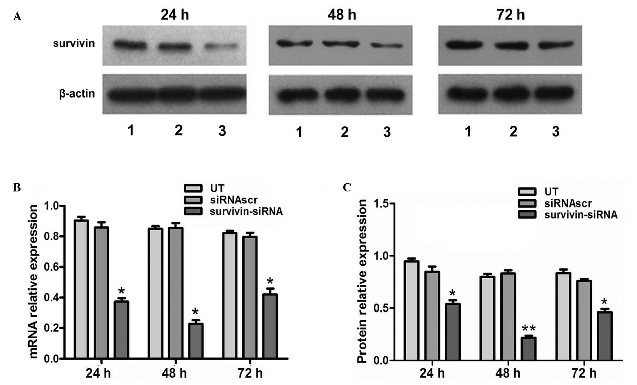
Survivin-siRNA inhibited survivin
expression in 4T1 cells
siRNA was transfected into 4T1 cells with high
efficiency. In the survivin-siRNA-transfected cells, the mRNA
levels of survivin were significantly reduced at 24, 48 and 72 h
post-transfection, compared with the UT and siRNAscr groups
(Fig. 1B). In order to confirm the
qPCR results, and better characterize the knocking down of survivin
at the protein level, western blot analysis was performed. The
results indicated that the protein expression levels of survivin
were significantly reduced at 24, 48 and 72 h post-transfection in
the survivin-siRNA group, compared with the UT and siRNAscr groups
(Fig. 1A and C).
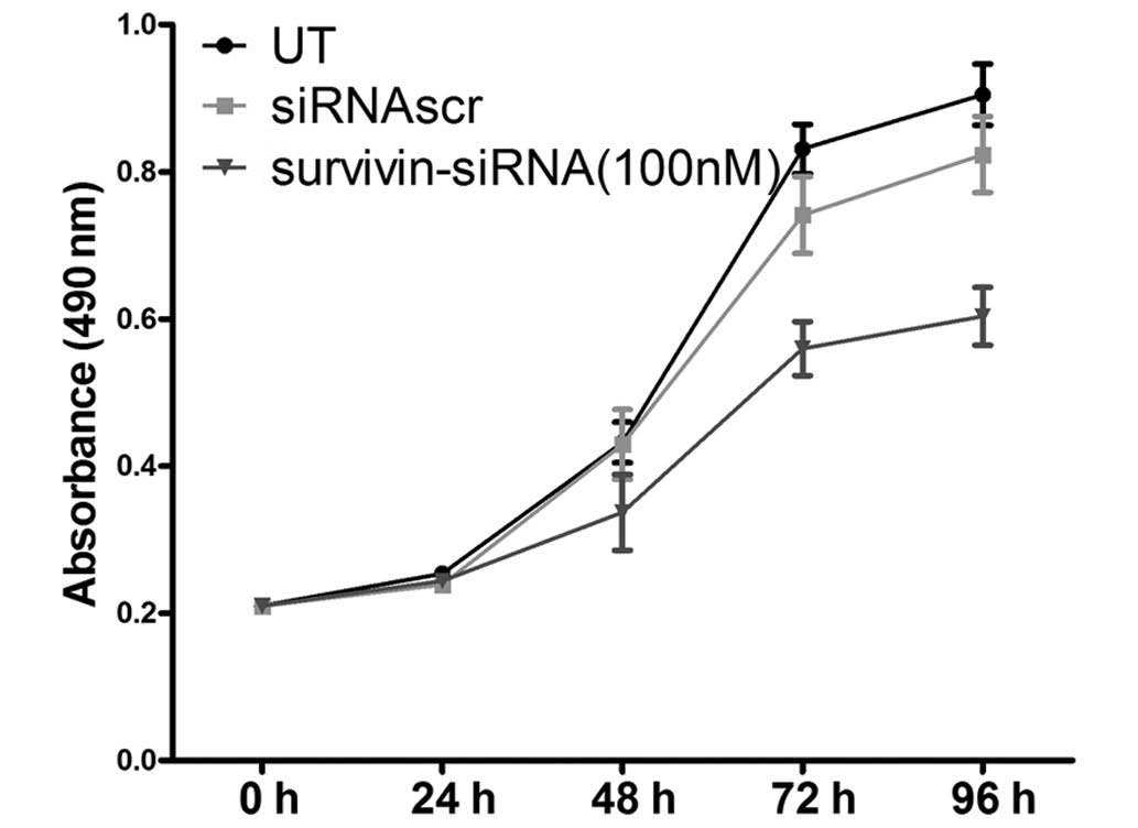
Antiproliferative effect of
survivin-siRNA on 4T1 cells
The effects of survivin-siRNA on the proliferation
ability of 4T1 cells were assessed by MTT assay. Inhibition of cell
proliferation was noticeable in 4T1 cells transfected with 100 nM
survivin-siRNA at 48 h post-transfection. The proliferation ability
of these cells was observed to be significantly suppressed in a
time-dependent manner, whereas no inhibitory effect was observed in
the UT or siRNAscr groups (Fig.
2).
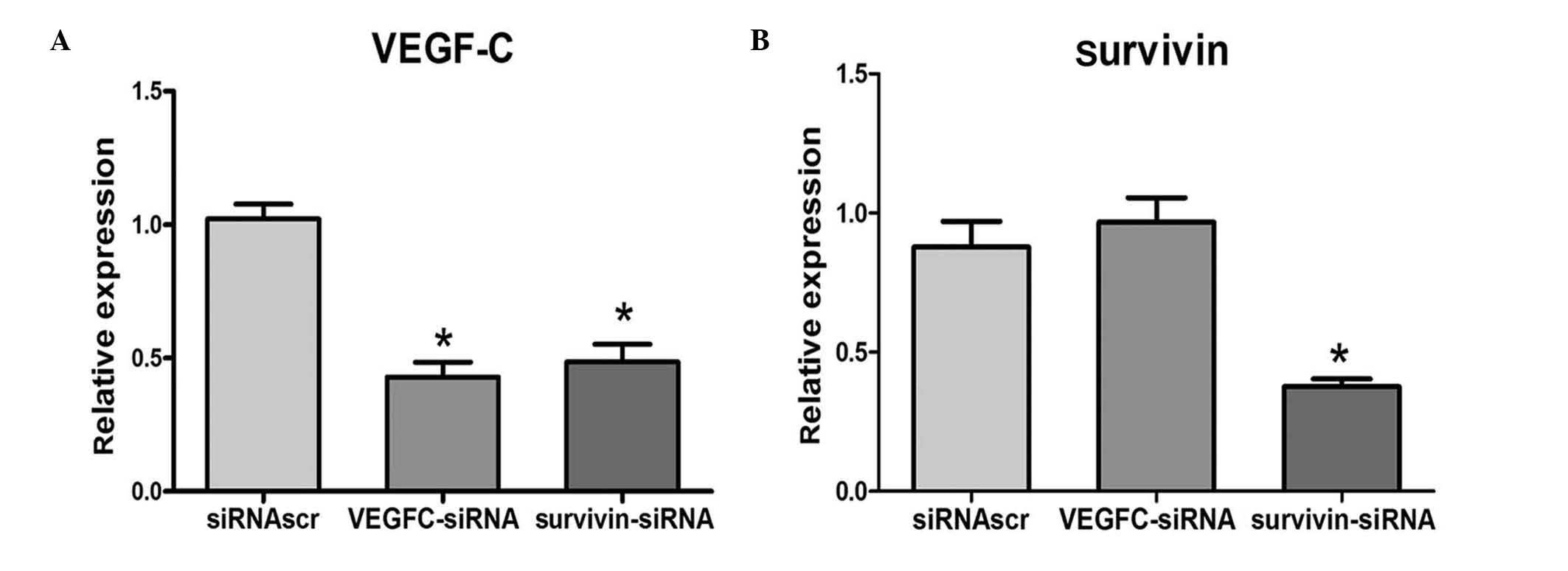
Positive correlation between the
expression levels of VEGF-C and survivin in 4T1 cells
Following the transfection of 4T1 cells with
survivin-siRNA, VEGF-C-siRNA or siRNAscr, the mRNA levels of
survivin and VEGF-C were examined by qPCR. The results indicated
that when survivin was downregulated in 4T1 cells, VEGF-C was also
downregulated. However, the expression levels of survivin were not
reduced when VEGF-C was downregulated. qPCR analysis revealed that
the mRNA levels of VEGF-C were positively correlated with the mRNA
levels of survivin (Fig. 3).
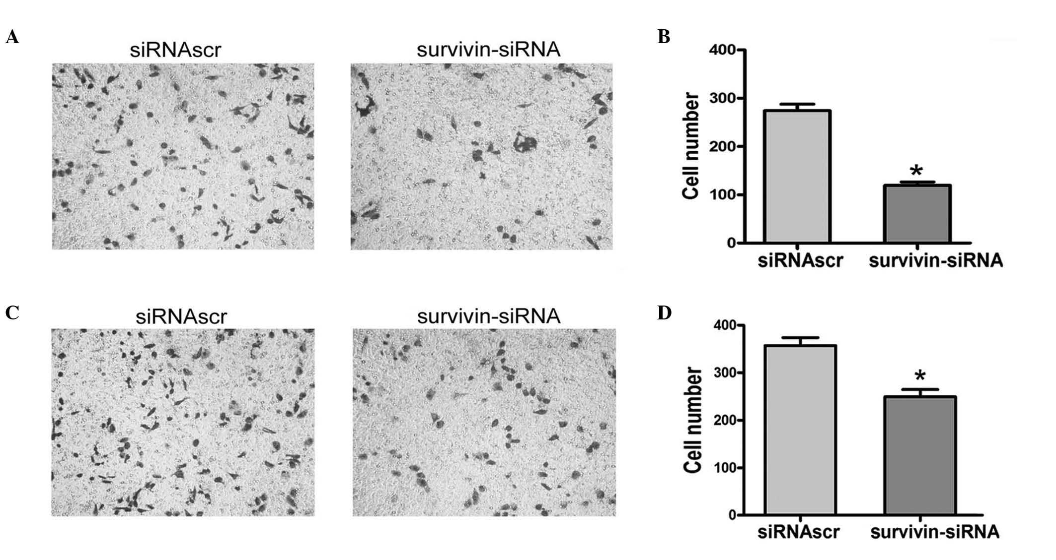
Survivin-siRNA inhibited the migration
and invasion abilities of 4T1 cells in vitro
To assess the role of endogenous survivin in the
invasion and migration abilities of 4T1 cells, the expression of
survivin was downregulated using siRNA. Following incubation for 24
h, the invasion ability of the survivin-siRNA-transfected cells was
inhibited, compared with the siRNAscr-transfected cells, indicating
that knocking down survivin in 4T1 cells resulted in retarded cell
invasion (Fig. 4A and B).
Furthermore, a significant difference was observed between the
number of migrated cells in the survivin-siRNA group, compared with
the siRNAscr group (Fig. 4C and D).
These results indicated that the expression of survivin in 4T1
cells was associated with the capacity of migration and invasion of
these cells.
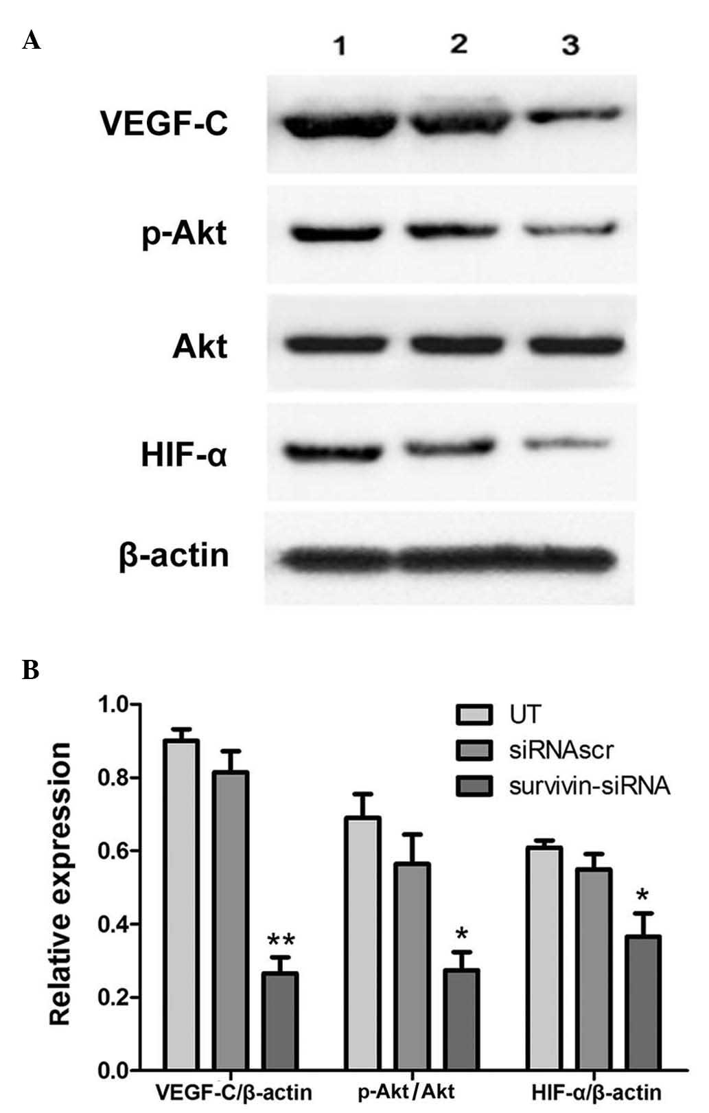
Effects of survivin-siRNA on the
VEGF-C and Akt/HIF-1α signaling pathways
The phosphoinositide 3-kinase (PI3K)/Akt signaling
pathway is known to participate in the malignant proliferation,
angiogenesis, metastasis and resistance to chemotherapy of tumor
cells. In various types of cancer, the expression of HIF-1α is
positively correlated with lymphatic metastasis. Therefore, to
investigate how survivin regulates VEGF-C, the expression levels of
p-Akt and HIF-1α were measured in the survivin-siRNA and control
groups. The results indicated that the expression levels of VEGF-C
and HIF-1α and the phosphorylation levels of Akt were reduced in
4T1 cells following transfection of survivin-siRNA (Fig. 5).
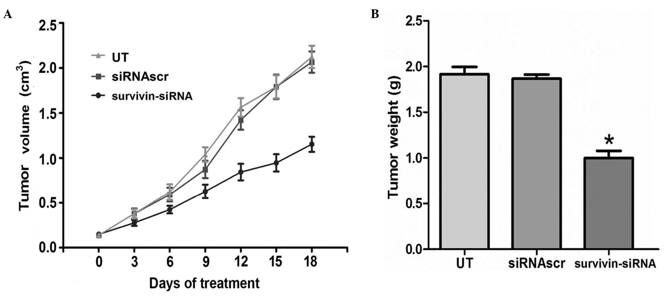
Survivin-siRNA inhibited tumor
proliferation in vivo
In the present study it was observed that
survivin-siRNA exhibited significant antitumor abilities in
vitro. Therefore, in order to explore the applicability of the
survivin-siRNA system in vivo, a murine metastatic breast
tumor model mimicking stage IV of human breast cancer was
established. Survivin-siRNA was able to reduce tumor volume and
weight in this xenograft model (Fig.
6). These results indicated that survivin-siRNA may exert its
antitumor effects on this model of breast cancer by inhibiting
tumor proliferation.
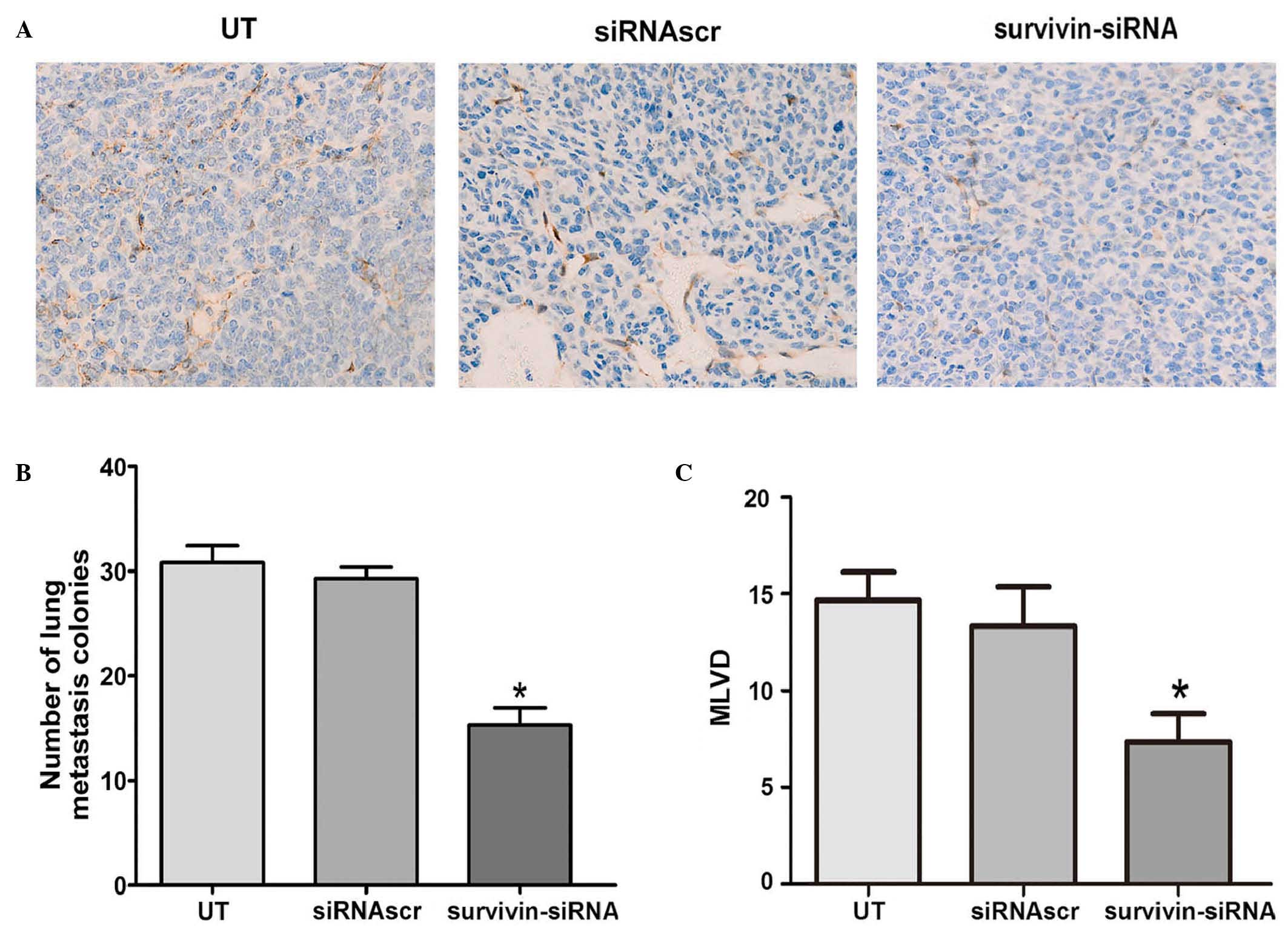
Survivin-siRNA inhibited
lymphangiogenesis and lung metastasis in vivo
To determine whether the treatment with
survivin-siRNA affected lymphangiogenesis in vivo, MLVD was
detected by podoplanin staining in the animal tumors. According to
the results obtained, the median numbers of MLVD were apparently
reduced in the tumors of the survivin-siRNA-treated group, compared
with the control groups (Fig. 7A and
C). Furthermore, to investigate the effect of survivin-siRNA on
metastasis, the lungs of the sacrificed mice were harvested, and
their metastatic nodules were counted. The results are presented as
the mean ± SD (n=10 tumors). Compared with the control mice, the
survivin-siRNA-treated mice exhibited significantly fewer lung
metastases (Fig. 7B). These results
indicated that survivin participates in the lymphangiogenesis and
metastasis of breast cancer tumors.
Discussion
Survivin is normally expressed in neoplasms and
fetal tissues, but is rarely present in normal adult tissues.
Survivin generally acts as an anti-apoptotic protein by inhibiting
the activation of caspases (28).
Previous studies have suggested that survivin participates in the
lymphatic metastasis of breast cancer by regulating the expression
of VEGF-C in tumor cells (13).
Lymphatic metastasis is the process by which detached tumor cells
may enter the lymph nodes located near the primary tumor via
lymphatic vessels, and is considered one of the most important
mechanisms of breast cancer systemic metastasis. VEGF-C is the most
important lymphangiogenic growth factor in the progression of
lymphogenesis and lymphatic metastasis (9). Downregulation of the expression or
function of VEGF-C has been previously demonstrated to supress
lymphogenesis and lymphatic metastasis in various tumor xenograft
models (29,30).
siRNA technology is a recently developed molecular
biological technique widely used in gene therapy and functional
genetics studies (31,32). In the present study, chemically
synthesized siRNAs specifically targeting mouse survivin inhibited
the expression of survivin at the mRNA and protein level in the
murine breast cancer cell line 4T1. The results of MTT assay
indicated that the proliferation ability of 4T1 cells was
significantly reduced in vitro following transfection with
survivin-siRNA, compared with the UT and siRNAscr groups.
Additionally, the migration and invasion assay revealed that the
motility of 4T1 cells was significantly inhibited upon transfection
with survivin-siRNA.
In the present study, the potential effects of
survivin on cellular signaling pathways that regulate the
expression of VEGF-C in tumor cells, were explored. The results
indicated that the PI3K/Akt signal transduction pathway may be
involved in the survivin-mediated regulation of the expression of
VEGF-C in breast cancer. Previous studies have suggested that
HIF-1α, a transcription factor involved in cellular adaptive
responses to hypoxia, may be responsible for the overexpression of
VEGF-C in cancer cells (33).
Previous studies have demonstrated that the activity of HIF-1α can
be blocked by the PI3K-inhibitor LY294002, indicating that the
activation of HIF-1α may be regulated by the PI3K/Akt pathway. Akt
is a serine/threonine protein kinase that functions as a critical
regulator of cell adhesion, migration and apoptosis (34). The activation of Akt has been
previously observed to contribute to tumorigenesis and tumor
metastasis in various types of human cancer (35,36).
McKenzie et al (18) reported
that survivin may trigger the activation of Akt, thus enhancing the
motility of melanoma cells. The activation of Akt is regulated by
phosphatase and tensin homolog (PTEN), which is reported to be
affected by survivin (18).
Therefore, it can be hypothesized that survivin may suppress
lymphatic metastasis in murine breast cancer by downregulating
VEGF-C through the Akt/HIF-1α pathway.
Previous studies have demonstrated that binding of
VEGF-C to VEGF receptor(R)-3 on the membrane of breast cancer and
endothelial cells results in the phosphorylation and subsequent
activation of the tyrosine kinase activity of VEGFR3, which
consequently stimulates lymphogenesis and mitosis in these cancer
cells (17,18). This ligand-induced phosphorylation of
VEGFR-3 has been previously suggested to activate downstream
signaling molecules such as Akt, by stimulating the kinase activity
of the receptor (17,18). Therefore, the knockdown of survivin
and the downregulation of VEGF-C may affect the phosphorylation of
Akt. In the present study, the results of western blotting revealed
that the knockdown of survivin in vitro led to the
downregulation of the expression of VEGF-C, HIF-1α and p-Akt at 48
h post-transfection, while the downregulation of survivin was
noticeable 24 h earlier. These findings indicated the existence of
a time window between the effects exerted by survivin-siRNA on the
expression of survivin and on the activation of VEGFR-3. Therefore,
it can be hypothesized that survivin suppresses the Akt/HIF-1α
signaling pathway, which results in the downregulation of the
expression of VEGF-C.
Furthermore, to evaluate the effects of
survivin-siRNA on breast cancer growth in vivo, the
antitumor efficacy of survivin-siRNA was examined in a mouse
metastatic breast cancer model. In this model, the intratumoral
injection of survivin-siRNA significantly inhibited the tumor
growth and pulmonary metastasis of the orthotopically implanted 4T1
cells. In addition, immunohistochemical staining, hematoxylin and
eosin staining and qPCR assays were performed on the tumor tissues
derived from the survivin-siRNA-injected mice. The data obtained
from these analyses suggested that the injection of survivin-siRNA
into the tumor exerted significant antitumor effects in
vivo, similar to those exhibited in vitro. According to
these results, it can be concluded that survivin participates in
cell proliferation and metastasis of mouse breast cancer.
In conclusion, the results of the present study
revealed that survivin-siRNA possessed potent in vitro and
in vivo antitumor and anti-metastatic abilities, by
inhibiting the proliferation and lymphogenesis of cancer cells.
Therefore, knocking down survivin by siRNA may be considered a
potential therapeutic approach for the treatment of breast cancer.
In addition, the present study has demonstrated that the ability of
survivin-siRNA to attenuate metastasis in murine breast cancer 4T1
cells may be due to the downregulation of VEGF-C, which is
associated with the PTEN/Akt/HIF-1α pathway in these cells.
However, the signals that affect metastasis function in complex
regulatory networks rather than a single pathway, and these signals
are differently activated in different types of tumors. Therefore,
the mechanisms by which survivin regulates metastasis remain to be
further investigated.
Acknowledgements
This study was supported by grants from Shandong
Science and Technology Development Planning (no.
2014GGH218023).
References
|
1
|
Jemal A, Bray F, Center MM, Ferlay J, Ward
E and Forman D: Global cancer statistics. CA Cancer J Clin.
61:69–90. 2011.(Erratum in CA Cancer J Clin 61: 134, 2011).
View Article : Google Scholar : PubMed/NCBI
|
|
2
|
Ali SM, Harvey HA and Lipton A: Metastatic
breast cancer: Overview of treatment. Clin Orthop Relat Res.
415(Suppl): S132–S137. 2003. View Article : Google Scholar : PubMed/NCBI
|
|
3
|
Bidard FC, Fehm T, Ignatiadis M, Smerage
JB, Alix-Panabières C, Janni W, Messina C, Paoletti C, Müller V,
Hayes DF, et al: Clinical application of circulating tumor cells in
breast cancer: Overview of the current interventional trials.
Cancer Metastasis Rev. 32:179–188. 2013. View Article : Google Scholar : PubMed/NCBI
|
|
4
|
Mackey JR, Kerbel RS, Gelmon KA, et al:
Controlling angiogenesis in breast cancer: A systematic review of
anti-angiogenic trials. Cancer Treat Rev. 38:673–688. 2012.
View Article : Google Scholar : PubMed/NCBI
|
|
5
|
Ran S, Volk L, Hall K and Flister MJ:
Lymphangiogenesis and lymphatic metastasis in breast cancer.
Pathophysiology. 17:229–251. 2010. View Article : Google Scholar : PubMed/NCBI
|
|
6
|
Carter CL, Allen C and Henson DE: Relation
of tumor size, lymph node status, and survival in 24,740 breast
cancer cases. Cancer. 63:181–187. 1989. View Article : Google Scholar : PubMed/NCBI
|
|
7
|
Timoshenko AV, Rastogi S and Lala PK:
Migration-promoting role of VEGF-C and VEGF-C binding receptors in
human breast cancer cells. Br J Cancer. 97:1090–1098. 2007.
View Article : Google Scholar : PubMed/NCBI
|
|
8
|
Sun P, Gao J, Liu YL, Wei LW, Wu LP and
Liu ZY: RNA interference (RNAi)-mediated vascular endothelial
growth factor-C (VEGF-C) reduction interferes with
lymphangiogenesis and enhances epirubicin sensitivity of breast
cancer cells. Mol Cell Biochem. 308:161–168. 2008. View Article : Google Scholar : PubMed/NCBI
|
|
9
|
Joukov V, Pajusola K, Kaipainen A, Chilov
D, Lahtinen I, Kukk E, Saksela O, Kalkkinen N and Alitalo K: A
novel vascular endothelial growth factor, VEGF-C, is a ligand for
the Flt4 (VEGFR-3) and KDR (VEGFR-2) receptor tyrosine kinases.
EMBO. 15:290–298. 1996.
|
|
10
|
Valtola R, Salven P, Heikkilä P, Taipale
J, Joensuu H, Rehn M, Pihlajaniemi T, Weich H, de Waal R and
Alitalo K: VEGFR-3 and its ligand VEGF-C are associated with
angiogenesis in breast cancer. Am J Pathol. 154:1381–1390. 1999.
View Article : Google Scholar : PubMed/NCBI
|
|
11
|
Kinoshita J, Kitamura K, Kabashima A,
Saeki H, Tanaka S and Sugimachi K: Clinical significance of
vascular endothelial growth factor-C (VEGF-C) in breast cancer.
Breast Cancer Res Treat. 66:159–164. 2011. View Article : Google Scholar
|
|
12
|
Chen Z, Varney ML, Backora MW, Cowan K,
Solheim JC, Talmadge JE and Singh RK: Down-regulation of vascular
endothelial cell growth factor-C expression using small interfering
RNA vectors in mammary tumors inhibits tumor lymphangiogenesis and
spontaneous metastasis and enhances survival. Cancer Res.
65:9004–9011. 2005. View Article : Google Scholar : PubMed/NCBI
|
|
13
|
Cai X, Ma S, Gu M, Zu C, Qu W and Zheng X:
Survivin regulates the expression of VEGF-C in lymphatic metastasis
of breast cancer. Diagn Pathol. 7:522012. View Article : Google Scholar : PubMed/NCBI
|
|
14
|
Ambrosini G, Adida C and Altieri DC: A
novel anti-apoptosis gene, survivin, expressed in cancer and
lymphoma. Nat Med. 3:917–921. 1997. View Article : Google Scholar : PubMed/NCBI
|
|
15
|
Suzuki A, Ito T, Kawano H, Hayashida M,
Hayasaki Y, Tsutomi Y, Akahane K, Nakano T, Miura M and Shiraki K:
Survivin initiates procaspase 3/p21 complex formation as a result
of interaction with Cdk4 to resist Fas-mediated cell death.
Oncogene. 19:1346–1353. 2000. View Article : Google Scholar : PubMed/NCBI
|
|
16
|
Altieri DC: Validating survivin as a
cancer therapeutic target. Nat Rev Cancer. 3:46–54. 2003.
View Article : Google Scholar : PubMed/NCBI
|
|
17
|
Altieri DC: Survivin, versatile modulation
of cell division and apoptosis in cancer. Oncogene. 22:8581–8589.
2003. View Article : Google Scholar : PubMed/NCBI
|
|
18
|
McKenzie JA, Liu T, Goodson AG and
Grossman D: Survivin enhances motility of melanoma cells by
supporting Akt activation and {α}5 integrin upregulation. Cancer
Res. 70:7927–7937. 2010. View Article : Google Scholar : PubMed/NCBI
|
|
19
|
Mehrotra S, Languino LR, Raskett CM,
Mercurio AM, Dohi T and Altieri DC: IAP regulation of metastasis.
Cancer Cell. 17:53–64. 2010. View Article : Google Scholar : PubMed/NCBI
|
|
20
|
Marioni G, Bertolin A, Giacomelli L,
Marchese-Ragona R, Savastano M, Calgaro N, Marino F, De Filippis C
and Staffieri A: Expression of the apoptosis inhibitor protein
Survivin in primary laryngeal carcinoma and cervical lymph node
metastasis. Anticancer Res. 26:3813–3817. 2006.PubMed/NCBI
|
|
21
|
Kedinger V, Meulle A, Zounib O, et al:
Sticky siRNAs targeting survivin and cyclin B1 exert an antitumoral
effect on melanoma subcutaneous xenografts and lung metastases. BMC
Cancer. 13:3382013. View Article : Google Scholar : PubMed/NCBI
|
|
22
|
Ikehara M, Oshita F, Kameda Y, Ito H,
Ohgane N, Suzuki R, Saito H, Yamada K, Noda K and Mitsuda A:
Expression of survivin correlated with vessel invasion is a marker
of poor prognosis in small adenocarcinoma of the lung. Oncol Rep.
9:835–838. 2002.PubMed/NCBI
|
|
23
|
Li X, Dang X and Sun X: Expression of
survivin and VEGF-C in breast cancer tissue and its relation to
lymphatic metastasis. Eur J Gynaecol Oncol. 33:178–182.
2012.PubMed/NCBI
|
|
24
|
Xue M, Ge Y, Zhang J, Liu Y, Wang Q, Hou L
and Zheng Z: Fucoidan inhibited 4T1 mouse breast cancer cell growth
in vivo and in vitro via downregulation of
Wnt/β-catenin signaling. Nutr Cancer. 65:460–468. 2013. View Article : Google Scholar : PubMed/NCBI
|
|
25
|
National Institutes of Health, Department
of Health and Human Services. Guide for the Care and Use of
Laboratory Animals (Guide) (revised 1985). NIH Publication No.
85–23. (Bethesda). National Institutes of Health. 1989.
|
|
26
|
Xue M, Ge Y, Zhang J, Wang Q, Hou L, Liu
Y, Sun L and Li Q: Anticancer properties and mechanisms of fucoidan
on mouse breast cancer in vitro and in vivo. PloS
One. 7:e434832012. View Article : Google Scholar : PubMed/NCBI
|
|
27
|
Dong XP, Xiao TH, Dong H, Jiang N and Zhao
XG: Endostar combined with cisplatin inhibits tumor growth and
lymphatic metastasis of lewis lung carcinoma xenografts in mice.
Asian Pac J Cancer Prev. 14:3079–3083. 2013. View Article : Google Scholar : PubMed/NCBI
|
|
28
|
Li F, Ambrosini G, Chu EY, Plescia J,
Tognin S, Marchisio PC and Altieri DC: Control of apoptosis and
mitotic spindle checkpoint by survivin. Nature. 396:580–584. 1998.
View Article : Google Scholar : PubMed/NCBI
|
|
29
|
Hirakawa S, Brown LF, Kodama S, Paavonen
K, Alitalo K and Detmar M: VEGF-C-induced lymphangiogenesis in
sentinel lymph nodes promotes tumor metastasis to distant sites.
Blood. 109:1010–1017. 2007. View Article : Google Scholar : PubMed/NCBI
|
|
30
|
Skobe M, Hawighorst T, Jackson DG, Prevo
R, Janes L, Velasco P, Riccardi L, Alitalo K, Claffey K and Detmar
M: Induction of tumor lymphangiogenesis by VEGF-C promotes breast
cancer metastasis. Nat Med. 7:192–198. 2001. View Article : Google Scholar : PubMed/NCBI
|
|
31
|
Xia H, Mao Q, Paulson HL and Davidson BL:
siRNA-mediated gene silencing in vitro and in vivo.
Nat Biotechnol. 20:1006–1010. 2002. View
Article : Google Scholar : PubMed/NCBI
|
|
32
|
Shim MS and Kwon YJ: Efficient and
targeted delivery of siRNA in vivo. FEBS J. 23:4814–4827.
2010. View Article : Google Scholar
|
|
33
|
Brito LGO, Schiavon VF, Andrade JM, Tiezzi
DG, Peria FM and Marana HR: Expression of Hypoxia-inducible factor
1-α and vascular endothelial growth factor-C in locally advanced
breast cancer patients. Clinics. 66:1313–1320. 2011.PubMed/NCBI
|
|
34
|
Song G, Ouyang G and Bao S: The activation
of Akt/PKB signaling pathway and cell survival. J Cell Mol Med.
9:59–71. 2005. View Article : Google Scholar : PubMed/NCBI
|
|
35
|
West KA, Castillo SS and Dennis PA:
Activation of the PI3K/Akt pathway and chemotherapeutic resistance.
Drug Resist Updat. 5:234–248. 2002. View Article : Google Scholar : PubMed/NCBI
|
|
36
|
Goc A, Al-Husein B, Kochuparambil ST, Liu
J, Heston WW and Somanath PR: PI3 kinase integrates Akt and MAP
kinase signaling pathways in the regulation of prostate cancer. Int
J Oncol. 38:267–277. 2011.PubMed/NCBI
|