Introduction
Extramedullary plasmacytoma (EMP) is a rare
malignant tumor that is characterized by a malignant plasma cell
neoplasm and accounts for ~4% of all plasma cell malignancies
(1). Additionally, 80% of such
lesions are located in the head and neck region (2). A paucity of data exists concerning the
presentation, natural course and outcome of EMP, however, Xing
et al (3) reviewed 147 cases
of EMP of the larynx. It was identified that most common treatment
modality was radiotherapy alone. The mean survival duration was
~184 months, and the 5- and 10-year survival rates were 71.1% and
67.4%, respectively. As the disease is a rare tumor, little
information exists regarding the mortality rates. In addition,
although certain patients have been treated with surgical excision,
radiotherapy, chemotherapy, or combined surgery and radiotherapy,
the exact treatment strategy and outcomes of EMP remain unclear. It
also has no typical presentation or features, with diagnosis
predominantly dependent on pathological analysis. In a literature
search performed by a professional librarian using MEDLINE and
EMBASE, it was found that studies on such neoplasms in HIV-infected
patients are extremely rare, with only eight cases reported to date
(4–10). To the best of our knowledge, the
present study describes the first case of a solitary adrenal EMP in
a patient with HIV.
Case report
A 35-year-old male who had been diagnosed with HIV 3
months previously presented to the West China Hospital of Sichuan
University (Chengdu, China) in July 2013. The patient had a 2-week
history of intermittent right flank pain. The patient had a 2-year
history of anal sexual intercourse. There was no family history or
other medical history. Findings on physical examination were
unremarkable. Abdominal computed tomography revealed a soft-tissue
density shadow ~35 mm in diameter, without enhancement, in the
right adrenal gland area. Routine pre-operative examination showed
no apparent abnormalities. Endocrine tests for adrenal
hypersecretion were negative. The findings of routine blood tests,
blood biochemical examination, routine urinalysis and renal
function tests were also normal. A clinical diagnosis of a
non-functioning adrenal tumor was suspected. Following
communication with the patient and his family, a retroperitoneal
laparoscopic adrenalectomy was performed. However,
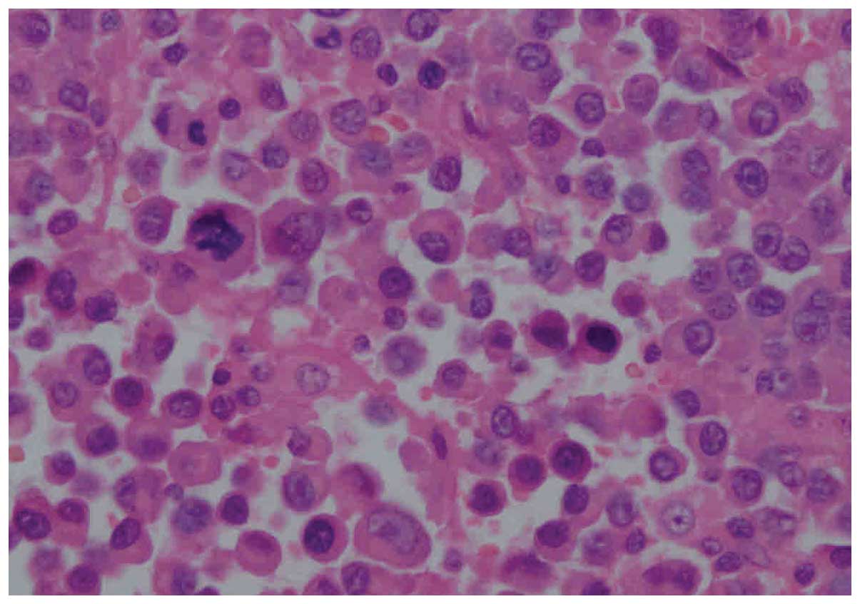
histopathological examination of the resected specimen suggested a
plasmacytoma. Microscopy showed that the mass was composed of a
dense and diffuse infiltrate of mature plasma cells with wheel
spoke-like nuclei. A few mitotic figures were also apparent
(Fig. 1). In addition, low-power
microscopy revealed a small amount of reticular fibers running
through the plasma cells (Fig.
2).
Subsequently, further post-operative investigations
were performed. The serum immunoglobulin (Ig)G level was 18.7 g/l
(normal range, 8.0–15.5 g/l), however, the serum IgA and IgM levels
were within the normal ranges. No Bence-Jones protein was detected
in the urine. Immunohistochemical analysis showed positive
cytoplasmic staining for λ-light chain. Subsequently, the bone
marrow biopsy demonstrated <3% plasma cells (diagnostic
criteria, <5%). A full-body bone scan revealed no anomalies.
Eventually, the patient was shown to meet all the required clinical
and laboratory criteria for a solitary EMP. The patient recovered
without complications and was discharged at 4 days post-surgery.
Subsequently, the patient was administered allopathic anti-acquired
immune deficiency syndrome (AIDS) drugs in the Sichuan Center for
Disease Control and Prevention (Sichuan, China). No recurrence was
detected after 2 years of follow-up.
Discussion
Plasmacytomas can be categorized as either EMP or
medullary plasmacytoma (9). Each type
is present in solitary and multiple forms. EMP is defined as an
extraosseous proliferation of neoplastic plasma cells. The study by
Juglard et al (11) suggested
that the incidence of plasmacytoma in HIV-positive patients is
greater than in non-infected patients. However, a solitary EMP
associated with AIDS involving the adrenal gland is extremely
uncommon. To the best of our knowledge, no case has been reported
in the literature to date. Among the reported case reports
involving EMP and infection by HIV, the locations of the tumors
include the gingiva, the larynx, the central nervous system and the
testes (4–10).
While the exact pathogenesis of EMP associated with
HIV remains unclear, Herranz et al (12) suggested that the immune alterations,
chronic viral infections and cytokine hyperexpression that exist in
patients infected by HIV may act as the triggering stimulus for
plasmacytoma to proliferate. The study also suggested that
plasmacytoma may be another malignant hematological process
associated with AIDS. In addition, a number of studies hypothesized
that infectious agents, such as HIV, hepatitis C virus and
Epstein-Barr virus, may play a role in the pathogenesis of
plasmacytoma (13–15). We agree with the current pathogenetic
hypotheses, however, further studies are required for
confirmation.
Although there are currently no clear treatment
guidelines for solitary EMP associated with HIV, thus far, these
patients have been treated with surgical excision, radiotherapy,
chemotherapy, or combined surgery and radiotherapy. Ramadan et
al (7) reported a patient with
solitary testicular EMP combined with AIDS who was treated with
combined surgical excision and radiotherapy. Hazarika et al
(8) reported a case of solitary
sinonasal EMP with concomitant HIV, wherein the tumor mass was
treated via radiotherapy. Wu et al (9) described the case of an HIV-positive
individual with an solitary EMP of the central nervous system that
was treated by chemotherapy. In addition, 2 other cases involving
AIDS combined with EMP in the head and neck were reported by
Bhattacharya et al (10),
where the patients were treated with chemotherapy. In the present
patient, retroperitoneal laparoscopic removal of the adrenal mass
was successfully performed. Although the patient refused to undergo
post-operative radiotherapy and chemotherapy, an uneventful
recovery was experienced and the patient was discharged at 5 days
post-surgery. The study suggest that the retroperitoneal
laparoscopic resection may be a good method to manage this
condition. Given the limited experience in treatment of EMP
infection following HIV infection, further study is required to
determine the appropriate regimen and duration of therapy.
Solitary EMP in HIV-positive patients tend to be
more aggressive, with a poor prognosis. This may be due to the poor
immunity of the patient. The study by Bhattacharya et al
(10) identified that the mean
survival time of two pa ti nets with HIV infection and EMP was
<6 months after the original diagnosis was made. The present
patient, however, was discharged after surgery and followed up for
2 years with no recurrence.
In conclusion, the present study illustrates the
fact that this rare type of solitary EMP associated with AIDS can
occur in the adrenal glands, and that retroperitoneal laparoscopic
resection may be a good method to manage this tumor. In addition,
although rare, solitary EMP should be considered in the
differential diagnosis of an adrenal mass in HIV-infected
patients.
Acknowledgements
The study was supported by the National Natural
Science Foundation of China (grant nos. 81200551, 81270841 and
81300627).
Glossary
Abbreviations
Abbreviations:
|
EMP
|
extramedullary plasmacytoma
|
|
HIV
|
human immunodeficiency virus
|
|
AIDS
|
acquired immune deficiency
syndrome
|
References
|
1
|
Galieni P, Cavo M, Awisati G, Pulsoni A,
Falbo R, Bonelli MA, Russo D, Petrucci MT, Bucalossi A and Tura S:
Solitary plasmacytoma of bone and extramedullary plasmacytoma: Two
different entities? Ann oncol. 6:687–691. 1995.PubMed/NCBI
|
|
2
|
Husarić S, Pašić J, Alić E and Kuljanin M:
Solitary extramedullary plasmacytoma of the liver. Acta Med Acad.
42:85–86. 2013. View Article : Google Scholar : PubMed/NCBI
|
|
3
|
Xing Y, Qiu J, Zhou ML, Zhou SH, Bao YY
and Zheng ZJ: Prognostic factors of laryngeal solitary
extramedullary plasmacytoma: A case report and review of
literature. Int J Clin Pathol. 8:2415–2435. 2015.
|
|
4
|
Gastaut J, Quilichini R and Horchowski N:
Localized extramedullary plasmacytomas (LEP) and HIV. IV
International Conference on AIDS Program and Abstract Book.
2:3271988.
|
|
5
|
Israel AM, Koziner B and Straus DJ:
Plasmacytoma and the acquired immunodeficiency syndrome. Ann Intern
Med. 99:635–636. 1983. View Article : Google Scholar : PubMed/NCBI
|
|
6
|
Vallisa D, Pagani L, Bertè R, Civardi G,
Viale P, Paties C and Cavanna L: Extramedullary plasmacytoma in a
patient with AIDS: Report of a case and review of the literature.
Tumori. 84:511–514. 1998.PubMed/NCBI
|
|
7
|
Ramadan A, Naab T, Frederick W and Green
W: Testicular plasmacytoma in a patient with the acquired
immunodeficiency syndrome. Tumori. 86:480–482. 2000.PubMed/NCBI
|
|
8
|
Hazarika P, Balakrishnan R, Singh R,
Pujary K and Aziz B: Solitary extramedullary plasmacytoma of the
sinonasal region. Indian J Otolaryngol Head Neck Surg. 63:33–35.
2011. View Article : Google Scholar : PubMed/NCBI
|
|
9
|
Wu W, Pasch W, Zhao X and Rezk SA:
Extraosseous plasmacytoma with an aggressive course occurring
solely in the CNS. Neuropathology. 33:320–323. 2013. View Article : Google Scholar : PubMed/NCBI
|
|
10
|
Bhattacharya AK, Han K and Baredes S:
Extramedullary plasmacytoma of the head and neck associated with
the human immunodeficiency virus. Ear Nose Throat J. 77:61–62.
1998.PubMed/NCBI
|
|
11
|
Juglard R, Vidal V, Calvet P, Dussaut JP,
Barea D, Colineau X, Tourrette JH and Solacroup JC: Plasmacytoma
and AIDS: Unusual duodenal localization. J Radiol. 82:1729–1731.
2001.(In French). PubMed/NCBI
|
|
12
|
Herranz S, Sala M, Cervantes M, Sasal M,
Soler A and Segura F: Neoplasia of plasma cells with atypical
presentation and infection by the human immunodeficiency virus. A
presentation of two cases. Am J Hematol. 65:239–242. 2000.
View Article : Google Scholar : PubMed/NCBI
|
|
13
|
Gold JE, Schwam L, Castella A, Pike SB,
Opfell R and Zalusky R: Malignant plasma cell tumors in human
immunodeficiency virus-infected patients. Cancer. 66:363–368. 1990.
View Article : Google Scholar : PubMed/NCBI
|
|
14
|
Montella M, Crispo A, Russo F, Ronga D,
Tridente V and Tamburini M: Hepatitis C virus infection and new
association with extrahepatic disease: Multiple myeloma.
Haematologica. 85:883–884. 2000.PubMed/NCBI
|
|
15
|
Sadeghian MH, Ayatollahi H, Keramati MR,
Memar B, Jamedar SA, Avval MM, Sheikhi M and Shaghayegh G: The
association of Epstein-Barr virus infection with multiple myeloma.
Indian J Pathol Microbiol. 54:720–724. 2011.PubMed/NCBI
|