Introduction
Worldwide, gastric cancer is the fourth most common
malignant disease and the second leading cause of cancer-associated
mortality (1). According to the
statistical data of the World Health Organization (2), there were 989,000 novel cases and
737,000 mortalities of gastric cancer in 2008, of which, the ratio
of men to women was 2:1. Surgical excision, chemotherapy and
radiotherapy are the basic treatment options for gastric cancer,
among which, surgical excision is currently the first choice
(3). However, the early diagnosis of
gastric cancer is difficult and the majority of cases have
progressed to a moderate or advanced stage prior to diagnostic
confirmation, therefore, the postoperative 5-year survival rate of
gastric cancer remains low at ~20–30%. Furthermore, elderly
patients are frequently unable to tolerate surgery (4,5). In recent
years, improvements to various resection methods, as well as
chemotherapeutic and radiotherapeutic plans has had little effect
(3). Surgical excision is incapable
of removing all tumor cells, and therefore the risk of
postoperative recurrence and metastasis remain (6). Furthermore, the major side effects of
chemotherapy and radiotherapy, particularly for elderly patients,
which include myelosuppression, immunosuppression and diarrhea, may
outweigh the curative effects (7,8).
Therefore, an effective therapy technique for the treatment of
gastric cancer is urgently required.
Photodynamic therapy (PDT) is a non-invasive
treatment method, used for a variety of malignant tumors (9,10). PDT has
been known to be effective for the treatment of gastric cancer for
a number of years (11,12). PDT typically involves the systemic
administration of a photosensitizer (PS), followed by its
activation by light at an appropriate wavelength, which results in
the generation of reactive oxygen species (ROS) and eventually
induces malignant cell death (13).
PDT has numerous advantages over typical cancer treatment
modalities, including surgery, chemotherapy and radiotherapy
(14): i) PDT is relatively
non-invasive, simply requiring illumination of the tumor site, and
therefore inducing minimal injury to the adjacent normal tissues
(15); ii) PDT does not induce
systemic immunosuppressive effects that may be translated into
clinical opportunistic infection (15); iii) PDT is able to be repeated without
detrimental consequences to the patient (16); and iv) PDT is simple to conduct, and
is particularly suitable for elderly patients who may be unable to
endure surgery, chemotherapy or radiotherapy (16). In addition, since the laser light
required for PDT may be delivered by an optical fiber, PDT is
particularly useful for the treatment of cavity-tumor gastric
cancer.
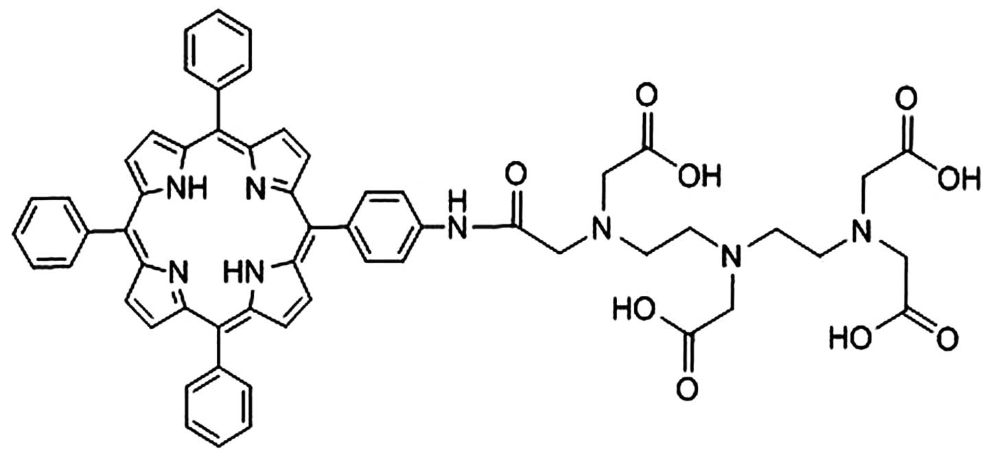
A novel porphyrin-based PS, named
meso-5-[ρ-diethylene triamine pentaacetic acid
(DTPA)-aminophenyl]-10,15,20-triphenyl-porphyrin (DTP), was
recently developed by the present group (17). This compound is a porphyrin derivative
and was designed to be a dissymmetrical DTPA-aminophenyl-based
porphyrin. DTP is a chemically pure compound and is synthesized
according to the patent specifications. The present study aimed to
investigate the effects and mechanism of DTP-mediated PDT against
human gastric cancer HGC27 and SNU-1 cells.
Materials and methods
Preparation of DTP
The synthetic process of DTP was obtained in detail
according to the patent specifications (CN 101805362) (17). Initially,
meso-5,10,15,20-tetraphenyl-porphyrin was synthesized in propionic
acid solvent (Meryer Chemical Technology Co., Ltd., Shanghai,
China), using the classical Adler method (18). Meso-5,10,15,20-tetraphenyl-porphyrin
(0.01 mol) was added and stirred into 20 ml dichloromethane
(Aladdin Industrial Inc., Shanghai, China) at −10°C until
dissolved. Subsequently, 65% concentrated nitric acid (1 ml;
Aladdin Industrial Inc.) was added to the solution. The nitrated
product was reduced with sodium nitrite (Aladdin Industrial Inc.)
in dichloromethane at room temperature, until a reduced mixture was
gained. The meso-5-(ρ-aminophenyl)-10,15,20-triphenyl-porphyrin was
obtained as the main product following separation of the mixture by
silica gel chromatography. Next, 0.2 g
meso-5-(ρ-aminophenyl)-10,15,20-triphenyl-porphyrin, 0.446 g
diethylenetriaminepentaacetic acid dianhydride, 0.046 g
dimethylaminopyridine (Aladdin Industrial Inc.) and 0.040 g
triethylamine were dissolved in 10 ml dimethyl sulfoxide (DMSO;
Beijing Solarbio Science & Technology Co., Ltd., Beijing,
China) in a 25 ml flask. Following agitation for 12 h at room
temperature, 5 ml H2O was added to the reaction solution
and a precipitate was formed. The final DTP product was then
obtained by crystallizing the precipitate of the mixed solution of
DMSO and H2O. The chemical structure of DTP is shown in
Fig. 1. DTP was dissolved in Roswell
Park Memorial Institute (RPMI)-1640 medium and stored at
12.5×10³ µM at 4°C, protected from visible light.
Cells and culture conditions
The PDT effect of DTP was studied using the HGC27
and SNU-1 human gastric cancer cell lines, which were purchased
from the Type Culture Collection of the Chinese Academy of Sciences
(Shanghai, China). The two types of cell were cultured in RPMI-1640
culture medium (Beijing Solarbio Science & Technology Co.,
Ltd.) with 10% Gibco fetal bovine serum (FBS; Thermo Fisher
Scientific, Waltham, MA, USA) at 37°C in a fully humidified
atmosphere with 5% CO2.
Spectral analysis of DTP
DTP solution was prepared in phosphate-buffered
saline (PBS; Beijing Solarbio Science & Technology Co., Ltd.)
to produce a solution of 3.12 µM concentration. Subsequently, 100
µl DTP solution was added into a 96-well plate. The ultraviolet
(UV)-visible absorption spectrum was recorded on a microplate
spectrophotometer (Thermo 3001; Thermo Fisher Scientific, Waltham,
MA, USA).
Photosensitization
HGC27 or SNU-1 cells were loaded into 96-well plates
(1×10⁴/well) and incubated in a humidified incubator
containing 95% air and 5% CO2 until cell attachment to
the substratum reached ~80% confluence. Subsequently, 100 µl of
various concentrations of DTP were added to the wells and incubated
for 24 h. Following incubation, the supernatant was replaced with
fresh culture medium supplemented with 10% FBS and the cells were
irradiated with 6 or 12 J/cm2 of laser light at a
wavelength of 650 nm, followed by incubation for an additional 3 h
at 37°C. The experiment was divided into groups, identical for
HGC27 and SNU-1 cells, as follows: Control groups, including
untreated cells, cells treated with PS alone and cells exposed to
laser light alone; and treatment groups, including cells treated
with DTP of various concentrations in combination with laser light
energy. The treatment groups comprised: Group 1, 0.78 µM DTP + 6/12
J/cm2; group 2, 1.56 µM DTP + 6/12 J/cm2;
group 3, 3.125 µM DTP + 6/12 J/cm2; group 4, 6.25 µM DTP
+ 6/12 J/cm2 and group 5 12.5 µM DTP + 6/12
J/cm2.
Colorimetric
3-(4,5-dimethyl-2-thiazolyl)-2, 5-diphenyl-2H-tetrazolium bromide
(MTT) assay
Cell viability was assessed using the MTT Cell
Proliferation and Cytotoxicity Assay Kit’ (Amresco, Solon, OH,
USA). Following photosensitization, 100 µg MTT in 20 µl PBS was
added into each well (96-well plates) and the cells
(1×10⁴/well) were then incubated for a further 3 h. The
reaction was stopped by the addition of 180 µl DMSO. The optical
density (OD) of each sample was subsequently measured at a
wavelength of 490 nm using a microplate spectrophotometer (Thermo
3001; Thermo Fisher Scientific). Cell survival rate (%) =
(ODtreated/ODcontrol) × 100%.
Cell apoptosis
Annexin V-fluorescein isothiocyanate (FITC) and
propidium iodide (PI) (Beyotime Institute of Biotechnology, Haimen,
China) were used to detect cell apoptosis induced by DTP-PDT. HGC27
and SNU-1 cells were seeded in 6-well plates, followed by
incubation with 6.25 µM DTP for 24 h. The cells were subsequently
irradiated at 650 nm with 12 J/cm2 laser light. Three
hours later, the irradiated cells were collected, rinsed with PBS
and stained with Annexin V-FITC (200 µg/ml) for 10 min and 30 µg/ml
PI in the dark at room temperature, successively. Immediately, the
fluorescence was analyzed in 10,000 cells/sample using a FACScan
flow cytometer (FCM; Beckman Coulter, Fullerton, CA, USA). The
results were expressed as the percentage of cells exhibiting
apoptosis relative to the total number of cells analyzed. Blank
control and treatment with DTP or 6 J/cm2 laser light
alone were included as control groups.
Morphological observations
HGC27 and SNU-1 cells were seeded in 24-well plates.
Following attachment to the substratum to ~80% confluence, 6.25 µM
DTP was added into each well for 24 h, followed by irradiation by a
laser at 650 nm with 12 J/cm2. The cells were
subsequently stained with Hoechst 33342 (Beijing Solarbio Science
& Technology Co., Ltd.) for 30 min at room temperature, washed
twice with PBS and exposed to UV illumination using a fluorescent
microscope (Leica DMIRE 2; Leica Microsystems GmbH, Wetzlar,
Germany) to detect the differences in chromatin condensation and
fragmentation between the control and treatment groups. The
staining was evaluated by three investigators.
Intracellular distribution of PS by
confocal laser scanning microscopy (CLSM)
HGC27 or SNU-1 cells were cultured on coverslips
(Citoglas; Citotest Labware Manufacturing Co., Ltd., Jiangsu,
China) placed in Petri dishes (Citoglas; Citotest Labware
Manufacturing Co., Ltd.) with RPMI-1640 culture medium. Once the
cells reached 85% confluence, 6.25 µM DTP in serum-free cell
culture medium was added to the dishes and cells were incubated for
6 h, followed by incubation with: i) 150 nM MitoTracker Green FM
for an additional 30 min or ii) 1.5 µM LysoSensor™ Green DND-189
(Invitrogen; Thermo Fisher Scientific, Waltham, MA, USA) for 1 h at
culture temperature. Following incubation, the staining solution
was replaced with fresh culture media and the cells were observed
by CLSM (TCS SP8; Leica Heidelberg GmbH, Heidelberg, Germany). The
excitation wavelength used for DTP was 405 nm, and the fluorescence
excitation and emission wavelengths for MitoTracker Green FM or
LysoSensor Green DND-189 were 488 and 520 nm, respectively.
Statistical analysis
Statistical analysis was performed based on one way
analysis of variance. All statistical analyses were performed using
SPSS 11.0 software (SPSS, Inc., Chicago, IL, USA). The results were
recorded as the mean ± standard error of the mean. P<0.05 was
considered to indicate a statistically significant difference and
all experiments were conducted at least 3 times.
Results
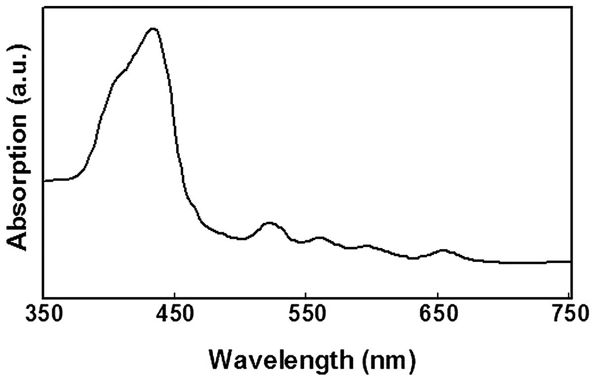
Spectral analysis of DTP
Initially, a spectral analysis of DTP was conducted
(Fig. 2). The absorption spectrum
indicated that DTP had significant absorption peaks at wavelengths
of 432, 522, 560, 596 and 652 nm, respectively. Since the
absorption was strongest at 432 nm, absorption at 405 nm was
evaluated, as this was the closest wavelength which was able to be
selected as the excitation laser channel during CLSM analysis.
Furthermore, considering the penetration depth of light in PDT
proportional to the wavelength, a long wavelength proximal to the
red region of the spectrum should be used for laser illumination
during treatment with PDT, and 650 nm is a suitable wavelength for
the MTT and flow cytometry assays (19).
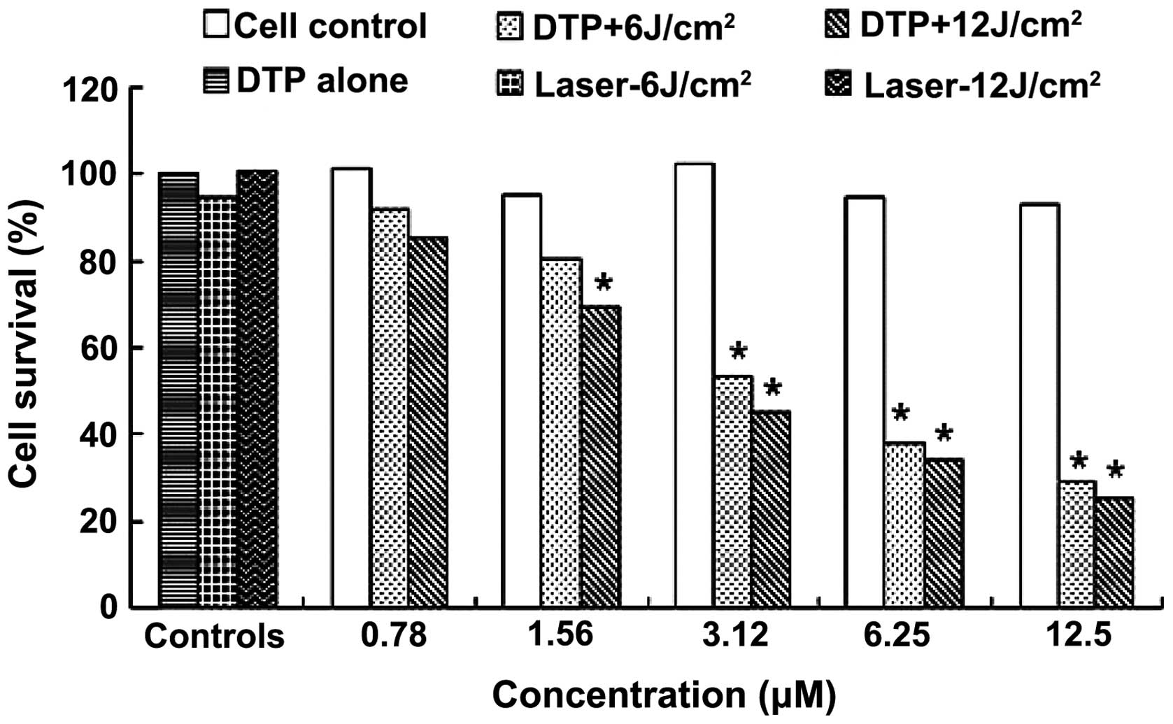
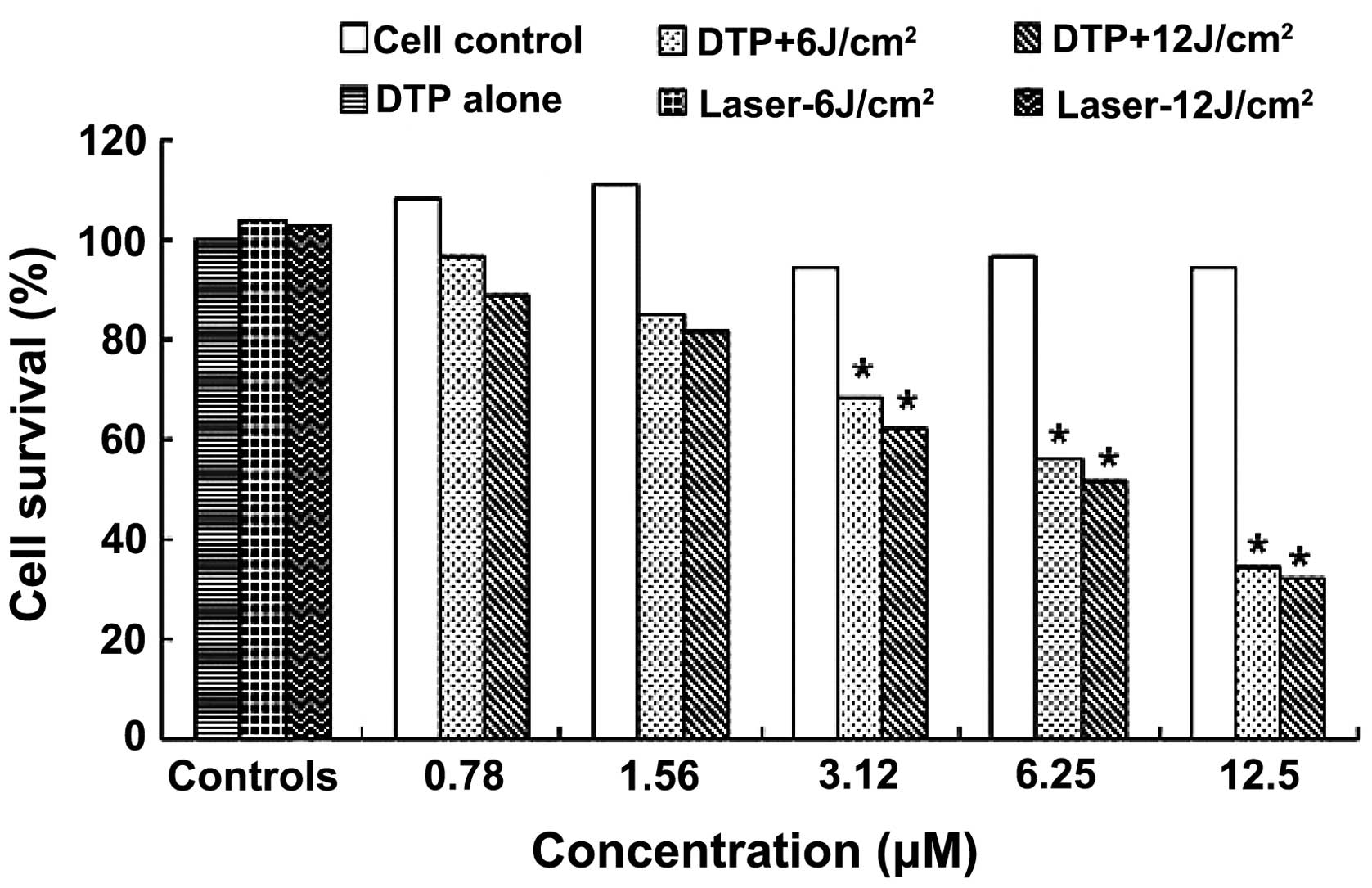
DTP combined with laser light induces
gastric cancer cell death
The photodynamic effect of DTP against human HGC27
and SNU-1 cells was evaluated by MTT assay. Table IA and B list the OD values of each
group and Figs. 3 and 4 demonstrate the corresponding cell survival
rates following PDT for HGC27 and SNU-1 cells, respectively.
 | Table I.OD values of each group following
treatment with DTP alone or in combination with laser light. |
Table I.
OD values of each group following
treatment with DTP alone or in combination with laser light.
| A, HGC27 cells |
|
|
|
|
|---|
|
|---|
|
|
|
| DTP + laser light,
OD |
|---|
|
|
|
|
|
|---|
| Group | DTP concentration,
µM | DTP alone, OD | 6
J/cm2 | 12
J/cm2 |
|---|
| 1 | 0.78 |
0.8741±0.1365a |
0.7946±0.0895a |
0.7368±0.1305a |
| 2 | 1.56 |
0.8249±0.0549a |
0.6912±0.6521a |
0.5962±0.0895b |
| 3 | 3.125 |
0.8846±0.0745a |
0.4595±0.0352b |
0.3892±0.0421b |
| 4 | 6.25 |
0.8186±0.1026a |
0.3265±0.0451b |
0.2956±0.0512b |
| 5 | 12.5 |
0.8018±0.0982a |
0.2498±0.0213b |
0.2228±0.0231b |
| 6 | 0.0 |
|
0.8165±0.0951a |
0.8694±0.1543a |
| 7 | Control |
| 0.8741±0.1116 |
|
|
|---|
| B, SNU-1 cells |
|
|
|
|
|
|---|
|
|
|
| DTP + laser light,
OD |
|
|
|
|
|
| Group | DTP concentration,
µM | DTP alone, OD | 6
J/cm2 | 12
J/cm2 |
|
| 1 | 0.78 |
0.8315±0.0865a |
0.7425±0.0745a |
0.6815±0.0351a |
| 2 | 1.56 |
0.8513±0.1065a |
0.6522±0.0534a |
0.6258±0.0541a |
| 3 | 3.125 |
0.7231±0.0256a |
0.5236±0.0982b |
0.4788±0.0255b |
| 4 | 6.25 |
0.7452±0.5614a |
0.4322±0.0415b |
0.3975±0.0413b |
| 5 | 12.5 |
0.7259±0.0635a |
0.2655±0.0265b |
0.2469±0.0215b |
| 6 | 0.0 |
|
0.7985±0.0261a |
0.7865±0.0452a |
| 7 | Control |
| 0.7656±0.0915 |
|
Table IA and Fig. 3 exhibit the MTT assay results for
DTP-PDT on HGC27 cells. The cell survival rate following DTP-PDT
declined with increasing PS concentration and light dose (Fig. 3). Statistically significant
differences were observed between treatment groups 3–5 and the cell
control group (P<0.01) following exposure to 6 J/cm2
illumination, while following 12 J/cm2 treatment, a
significant difference was identified between treatment groups 2–5
and the cell control group (P<0.01) (Table IA). Fig.
3 also demonstrated that 0.78 µM PS-mediated PDT only induced
slight HGC27 cell death, with ~7.75 and 14.46% cell death at 6 and
12 J/cm2 illumination, respectively. However, treatment
with 3.12 µM PS was able to induce 46.65 and 54.82% cell death. The
highest concentration of DTP (12.5 µM) was found to be highly
effective, inducing 70.99 and 74.14% mortality at light doses of 6
and 12 J/cm2, respectively. The IC50 of DTP
was 4.75 and 2.98 µM at light doses of 6 and 12 J/cm2,
respectively. DTP alone exerted no marked cytotoxicity on HGC27
cells at any concentration. In addition, laser light alone also did
not exert any effects on the growth of HGC27 cells.
SNU-1 cells demonstrated similar MTT results to
those observed in HGC27 cells (Table
IB; Fig. 4). DTP exerted
significant apoptosis induction in SNU-1 cells. A significant
difference was observed between treatment groups 3–5 and the cell
control group (P<0.01) for light doses of 6 and 12
J/cm2, while no difference was detected between
treatment groups 1 and 2 and the cell control (P>0.05) (Table IB). The death of SNU-1 cells induced
by DTP-PDT was proportional to PS concentration and light dose
(Fig. 4). The lower concentrations of
0.78 and 1.56 µM resulted in the death of a small percentage cells
at light doses of 6 and 12 J/cm2, respectively; however,
12.5 µM DTP induced 65.33 and 67.76% mortality and an
IC50 of 4.75 and 6.38 µM, respectively. The data also
revealed that DTP or laser treatment alone were nontoxic to SNU-1
cells.
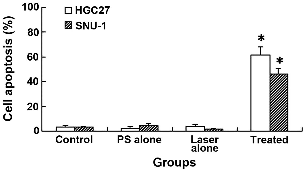
DTP-PDT induces gastric cancer cell
apoptosis
DTP-PDT induced significant cell apoptosis in HGC27
and SNU-1 cells, as determined by FCM analysis (Fig. 5). The apoptotic rates of HGC27 and
SNU-1 cells were 61.5±6.56 and 45.9±4.62%, respectively. These
values were significantly higher than those of the control groups
(P<0.01), which demonstrated 3.1±1.25 and 2.9±0.85% apoptosis in
HGC27 and SNU-1 cells, respectively. In the DTP alone group, only
2.5±0.98 and 4.3±1.25% cell apoptosis was detected, while 3.7±1.64
and 1.8±0.75% cell apoptosis were detected in the laser light alone
group for HGC27 and SNU-1 cells, respectively. Therefore, no
significant differences were detected, in comparison with the cell
control (P>0.05).
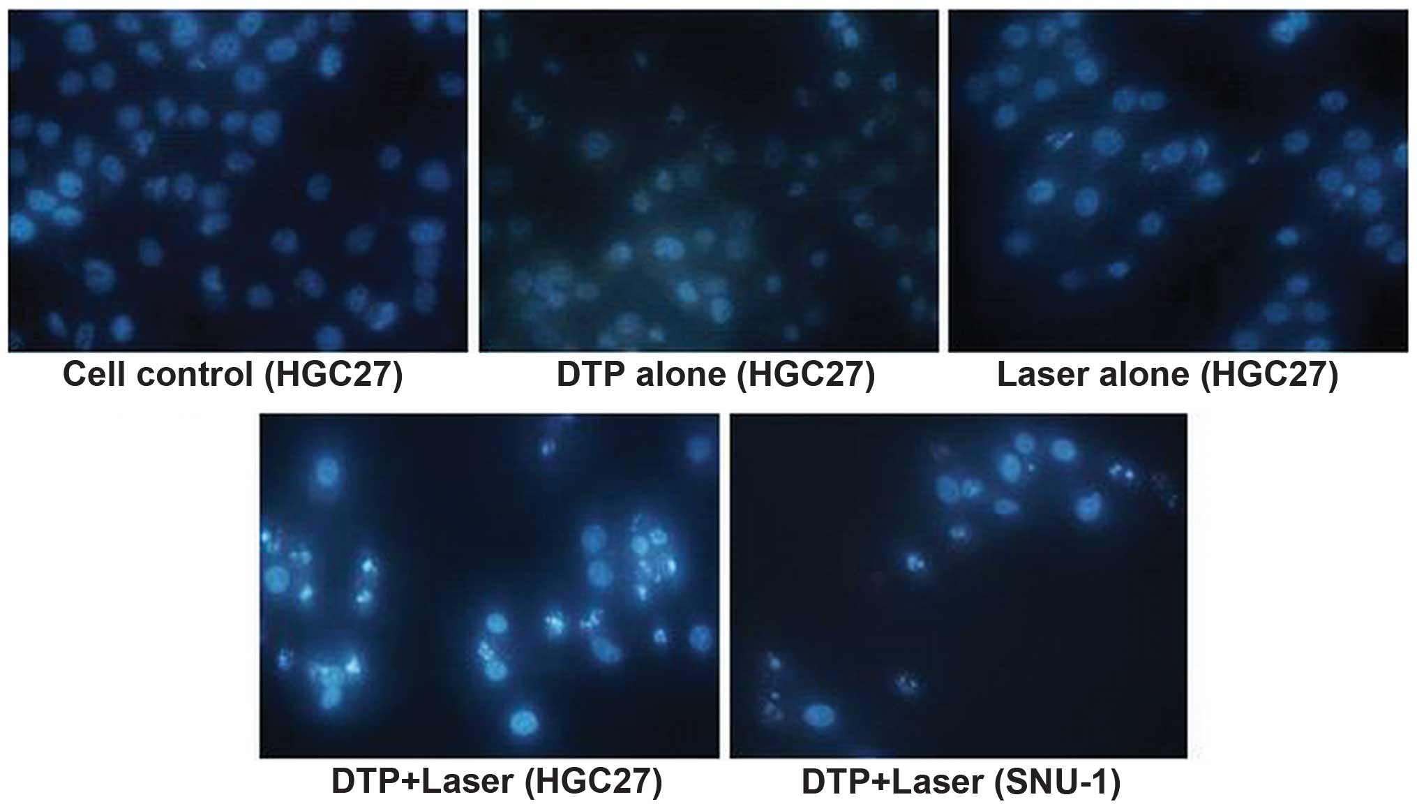
Morphological changes occur in gastric
cancer cells following PDT
Morphological changes in the cellular nuclei of each
group were observed using fluorescence microscopy (Fig. 6). A large quantity of apoptotic cells
were detected following PS-PDT, demonstrating significant
characteristics of apoptosis, including chromatin condensation,
nucleus fragmentation and apoptotic bodies in HGC27 and SNU-1
cells. By contrast, in the control cell, DTP-alone or light-alone
groups, the majority of the cells demonstrated intact,
rounded/elliptical nuclei and demonstrated no morphological changes
in either of the cell types.
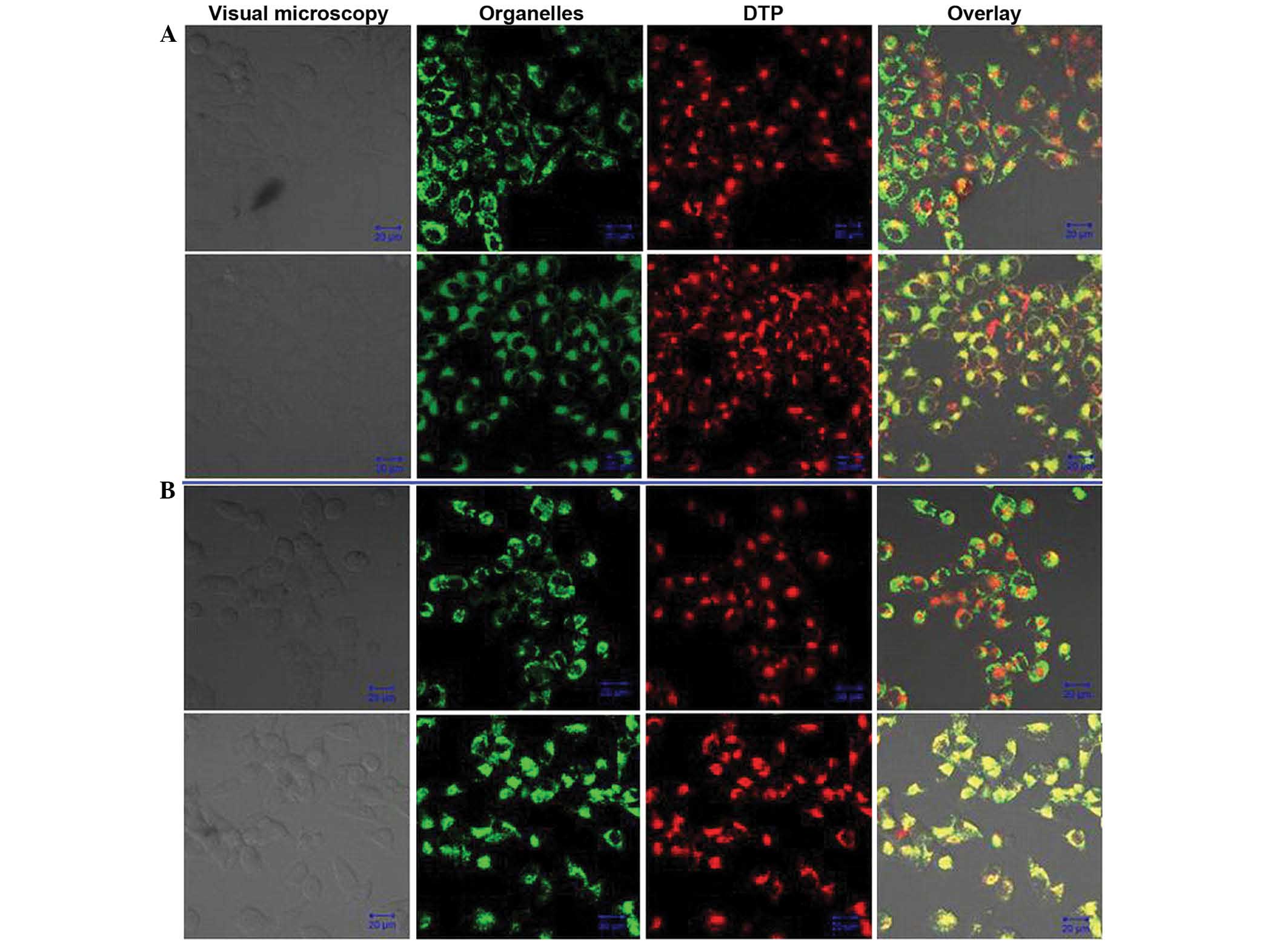
DTP is localized to the lysosomes
The distribution of DTP within HGC27 and SNU-1 cells
was evaluated via fluorescent observation by CLSM (Fig. 7). Following the incubation of cells
with DTP and MitoTracker Green Probe or LysoSensor Green Probe, the
PS emitted red fluorescence that was excited by the 405 nm
wavelength and the MitoTracker Green Probe and LysoSensor Green
Probe emitted green fluorescence that was excited by the 488 nm
wavelength in the same view. The lysosomal overlay images for HGC27
and SNU-1 cells revealed a large quantity of yellow-green
fluorescent spots (Fig. 7, lower
panels), indicating co-localization of DTP and the lysosomal probe.
Conversely, no overlap was detectable between DTP and the
mitochondrial probe for the two types of cell (Fig. 7, upper panels). The distribution of
DTP in HGC27 and SNU-1 cells was analogous.
Discussion
PDT is a novel treatment for malignant tumors, which
is derived from ‘photodynamic action’ (20). In 1900, Raab (21) discovered that paramecium, having
absorbed acridines, were able to be killed by light illumination;
however, light or acridines alone did not produce this effect. This
phenomenon was subsequently known as ‘photodynamic action’. In
1960, Lipson et al (22)
prepared the PS, hematoporphyrin derivative and evaluated its
effects in the treatment of malignant tumors. Kato et al
(23), in 1980, successfully
introduced the application of an endoscope to deliver laser light
during PDT treatment of lung cancer, which initiated the
application of PDT in the therapy of cavity tumors. To date,
numerous studies and practices have been focused on the PDT
treatment of gastric cancer and have proven that this method is an
effective technology for use in the treatment of gastric cancer
(11,24).
PDT consists of a PS, laser light and oxygen, among
which, PS is the most important component. The four main classes of
PS are: Porphyrin derivatives, chlorins, phthalocyanines and
porphycenes. These each possess distinct photochemical and
photophysical properties, underlying their mechanisms of action and
light activation (25). DTP,
evaluated in the present study, belongs to the porphyrin
derivatives family. The advantages of DTP when compared with other
porphyrin derivatives were as follows: i) DTP is a pure compound
with clear chemical and optical properties, and was able to be
synthesized relatively easily, according to the patent
specifications; ii) DTP exhibited improved chemical and physical
stability; iii) DTP demonstrated little toxicity in dark conditions
and is relatively safe for PDT treatment; iv) DTP has an absorption
peak of 652 nm, which is optimal for PDT use; and v) the authors of
the present study have applied for patent protection of the
compound. These advantages make the novel photosensitizer DTP a
promising potential PDT drug for use in the treatment of gastric
cancer, and therefore prompted initiation of the present study.
Initially, the photodynamic effects of DTP on HGC27
and SNU-1 human gastric cancer cells were evaluated by MTT assay.
The results revealed that DTP exerted marked phototoxicity on the
two types of cell. In HGC27 cells, a concentration of 6.25 µM
DTP-PDT resulted in 62.09 and 65.68% cell death at light doses of 6
and 12 J/cm2, respectively. Furthermore, treatment with
12.5 µM induced 70.99 and 74.14% mortality, respectively. Similar
phototoxicity was observed in SNU-1 cells. A concentration of 12.5
µM DTP-PDT induced 65.33 and 67.74% cell death at light doses of 6
and 12 J/cm2, respectively, and the corresponding
IC50 was 4.75 and 6.38 µM, respectively. However, in the
absence of light stimulation, DTP did not exert any significant
damage on the cells, even at the highest concentration of 12.5 µM.
These data indicate that this PS is a safe and effective PDT drug
for the treatment of gastric cancer cells, and the therapeutic
effects were associated with PS concentration and light dose.
To further investigate the mechanism of DTP-mediated
PDT on HGC27 and SNU-1 cells, flow cytometry and Hoechst 33342
staining were used to analyze cell apoptosis. Flow cytometric
analysis revealed that DTP-PDT was able to induce marked apoptosis
of HGC27 and SNU-1 cells, and that the apoptotic rate was 61.5±6.56
and 45.9±4.62%, respectively, significantly higher than that of the
control group. Additionally, fluorescent microscope observation
following Hoechst 33342 staining identified a large number of
apoptotic cells in the DTP-PDT treatment group. By contrast, the
majority of cells in the control groups remained rounded and
intact. The above results supported the hypothesis that apoptosis
is involved in the death process triggered by DTP-PDT in HGC27 and
SNU-1 cells.
The intracellular localization of a PS is crucial
for photodynamic damage. It is the ROS generated by PS that induce
cell death during PDT. Since ROS have a short half-life and are
only able to act close to the site of generation (26), photodynamic damage is closely
associated with the generation of ROS within cells, and therefore
to the precise intracellular localization of PS. PSs are able to
localize various organelles within cells, including the
mitochondria, lysosomes, endoplasmic reticulum, Golgi apparatus and
plasma membranes (27). Generally, PS
distribution in mitochondria commonly activates cell apoptosis
through various pathways, while PS distribution in lysosomes
frequently leads to cell necrosis (28). The present study showed that DTP
localized within the lysosomes of HGC27 and SNU-1 cells, but
induced apoptosis as the major death mode. This phenomenon may be
due to the indirect mitochondrial injury induced by direct lysosome
damage by DTP-PDT. It has been reported that the permeability of
the mitochondrial membrane was increased by PDT-mediated lysosomal
damage, which indirectly results in apoptosis of tumor cells
(29). Photodynamically-produced ROS
by lysosomal PS, induced rapid destruction of lysosomes, resulting
in the release of cathepsins, inducing the following spatiotemporal
sequence of cellular events: Bid/Bax activation, cytochrome
c release from the mitochondria to cytosol, activation of
downstream caspases and subsequent apoptisis (30,31).
Nagata et al (32) revealed
that the PS ATX-S10 (Na) had a primary site of accumulation in
lysosomes, however cells underwent apoptosis following
illumination, leading to 70% cell death. The results of these
studies were in agreement with those of the present study.
In conclusion, the results of the present study
indicate that the novel porphyrin-based PS, DTP, exhibited
significant PDT effects on HGC27 and SNU-1 human gastric cancer
cells in vitro, and that DTP is safe for use in the PDT
process. This compound localized in the lysosomes of HGC27 and
SNU-1 cells, but indirectly induced the mitochondrial apoptotic
pathway. Further investigations, including animal experiments and
additional molecular mechanism of action investigations should be
conducted for this compound in order to verify its potential
clinical application for the treatment of human gastric cancer.
References
|
1
|
Wong JE, Ito Y, Correa P, Peeters KC, van
de Velde CJ, Sasako M and Macdonald J: Therapeutic strategies in
gastric cancer. J Clin Oncol. 21(23 Suppl): 267s–269s. 2003.
View Article : Google Scholar : PubMed/NCBI
|
|
2
|
Ferlay J, Shin HR, Bray F, Forman D,
Mathers C and Parkin DM: Estimates of worldwide burden of cancer in
2008: GLOBOCAN 2008. Int J Cancer. 127:2893–2917. 2010. View Article : Google Scholar : PubMed/NCBI
|
|
3
|
Gu JR and Cao XT: Conceptual consideration
of cancer, challenges and opportunities for cancer biotherapy.
Zhong Guo Zhong Liu Sheng Wu Zhi Liao Za Zhi Bian Ji Bu. 15:2–7.
2008.(In Chinese).
|
|
4
|
Liu X, Ru J, Zhang J, Zhu LH, Liu M, Li X
and Tang H: miR-23a targets interferon regulatory factor 1 and
modulates cellular proliferation and paclitaxel-induced apoptosis
in gastric adenocarcinoma cells. PLoS One. 8:e647072013. View Article : Google Scholar : PubMed/NCBI
|
|
5
|
Li H, Liu Z, Xu C, Chen Y, Zhang J, Cui B,
Chen X, An G, She X, Liu H, et al: Overexpression of S100A4 is
closely associated with the progression and prognosis of gastric
cancer in young patients. Oncol Lett. 5:1485–1490. 2013.PubMed/NCBI
|
|
6
|
Zhang ZQ, Yao HL, Wen Y, Miao XY and Xiong
L: Advances in photodynamic therapy for gastric cancer. Ji Guang
Sheng Wu Xue Bao Bian Ji Bu. 21:289–293. 2012.(In Chinese).
|
|
7
|
Pan XR and Chen XX: Recent development on
adverse reactions induced by cancer chemotherapy treated with
traditional Chinese medicine. Yi Yao Yu Bao Jian Bian Ji Bu.
16:2358–2360. 2010.(In Chinese).
|
|
8
|
Ma XQ, He TQ, Zheng Tao, Xu BX, Xiao CR,
Zheng SX, Lin XR, Gao PY, Shen G, Bao YH, et al: Clinical study on
protective effect of diltiazem against radiation injuries on
cancer. Zhong Guo Yao Xue Za Zhi Bian Ji Bu. 39:231–232. 2004.(In
Chinese).
|
|
9
|
Vrouenraets MB, Visser GWM, Snow GB and
van Dongen GA: Basic principles, applications in oncology and
improved selectivity of photodynamic therapy. Anticancer Res.
23:505–522. 2003.PubMed/NCBI
|
|
10
|
Nowak-Stepniowska A, Pergoł P and
Padzik-Graczyk A: Photodynamic method of cancer diagnosis and
therapy - mechanisms and applications. Postepy Biochem. 59:53–63.
2013.(In Polish). PubMed/NCBI
|
|
11
|
Karanov SI: Long-term results after
photodynamic therapy (PDT) of T1N0M0 gastrointestinal (GI) tumors.
Hepatogastroenterology. 49:1579–1581. 2002.PubMed/NCBI
|
|
12
|
Ell C, Gossner L, May A, Schneider HT,
Hahn EG, Stolte M and Sroka R: Photodynamic ablation of early
cancers of the stomach by means of mTHPC and laser irradiation:
Preliminary clinical experience. Gut. 43:345–349. 1998. View Article : Google Scholar : PubMed/NCBI
|
|
13
|
Grimm S, Mvondo D, Grune T and Breusing N:
The outcome of 5-ALA-mediated photodynamic treatment in melanoma
cells is influenced by vitamin C and heme oxygenase-1. Biofactors.
37:17–24. 2011. View
Article : Google Scholar : PubMed/NCBI
|
|
14
|
Sibata CH, Colussi VC, Oleinick NL and
Kinsella TJ: Photodynamic therapy in oncology. Expert Opin
Pharmacother. 2:917–927. 2001. View Article : Google Scholar : PubMed/NCBI
|
|
15
|
Selbo PK, Høgset A, Prasmickaite L and
Berg K: Photochemical internalisation: A novel drug delivery
system. Tumour Biol. 23:103–112. 2002. View Article : Google Scholar : PubMed/NCBI
|
|
16
|
Rockson SG, Lorenz DP, Cheong WF and
Woodburn KW: Photoangioplasty. An emerging clinical cardiovascular
role for photodynamic therapy. Circulation. 102:591–596. 2000.
View Article : Google Scholar : PubMed/NCBI
|
|
17
|
Liu TJ, Wang YM, Wu L and Lv F:
[Diethylenetriaminepentaacetaacetato-N,N',N”]gadolinium-modified
porphyrins, and preparation method and application thereof. 0086.
CN 101805362 Filed. March 30–2010.issued August 18, 2010.
|
|
18
|
Adler AD, Longo FR, Finarelli JD,
Goldmacher J, Assour J and Korsakoff L: A simplified synthesis for
meso-tetraphenylporphine. J Org Chem. 32:467–477. 1967. View Article : Google Scholar
|
|
19
|
Yan YJ, Zheng MZ, Chen ZL, Yu XH, Yang XX,
Ding ZL and Xu L: Studies on preparation and photodynamic mechanism
of chlorin P6-13,15-N-(cyclohexyl)cycloimide (Chlorin-H) and its
antitumor effect for photodynamic therapy in vitro and in vivo.
Bioorg Med Chem. 18:6282–6291. 2010. View Article : Google Scholar : PubMed/NCBI
|
|
20
|
Juarranz A, Jaén P, Sanz-Rodríguez F,
Cuevas J and González S: Photodynamic therapy of cancer. Basic
principles and applications. Clin Transl Oncol. 10:148–154. 2008.
View Article : Google Scholar : PubMed/NCBI
|
|
21
|
Raab O: The effect of fluorescent
substances on infusoria. Z Biol. 39:524–526. 1900.
|
|
22
|
Lipson RL, Baldes EJ and Olsen AM:
Hematoporphyrin derivative: A new aid for endoscopic detection of
malignant disease. J Thorac Cardiovasc Surg. 42:623–629.
1961.PubMed/NCBI
|
|
23
|
Kato H, Konaka C, Saito M, Nishimiya K,
Kawate N, Aizawa K and Hayata Y: Laser photodynamic therapy with
hematoporphyrin derivative in lung cancer. Nihon Geka Gakkai
Zasshi. 86:1059–1063. 1985.(In Japanese). PubMed/NCBI
|
|
24
|
Nakamura H, Yanai H, Nishikawa J, Okamoto
T, Hirano A, Higaki M, Omori K, Yoshida T and Okita K: Experience
with photodynamic therapy (endoscopic laser therapy) for the
treatment of early gastric cancer. Hepatogastroenterology.
48:1599–1603. 2001.PubMed/NCBI
|
|
25
|
De Rosa FS and Bentley MVLB: Photodynamic
therapy of skin cancers: Sensitizers, clinical studies and future
directives. Pharm Res. 17:1447–1455. 2000. View Article : Google Scholar : PubMed/NCBI
|
|
26
|
Waidelich R: Laser-induced lithotripsy and
photodynamic therapy in urology - A short introduction to current
laser applications. Med Laser Appl. 25:14–19. 2010. View Article : Google Scholar
|
|
27
|
Pazos M and Nader HB: Effect of
photodynamic therapy on the extracellular matrix and associated
components. Braz J Med Biol Res. 40:1025–1035. 2007. View Article : Google Scholar : PubMed/NCBI
|
|
28
|
Ali SM and Olivo M: Bio-distribution and
subcellular localization of Hypericin and its role in PDT induced
apoptosis in cancer cells. Int J Oncol. 21:531–540. 2002.PubMed/NCBI
|
|
29
|
Garg AD, Nowis D, Golab J and Agostinis P:
Photodynamic therapy: Illuminating the road from cell death towards
anti-tumour immunity. Apoptosis. 15:1050–1071. 2010. View Article : Google Scholar : PubMed/NCBI
|
|
30
|
Liu L, Zhang Z and Xing D: Cell death via
mitochondrial apoptotic pathway due to activation of Bax by
lysosomal photodamage. Free Radic Biol Med. 51:53–68. 2011.
View Article : Google Scholar : PubMed/NCBI
|
|
31
|
Chiu SM, Xue LY, Lam M, Rodriguez ME,
Zhang P, Kenney ME, Nieminen AL and Oleinick NL: A requirement for
bid for induction of apoptosis by photodynamic therapy with a
lysosome- but not a mitochondrion-targeted photosensitizer.
Photochem Photobiol. 86:1161–1173. 2010. View Article : Google Scholar : PubMed/NCBI
|
|
32
|
Nagata S, Obana A, Gohto Y and Nakajima S:
Necrotic and apoptotic cell death of human malignant melanoma cells
following photodynamic therapy using an amphiphilic
photosensitizer, ATX-S10(Na). Lasers Surg Med. 33:64–70. 2003.
View Article : Google Scholar : PubMed/NCBI
|