Introduction
Malignant pleural mesothelioma (MPM) is an
aggressive tumor with a poor prognosis, arising from mesothelial
cells in the pleural cavity (1).
Exposure to asbestos is closely associated with the development of
MPM (2). MPM is historically a rare
disease; however, the incidence of MPM is predicted to increase due
to the use of asbestos worldwide and the long latency period for
the development of MPM following exposure (3). Multimodality therapy that is centered on
surgical resection is indicated for the treatment of MPM in the
early stage (4). However, MPM is
often diagnosed at an advanced stage, and it is known to be
refractory to conventional therapies, such as surgery,
chemotherapy, and radiotherapy (1).
Therefore, novel strategies for the diagnosis and treatment of MPM
are needed. However, much less information is available for MPM
compared with other solid neoplasms. Thus, it is of great
importance to investigate the biological behaviors of MPM.
In the current study, 4 MPM cell lines were
established from Japanese patients and the methylation status,
presence of mutations, copy number and protein expression of
representative genes, including NF2, P16, and
RASSF1A, which have been reported to be frequently altered
in MPM, were characterized (5–7). In
addition, the status of the microRNA (miR) 34 family, which we have
previously reported to be important in the pathogenesis of MPM,
were analyzed (8). MiR-34b/c is the
direct transcriptional target of p53, is reported to serve an
important role in in the pathogenesis of MPM and to be frequently
downregulated in MPM by promoter methylation (8). Furthermore, the sensitivity of the
established cell lines were examined in response to various drugs,
including conventional chemotherapeutic drugs, histone deacetylase
inhibitors, and heat shock protein 90 inhibitors.
Materials and methods
Patients and establishment of the cell
lines
A total of 4 Japanese patients with pleural
thickening and/or pleural effusion were diagnosed with MPM through
histopathological examinations of hematoxylin-eosin and
immunocytochemical staining of the specimens that were obtained
from pleural biopsies at Yamaguchi-Ube Medical Center (Ube, Japan).
YUMC8 was established from the pleural effusion of a 76-year-old
man; YUMC44 was established from the tumor tissue of a 67-year-old
man; YUMC63 was established from the pleural effusion of a
72-year-old man; and YUMC64 was established from the pleural
effusion of a 58-year-old man. After the collection of the
materials, the pleural effusion or dissected tumor samples were
transferred into Primaria™ culture dishes (BD Biosciences, Franklin
Lakes, NJ, USA). The samples were incubated in AmnioMAX-C100 medium
(Life Technologies, Carlsbad, CA) at 37°C in a humidified
atmosphere with 5% CO2. To eliminate fibroblasts, the
medium was replaced with serum-free medium GIT (Nihon
Pharmaceuticals Co, Ltd., Tokyo, Japan) accordingly. After stable
growth of the tumor cells was observed, the medium was replaced
with RPMI 1640 medium (Sigma-Aldrich, St. Louis, MO, USA) with 10%
fetal bovine serum (FBS). To confirm the uniqueness and absence of
cross-contamination of the established cell lines, DNA
fingerprinting was performed with a Cell ID™ System (Promega
Corporation, Madison, WI, USA). The resulting electropherogram was
compared to the fingerprint results of hundreds of cell lines that
were obtained by Gazdar et al (9). The present study was approved by the
Ethical Committee of Yamaguchi-Ube Medical Center (Ube, Japan), and
written informed consent was obtained from all of the patients.
Additional cell lines used in the
present study
A total of 4 MPM cell lines, NCI-H28, NCI-H290,
NCI-H2052, and NCI-H2452, and one human bronchial epithelial cell
line, HBEC-5KT, were kindly provided by Dr. Adi F. Gazdar (Hamon
Center for Therapeutic Oncology Research and Department of
Pathology, University of Texas Southwestern Medical Center, Dallas,
TX, USA). The lung cancer cell line A549 and the nonmalignant
transformed human pleural mesothelial cell line MeT-5A were
purchased from the American Type Culture Collection (Manassas, VA,
USA). The MPM cell lines and A549 were cultured in RPMI 1640 medium
that was supplemented with 10% FBS, 100 U/ml of penicillin, and 100
mg/ml of streptomycin. The MeT-5A cell line was cultured in Medium
199 (Life Technologies, Grand Island, NY, USA), which was
supplemented with 20 mM of HEPES, 24 mM of sodium bicarbonate, 3.3
nM of epidermal growth factor (EGF; Sigma-Aldrich), 100 nM of
hydrocortisone (Sigma-Aldrich), 4 mg/l of insulin (Waka, Osaka,
Japan), 2 mM of L-glutamine (Life Technologies, Inc.), 100 U/ml of
penicillin (Life Technologies, Inc.), 100 mg/ml of streptomycin
(Life Technologies, Inc.), and 10% FBS (Life Technologies, Inc.).
The HBEC-5KT cell line was maintained in Keratinocyte serum-free
medium (Life Technologies, Inc.) with bovine pituitary extract and
human recombinant EGF. They were grown in a humidified incubator
with 5% CO2 at 37°C.
Cytological examination and
immunocytochemistry
The established cell lines were seeded onto
autoclaved glass slides in culture dishes. After growing on the
glass slides, the cells were immediately fixed in ice-cold 70%
ethanol and then stained with hematoxylin-eosin (Sakura Finetek,
Tokyo, Japan), Papanicolaou, and monoclonal antibodies against
cytokeratin (CAM5.2; BD Biosciences; ready to use), pan-cytokeratin
(AE1/AE3; Dako Denmark A/S, Glostrup, Denmark; diluted 1:100),
Calretinin (Life Technologies; diluted 1:100), podoplanin (D2–40;
Dako Denmark A/S; diluted 1:100), cytokeratin 5/6 (CK5/6; Dako
Denmark A/S; diluted 1:50), Wilms' Tumor-1 (WT-1; Leica Biosystems
Newcastle Ltd., Newcastle upon Tyne, UK; diluted 1:40),
carcinoembryonic antigen (CEA; Biocare Medical, Concord, CA, USA;
diluted 1:100), thyroid transcription factor-1 (TTF-1; Dako Denmark
A/S; diluted 1:100), epithelium (BerEP4; Dako Denmark A/S; diluted
1:400), epithelial-related antigen (MOC31; Dako Denmark A/S;
diluted 1:50), desmin (Dako Denmark A/S; diluted 1:100), S-100
(Dako Denmark A/S; diluted 1:1,000), α-smooth muscle actin (α-SMA;
Dako Denmark A/S; diluted 1:100), glucose transporter 1 (GLUT1;
Spring Bioscience, Inc., Pleasanton, CA; ready to use), and p53
(Dako Denmark A/S; diluted 1:50).
For immunocytochemistry, the specimens were heated
to 110°C in a Decloaking Chamber (Biocare Medical) at a pH of 6 or
9 or incubated with trypsin at 37°C to facilitate antigen
retrieval. Before applying the antibodies, internal peroxidase
activity was inhibited with 3% H2O2. The
specimens were incubated with each antibody, and a secondary
antibody was then applied (Histofine Simple Stain MAX PO, Nichirei
Biosciences Inc., Tokyo, Japan). Finally, diaminobenzidine was
applied to the specimens and counterstained with hematoxylin.
DNA extraction
Genomic DNA was obtained from the cell lines with a
DNeasy Tissue Kit (Qiagen Inc., Valencia, CA, USA) according to the
manufacturer's instructions.
Gene mutational analyses with SNaPShot
and polymerase chain reaction (PCR)-based sizing assays
Mutational status was determined for EGFR,
KRAS, BRAF, AKT1, MEK1, NRAS,
PIK3CA, and PTEN with a SNaPShot assay, which is a
targeted mutational analysis assay that was designed by Su et
al (10). The platform involves 2
methods: A screen (SNaPShot) that is based on the multiplex PCR,
primer extension, and capillary electrophoresis of 38 somatic
mutations in the eight genes and a PCR-based sizing assay that
assessed EGFR exon 19 deletions, EGFR exon 20
insertions, and HER2 exon 20 insertions. The Snapshot assay
reagents were purchased from Applied Biosystems (Foster City, CA,
USA), and performed as described previously (10).
Copy number analysis by quantitative
PCR (qPCR)
Copy number gains of EGFR, HER2, and
MET genes were determined by qPCR with Power SYBR Green PCR
Master Mix (Life Technologies), as previously reported (8,11). In
brief, the LINE-1 gene was used as a reference gene for each
copy number analysis. The gene dosage of each target and reference
gene was calculated by the standard curve method. Relative copy
number of each sample was determined by comparing the ratio of the
target gene to LINE-1 in each sample with the ratio of these
genes in human genomic DNA (Merck KGaA, Darmstadt, Germany).
Copy number loss of the P16 and NF2
genes in the genomic DNA of MPM cells was analyzed by qPCR and
determined by the comparative threshold cycle method using the
RPS6 gene as a control and the genomic DNA of MeT-5A, which
are human pleural mesothelial cells, as a reference, as described
previously (12). In the comparative
threshold cycle method, a calculated value of 1.0 indicates no
deletion, a value of 0.5 indicates a one-allele deletion, and a
value of 0 indicates a two-allele deletion.
Methylation-specific PCR assay
Genomic DNA was subjected to bisulfite treatment
with an Epitect Bisulfite Kit (Qiagen Inc.), according to the
manufacturer's protocol. The DNA methylation status of P16,
RASSF1A, and miR-34s was examined by bisulfite genomic
sequencing and methylation-specific PCR, as previously reported
(13).
MiR-34s expression analysis with
reverse transcription-quantitative PCR (RT-qPCR)
MicroRNA were isolated using TaqMan MicroRNA
Cells-to-CT Kit (Life Technologies), and reverse transcription was
conducted using TaqMan MicroRNA Reverse Transcriptional Kit system
(Life Technologies) and TaqMan primers for each miR as to the
manufacturer's instructions. The primer and probe sets were
purchased from Life Technologies. PCR amplification was conducted
on an ABI StepOne Real-Time PCR Instrument (Life Technologies), and
gene expression was calculated with the comparative Cq method. To
quantify the relative changes in the gene expression, the ∆∆Cq
method was used, and the reactions were normalized to miR-374
expression level. As miR-34b/c was partially methylated in normal
mesothelial cells (MeT-5A), normal bronchial cells (HBECK-5KT) were
used as a reference for each analysis of miR-34s expression.
miR-34b/c expression values were relative expression values that
were compared to those of HBECK-5KT, which was defined as 100.
Western blotting
Preparation of total cell lysates and western
blotting were performed as described previously (8). The primary antibodies that were used for
western blotting were as follows: rabbit monoclonal
anti-E-cadherin, anti-N-cadherin, and anti-vimentin antibody from
Cell Signaling Technology, Inc. (Danvers, MA) and rabbit polyclonal
anti-NF2 and mouse monoclonal anti-p16 antibody from Santa Cruz
Biotechnology, Inc. (Santa Cruz, CA). Mouse monoclonal anti-actin
antibody, which was used as an equal loading control, was purchased
from Merck KGaA. The following secondary antibodies were used:
horseradish peroxidase-conjugated goat anti-rabbit or anti-mouse
IgG (Santa Cruz Biotechnology, Inc.). To detect specific signals,
the membranes were examined with an ECL Prime Western Blotting
Detection System (GE Healthcare Life Sciences, Chalfont, UK).
Cell growth inhibition assay
The drugs were obtained from the following sources.
The chemotherapeutic agents cisplatin and docetaxel were purchased
from Wako Pure Chemical Industries, Ltd. (Osaka, Japan).
Vinorelbine was purchased from Kyowa Hakko Kirin Co., Ltd. (Tokyo,
Japan). Pemetrexed was purchased from Eli Lilly Japan K.K. (Kobe,
Japan). The histone deacetylase inhibitors Trichostatin A and
Vorinostat were purchased from Sigma-Aldrich Co. LLC and Cayman
Chemical Company (Ann Arbor, MI), respectively. The heat shock
protein 90 inhibitors
17-dimethylaminoethylamino-17-demethoxygeldanamycin (17-DMAG) and
NVP-AUY922 (AUY-922) were obtained from LC Laboratories (Woburn,
MA) and Novartis (Nuremberg, Germany), respectively.
The sensitivities of the MPM cell lines to the drugs
above were determined by a modified MTS assay with CellTiter® 96
Aqueous One Solution Reagent (Promega Corporation), as described
previously (14). The
anti-proliferative activities of each drug are presented as
IC50, which is the concentration of the drug required to
inhibit cell proliferation by 50%.
Results
Establishment of the MPM cell
lines
The 4 cell lines that were designated as YUMC8,
YUMC44, YUMC63, and YUMC64 were established from pleural effusion
or tumor tissue of Japanese patients with MPM by Dr Hirosoma
Yamamoto. The patients' backgrounds and the immunocytochemical
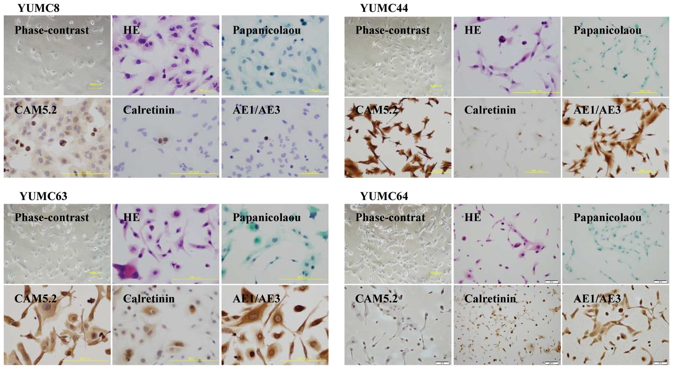
results of each cell line are summarized in Table I. Representative images of these cells
stained with immunocytochemical staining are presented in Fig. 1. The cytological examinations and
immunocytochemistry that were performed in the established cell
lines demonstrated that all of the established cell lines were
derived from MPM and that the immunocytochemical characteristics
were concordant with those of each original specimen. In YUMC8
cells, the immunocytochemical staining for desmin in the original
tissue was partially positive: Only some components of the tumor
cells were stained positively. It is possible that the
desmin-positive cells were not tumor cells but instead normal
spindle mesothelial cells that were mixed in the tumor tissue. As
for the histological subtypes, YUMC8 and YUMC44 cells were
diagnosed as the sarcomatoid type, and YUMC63 and YUMC64 cells were
diagnosed as the epithelioid type. The results of DNA
fingerprinting revealed that each established cell line was unique,
that there was no cross-contamination of cell lines, and that none
of the cell lines were contaminated by mycoplasma (data not
shown).
 | Table I.Characteristics and immunocytochemical
findings of 4 established MPM cell lines. |
Table I.
Characteristics and immunocytochemical
findings of 4 established MPM cell lines.
| Cell line name | YUMC8 | YUMC44 | YUMC63 | YUMC64 |
|---|
| Sex | M | M | M | M |
| Age | 76 | 67 | 72 | 58 |
| Histological
subtype | Sarcomatoid | Sarcomatoid | Epithelioid | Epithelioid |
| Asbestos
exposure | No | Yes | No | Yes |
| Smoking status
(pack/year) | 90 | 10 | 100 | 120 |
| Origin of
culture | Pleural
effusion | Tumor | Pleural
effusion | Pleural
effusion |
| Pretreatment at the
time of primary culture | No | No | No | No |
| Immunocytochemical
profile | Cell line | Original
tissue | Cell line | Original
tissue | Cell line | Original
tissue | Cell line | Original
tissue |
|
CAM5.2 | + | ND | + | + | + | ND | + | ND |
|
AE1/AE3 | + | + | + | + | + | ND | + | ND |
|
Calretinin | + | + | + | + | + | + | + | + |
|
D2–40 | − | ND | + | + | − | ND | − | + |
|
CK5/6 | ND | + | − | − | + | ND | − | + |
|
WT-1 | − | ND | − | − | + | ND | + | ND |
|
HBME1 | ND | − | ND | − | − | + | − | + |
|
Thrombomodulin | ND | ND | ND | + | − | + | + | + |
|
CEA | − | ND | − | − | − | − | − | ND |
|
TTF-1 | − | ND | − | − | − | ND | − | ND |
|
BerEP4 | ND | ND | ND | − | − | − | − | − |
|
MOC31 | ND | ND | − | − | − | − | + | − |
|
Desmin | − | partially + | − | − | − | ND | − | ND |
|
S-100 | − | − | − | ND | ND | ND | ND | ND |
|
α-SMA | − | − | − | ND | ND | ND | − | ND |
|
GLUT1 | ND | ND | ND | ND | + | ND | + | ND |
|
p53 | ND | ND | ND | ND | − | ND | + | ND |
Profiling of the molecular
characteristics of the established MPM cell lines
The genetic and epigenetic characteristics of
several molecules were analyzed in each established cell line.
NF2 deletion was observed in YUMC44 cells, and P16
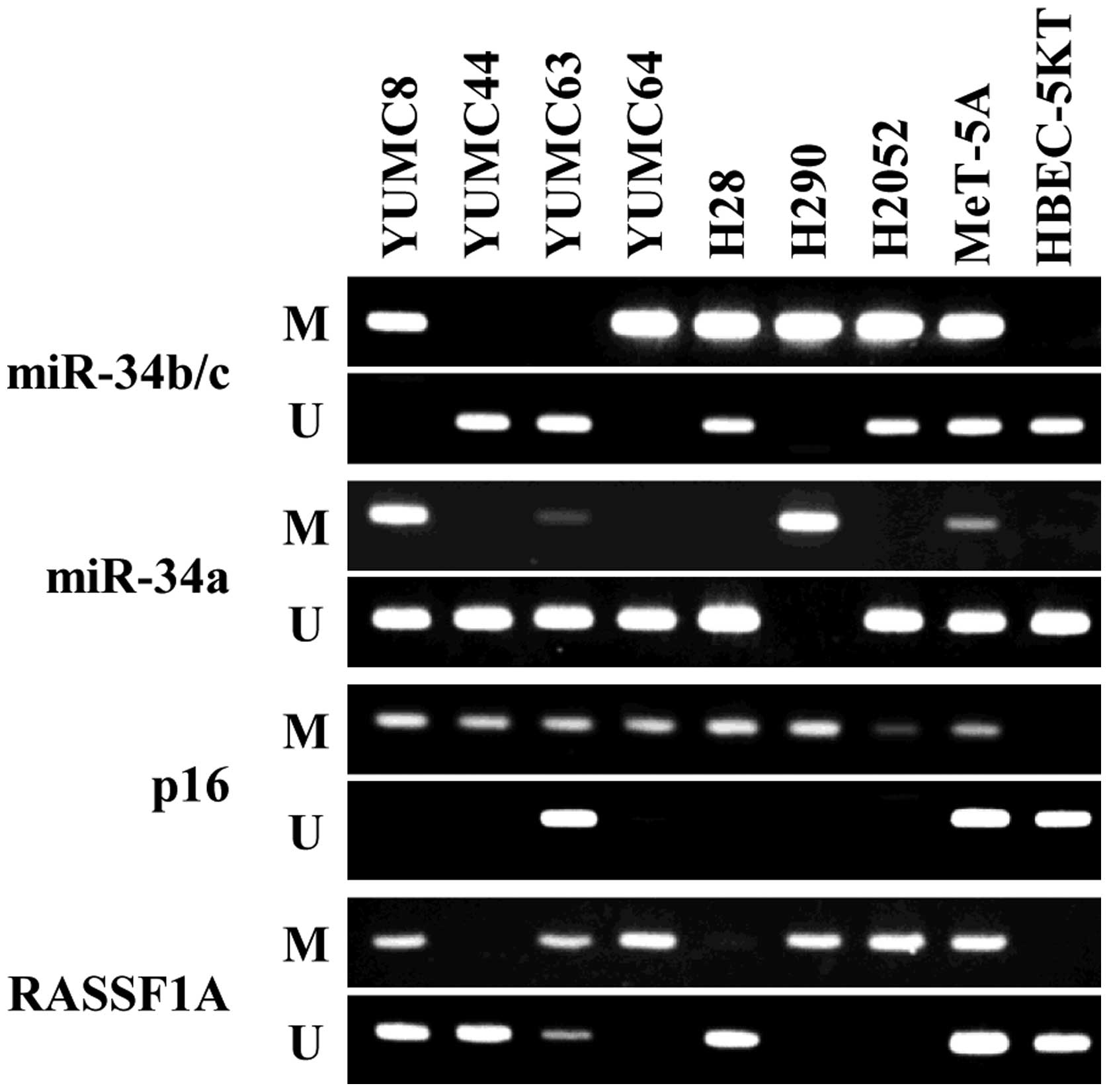
deletion was observed in YUMC44 and YUMC64 cells (Table II). As for methylation, P16
was heavily methylated in YUMC8, YUMC44, and YUMC64 cells and
partially methylated in YUMC63 cells. RASSF1A was heavily
methylated in YUMC64 cells and partially methylated in YUMC8 and
YUMC63 cells. miR-34b/c was heavily methylated in YUMC8 and YUMC64
cells, and miR-34a was partially methylated in YUMC8 and YUMC63
cells (Fig. 2).
| Table II.Genetic and epigenetic alterations in
MPM cell lines. |
Table II.
Genetic and epigenetic alterations in
MPM cell lines.
| Cell line | YUMC8 | YUMC44 | YUMC63 | YUMC64 | NCI-H28 | NCI-H290 | NCI-H2052 |
|---|
| Copy number
change |
|
|
|
|
|
|
|
|
NF2 | 1.3 | 0 | 1.12 | 1.44 | 1.66 | 1.33 | 2.52 |
|
p16 | 1.57 | 0 | 1.7 | 0 | 0 | 0 | 0 |
|
EGFR | 3.91 | 1.77 | 2.71 | 1.65 | 2.67 | 2.41 | 1.19 |
|
HER2 | 2.87 | 3.32 | 3.2 | 2.72 | 2.85 | 3.17 | 2.5 |
|
MET | 2.35 | 2 | 2.62 | 1.67 | 0.97 | 1.72 | 1.1 |
| Methylation |
|
|
|
|
|
|
|
|
p16 | M | M | M/U | M | M | M | M |
|
RASSF1A | M/U | U | M/U | M | U | M | M |
|
miR-34b/c | M | U | U | M | M/U | M | M/U |
| Mutational
status |
|
|
|
|
|
|
|
|
EGFR | WT | WT | WT | WT | WT | WT | WT |
|
KRAS | WT | WT | WT | WT | WT | WT | WT |
|
HER2 | WT | WT | WT | WT | WT | WT | WT |
|
BRAF | WT | WT | WT | WT | WT | WT | WT |
|
AKT1 | WT | WT | WT | WT | WT | WT | WT |
|
MEK1 | WT | WT | WT | WT | WT | WT | WT |
|
NRAS | WT | WT | WT | WT | WT | WT | WT |
|
PIK3CA | WT | WT | WT | WT | WT | WT | WT |
|
PTEN | WT | WT | WT | WT | WT | WT | WT |
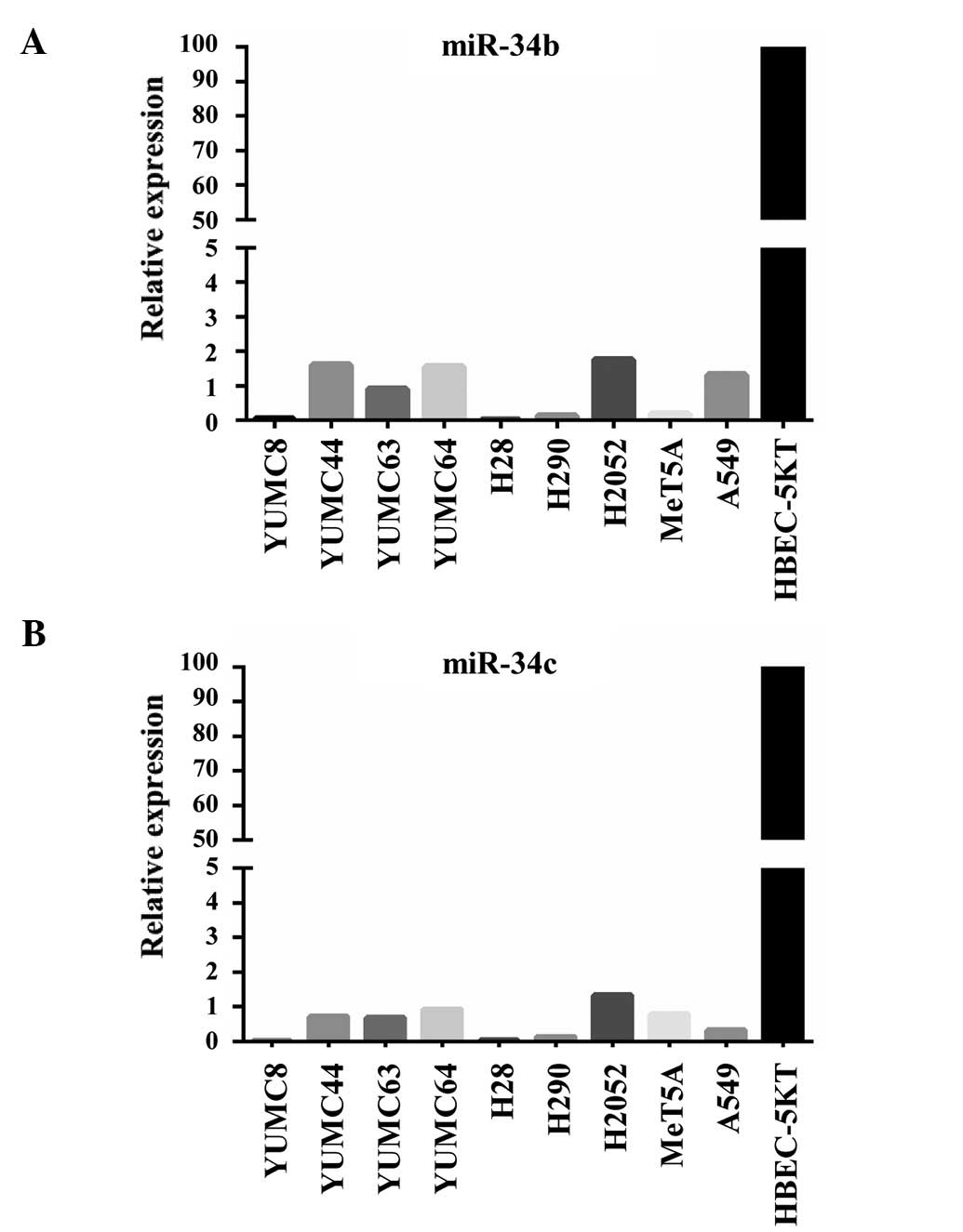
The expression of miR-34b and miR-34c was examined
with RT-qPCR. As miR-34b/c was partially methylated in normal
mesothelial cells (MeT-5A), the expression levels of these miRs
were examined in established MPM cells to those of normal bronchial
cells (HBECK-5KT). In all of the MPM cell lines, including
unmethylated cells (YUMC44 and YUMC63), the expression of miR-34b/c
was suppressed (Fig. 3).
The copy number gains for EGFR, HER2,
and MET were analyzed, which are often altered in non-small
cell lung cancer (15). These genes
were not amplified in the MPM cell lines that were established.
Although the mutational statuses of EGFR, KRAS,
HER2, BRAF, and PIK3CA were also studied,
mutations were not detected in these genes (Table II). These results were consistent
with those of previous reports regarding MPMs (16,17).
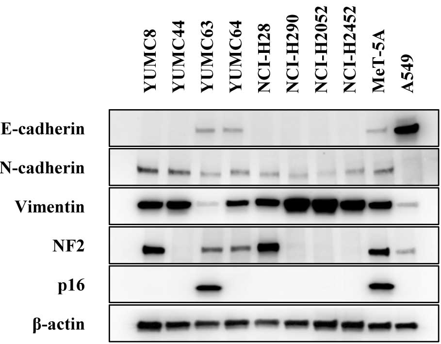
Western blotting revealed that E-cadherin was
expressed in the epithelioid MPM cell lines (YUMC63 and YUMC64).
N-cadherin and vimentin were expressed in all of the MPM cell
lines. NF2 was not expressed in YUMC44 cells. p16 was expressed
only in YUMC63 cells (Fig. 4).
Association between the molecular
characteristics and drug sensitivity
The drug sensitivities against various agents are
presented as IC50 in Table
III. Associations between the drug sensitivities and the
molecular profile were not observed in this study.
| Table III.Sensitivity (IC50 values)
to various drugs in 4 MPM cell lines. |
Table III.
Sensitivity (IC50 values)
to various drugs in 4 MPM cell lines.
| Cell line | YUMC8 | YUMC44 | YUMC63 | YUMC64 |
|---|
| Drugs |
|
|
|
|
|
Cisplatin | 5.9±2.4 | 19.6±5.9 | 12.6±2.9 | 4.8±0.13 |
|
Pemetrexed | >100 | >160 | >160 | >160 |
|
Vinorelbine | 0.0078±0.0026 | 1.1±0.9 | >10 | 0.020±0.016 |
|
NVP-AUY922 | 0.019±0.010 | 1.7±0.3 | >10 | 3.2±2.0 |
|
17-DMAG | 0.056±0.026 | 0.19±0.12 | >10 | 4.3±2.5 |
|
Trichostatin A | 0.060±0.012 | 0.20±0.04 | 0.15±0.05 | 0.18±0.01 |
|
Vorinostat | 5.1±3.6 | 5.6±1.5 | 7.5±5.8 | 10.3±4.5 |
Discussion
In the current study, 4 MPM cell lines were newly
established that consisted of 2 sarcomatoid and 2 epithelioid cell
lines, and their immunocytochemical characteristics were concordant
with those of the original tumors. Established tumor cell lines are
useful for elucidating the molecular characteristics of the tumors
that they are derived from, which results in substantial
contributions to tumor translational research. For example, >200
lung cancer cell lines have been established and used for research,
which has led to the discovery of intriguing properties that are
linked with treatment, such as EGFR-activating mutations and
EML-ALK fusion genes (9). In
contrast, a relatively small number of MPM cell lines have been
established compared with those for lung cancer (12,17), and
the number of cell lines that have been deposited in publicly open
banks is limited. Thus, we plan to deposit them with the molecular
profiles that we determined in a public cell bank for the
convenience of other researchers.
The molecular statuses of representative
tumor-associated genes or proteins of these established cell lines.
Among the examined molecules, the P16INK4a gene, which is
located at chromosome 9p21, is frequently deleted in MPM, and it
has been demonstrated that its deletion results in the
dysregulation of cell-cycle control through the Rb pathway and
malignant transformation (18,19). In
addition, the present authors have previously reported that DNA
methylation of P16 is another mechanism that leads to the
loss of p16 expression (7). In the
present study, P16 deletion was observed in YUMC44 and
YUMC64 cells, and P16 was methylated completely in YUMC8,
YUMC44, and YUMC64 cells and partially in YUMC63 cells. Both
deletion and methylation were observed as the cause of the loss of
protein expression.
The status of the other molecules, such as the
methylation status of RSSF1A and miR-34b/c, the expression of
miR-34b/c, the mutational status of 8 cancer-related genes and the
expression of EMT markers, were similar to those of previous
reports (6,8,17,20). The sensitivity of the cell lines to a
number of therapeutic drugs and their associations with the
molecular profile were examined, however no significant effects
were observed. Taken together, the molecular characteristics of
these cell lines appeared to be similar to those of previous
reports, which indicated that the characteristics of the
established cell lines were representative of MPM and thus they may
be considered useful for studying the biological behavior of
MPM.
To determine the anchorage-independent growth of the
4 established MPM cell lines, a soft agar assay was performed.
However, a significant number of colonies were not detected (data
not shown). In vivo experiments of the tumorigenicity of the
established MPM cell lines were not performed, because these cell
lines did not demonstrate anchorage-independent growth.
In conclusion, novel MPM cell lines were established
that were named YUMC8, YUMC44, YUMC63, and YUMC64. Since MPM cell
lines are less established compared with other malignancies, these
established cell lines are valuable for analyzing the molecular and
biological characteristics of MPMs, which may lead to the
development of novel therapeutic strategies.
Acknowledgements
The authors wish to thank Ms. Fumiko Isobe for her
technical assistance. The present study was supported by a
Grant-in-Aid for Scientific Research from the Ministry of
Education, Culture, Sports, Science and Technology of Japan (grant
no. 22591566 to Dr Shinichi Toyooka).
Glossary
Abbreviations
Abbreviations:
|
MPM
|
malignant pleural mesothelioma
|
|
PCR
|
polymerase chain reaction
|
|
qPCR
|
quantitative PCR
|
References
|
1
|
Robinson BW and Lake RA: Advances in
malignant mesothelioma. N Engl J Med. 353:1591–1603. 2005.
View Article : Google Scholar : PubMed/NCBI
|
|
2
|
Spirtas R, Heineman EF, Bernstein L, Beebe
GW, Keehn RJ, Stark A, Harlow BL and Benichou J: Malignant
mesothelioma: Attributable risk of asbestos exposure. Occup Environ
Med. 51:804–811. 1994. View Article : Google Scholar : PubMed/NCBI
|
|
3
|
Robinson BM: Malignant pleural
mesothelioma: An epidemiological perspective. Ann Cardiothorac
Surg. 1:491–496. 2012.PubMed/NCBI
|
|
4
|
Flores RM, Riedel E, Donington JS, Alago
W, Ihekweazu U, Krug L, Rosenzweig K, Adusumilli PS, Carbone M and
Pass HI: Frequency of use and predictors of cancer-directed surgery
in the management of malignant pleural mesothelioma in a
community-based (Surveillance, Epidemiology, and End Results
[SEER]) population. J Thorac Oncol. 5:1649–1654. 2010. View Article : Google Scholar : PubMed/NCBI
|
|
5
|
Sekido Y, Pass HI, Bader S, Mew DJ,
Christman MF, Gazdar AF and Minna JD: Neurofibromatosis type 2
(NF2) gene is somatically mutated in mesothelioma but not in lung
cancer. Cancer Res. 55:1227–1231. 1995.PubMed/NCBI
|
|
6
|
Toyooka S, Carbone M, Toyooka KO,
Bocchetta M, Shivapurkar N, Minna JD and Gazdar AF: Progressive
aberrant methylation of the RASSF1A gene in simian virus 40
infected human mesothelial cells. Oncogene. 21:4340–4344. 2002.
View Article : Google Scholar : PubMed/NCBI
|
|
7
|
Kobayashi N, Toyooka S, Yanai H, Soh J,
Fujimoto N, Yamamoto H, Ichihara S, Kimura K, Ichimura K, Sano Y,
et al: Frequent p16 inactivation by homozygous deletion or
methylation is associated with a poor prognosis in Japanese
patients with pleural mesothelioma. Lung Cancer. 62:120–125. 2008.
View Article : Google Scholar : PubMed/NCBI
|
|
8
|
Kubo T, Toyooka S, Tsukuda K, Sakaguchi M,
Fukazawa T, Soh J, Asano H, Ueno T, Muraoka T, Yamamoto H, et al:
Epigenetic silencing of microRNA-34b/c plays an important role in
the pathogenesis of malignant pleural mesothelioma. Clin Cancer
Res. 17:4965–4974. 2011. View Article : Google Scholar : PubMed/NCBI
|
|
9
|
Gazdar AF, Girard L, Lockwood WW, Lam WL
and Minna JD: Lung cancer cell lines as tools for biomedical
discovery and research. J Natl Cancer Inst. 102:1310–1321. 2010.
View Article : Google Scholar : PubMed/NCBI
|
|
10
|
Su Z, Dias-Santagata D, Duke M, Hutchinson
K, Lin YL, Borger DR, Chung CH, Massion PP, Vnencak-Jones CL,
Iafrate AJ and Pao W: A platform for rapid detection of multiple
oncogenic mutations with relevance to targeted therapy in
non-small-cell lung cancer. J Mol Diagn. 13:74–84. 2011. View Article : Google Scholar : PubMed/NCBI
|
|
11
|
Soh J, Okumura N, Lockwood WW, Yamamoto H,
Shigematsu H, Zhang W, Chari R, Shames DS, Tang X, MacAulay C, et
al: Oncogene mutations, copy number gains and mutant allele
specific imbalance (MASI) frequently occur together in tumor cells.
PLoS One. 4:e74642009. View Article : Google Scholar : PubMed/NCBI
|
|
12
|
Sato A, Torii I, Tao LH, Song M, Kondo N,
Yoshikawa Y, Hashimoto-Tamaoki T, Hasegawa S, Nakano T and
Tsujimura T: Establishment of a cell line from a Japanese patient
useful for generating an in vivo model of malignant pleural
mesothelioma. Cancer Sci. 102:648–655. 2011. View Article : Google Scholar : PubMed/NCBI
|
|
13
|
Herman JG, Graff JR, Myöhänen S, Nelkin BD
and Baylin SB: Methylation-specific PCR: A novel PCR assay for
methylation status of CpG islands. Proc Natl Acad Sci USA.
93:9821–9826. 1996. View Article : Google Scholar : PubMed/NCBI
|
|
14
|
Shien K, Ueno T, Tsukuda K, Soh J, Suda K,
Kubo T, Furukawa M, Muraoka T, Maki Y, Tanaka N, et al: Knockdown
of the epidermal growth factor receptor gene to investigate its
therapeutic potential for the treatment of non-small-cell lung
cancers. Clin Lung Cancer. 13:488–493. 2012. View Article : Google Scholar : PubMed/NCBI
|
|
15
|
Zhang X and Chang A: Molecular predictors
of EGFR-TKI sensitivity in advanced non-small cell lung cancer. Int
J Med Sci. 5:209–217. 2008. View Article : Google Scholar : PubMed/NCBI
|
|
16
|
Taniguchi T, Karnan S, Fukui T, Yokoyama
T, Tagawa H, Yokoi K, Ueda Y, Mitsudomi T, Horio Y, Hida T, et al:
Genomic profiling of malignant pleural mesothelioma with
array-based comparative genomic hybridization shows frequent
non-random chromosomal alteration regions including JUN
amplification on 1p32. Cancer Sci. 98:438–446. 2007. View Article : Google Scholar : PubMed/NCBI
|
|
17
|
Usami N, Fukui T, Kondo M, Taniguchi T,
Yokoyama T, Mori S, Yokoi K, Horio Y, Shimokata K, Sekido Y and
Hida T: Establishment and characterization of four malignant
pleural mesothelioma cell lines from Japanese patients. Cancer Sci.
97:387–394. 2006. View Article : Google Scholar : PubMed/NCBI
|
|
18
|
Prins JB, Williamson KA, Kamp MM, Van
Hezik EJ, Van der Kwast TH, Hagemeijer A and Versnel MA: The gene
for the cyclin-dependent-kinase-4 inhibitor, CDKN2A, is
preferentially deleted in malignant mesothelioma. Int J Cancer.
75:649–653. 1998. View Article : Google Scholar : PubMed/NCBI
|
|
19
|
Lee AY, Raz DJ, He B and Jablons DM:
Update on the molecular biology of malignant mesothelioma. Cancer.
109:1454–1461. 2007. View Article : Google Scholar : PubMed/NCBI
|
|
20
|
Donninger H, Vos MD and Clark GJ: The
RASSF1A tumor suppressor. J Cell Sci. 120:3163–3172. 2007.
View Article : Google Scholar : PubMed/NCBI
|