Introduction
An epidermal inclusion cyst (EIC) of the breast is a
rare condition that develops due to the proliferation and
implantation of epidermal elements within a circumscribed space in
the dermis (1). Growth of the EIC
occurs through the accumulation of epithelial and keratinous
debris, and it is formed by the inclusion of keratinizing squamous
epithelium within the dermis, resulting in a lamellated
keratin-filled cyst (2).
Historically, EICs have been referred to using a number of
different terms, including follicular infundibular cysts, epidermal
cysts and epidermoid cysts. EICs may occur anywhere in the body,
although they are most prevalent on the face, trunk, neck,
extremities and scalp (1). EICs
rarely develop in the breastdevelops in the breast, it presents as
a lump that is primarily localized in the periareolar region
(3). The importance of this benign
lesion lies in the differentiation between other non-neoplastic and
neoplastic breast lesions (2).
Furthermore, an association between EIC and squamous cell carcinoma
has been reported (4). The incidence
of malignant potential is highly variable (0.045–19.0%) and the
true incidence remains uncertain (5).
The aim of the current literature review, with the
addition of an individual case reported in the present study, is to
identify and discuss the incidence, demographic occurrence, racial
differences, clinical characteristics, pathology, and diagnostic
and treatment options of this disease.
Materials and methods
Literature review
The literature search involved the use of the Scopus
(www.scopus.com), Embase (www.embase.com) and Medline (www.ncbi.nlm.nih.gov/pubmed) databases during
September 2014. Manual searching of reference lists of the relevant
studies and previous reviews was also performed. No language
restrictions were applied. The primary search term was conducted
for any combination of the words ῾epidermal inclusion cyst breast᾽.
Studies were included if they contained adequate information
regarding symptoms, EIC characteristics (size and location) and the
type of procedures performed. In the case of duplicate
publications, the latest and most complete study was included.
Studies that dealt with locations other than the breast were
excluded.
Data extraction
Two independent reviewers extracted data from each
study using a predefined database form, which resulted in high
interobserver agreement. The information included the names of the
authors, the title of the study, the journal in which the study was
published, the country and year of the study, the demographics of
the patients, anamnestic information, the diagnostic tools used,
the disease pathology and the treatment performed.
Following completion of data extraction from the
included studies, the two independent reviewers discussed the
results of the collected data and, if discrepancies were present, a
consensus was reached by mutual agreement on the accuracy of the
data.
Statistical analysis
The data were entered into a computer spreadsheet
and statistically analyzed through the use of SPSS software,
version 21.0 (IBM SPSS, Armonk, NY, USA). Data were expressed as
the mean ± standard deviation. Comparisons between groups were
tested with Pearson's χ2 test, either using Yates'
correction or Fisher's exact test when appropriate for categorical
variables, and Student's t-test for continuous variables. The
association between tumor size (cut-off at 4 cm) and malignant
transformation was analyzed and calculated using the Spearman's
rank correlation coefficient method. The r value is reported for
all linear regressions. P<0.05 was considered to indicate a
statistically significant difference.
Results
Study data
The search identified 35 studies (1–35),
describing the clinical course of 91 patients affected by EIC of
the breast. Following a review of the literature, 8 studies that
described 10 patients were excluded, as the complete studies could
not be found or only partial data was available (28–35). The
remaining studies fulfilled the inclusion criteria. A total of 81
patient cases were retrieved, to which an individual case reported
by the present study was added, creating the final total of 82
cases available for the present analysis.
Case report
A 70-year-old woman was referred to the Department
of Surgery, Policlinico Umberto I (Rome, Italy) in June 2013 due to
a painful and rapidly growing palpable mass in the left breast. The
medical history of the patient was notable due to previous
addictive breast plastic surgery and subsequent implant removal due
to rupture. Physical examination identified a round, firm lesion
that was non-adherent to the overlying skin in the lower outer
quadrant of the left breast. Ultrasonography exhibited a solid,
heterogeneously hypoechoic, well-circumscribed mass measuring
3.1×3.0 cm. An excisional biopsy was performed with an ellipse of
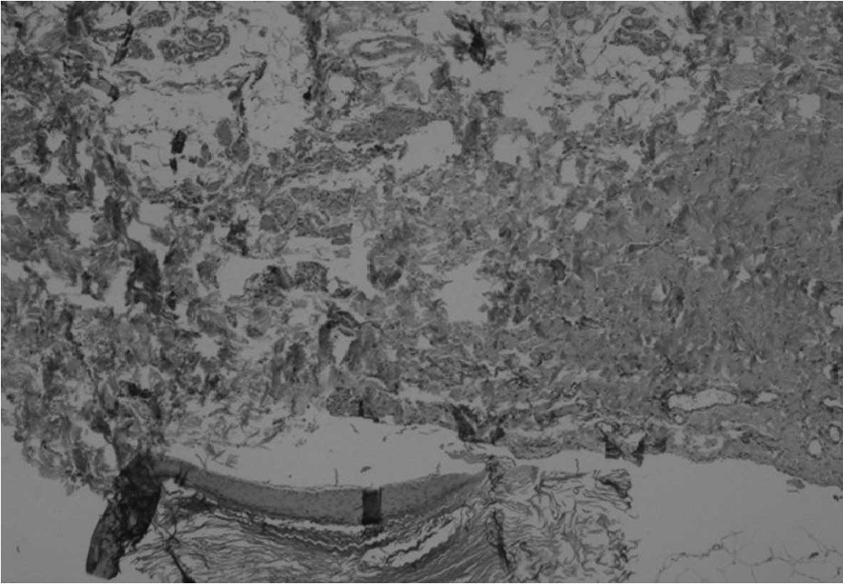
skin. Pathological examination of the mass detected breast lobules
and EIC (Fig. 1). The patient
provided written informed consent.
Demographics
Of the 82 patients, there were 60 females (73%) and
8 males (10%), while in 14 cases (17%) the gender was unspecified.
The mean age at presentation was 48±14 years (median, 47 years;
range, 20–85 years). EIC was identified in the majority of cases
within the Asian and Caucasian populations (28 and 22 cases,
respectively). The right side was affected in 24 patients (29%) and
the left in 32 (39%). Out of 68 patients from whom information was
retrieved, 11 (13%) had undergone previous cosmetic breast surgery
and 7 (19%) had experienced previous trauma. Neither a family
history of breast disease or previous/current hormonal medication
was noted. Furthermore, a predisposition to develop EIC of the
breast was not observed with oral estroprogestinic therapy or
genetic patterns. No specific information regarding weight, body
mass index and biochemical tests were available, and no patients
had associated EIC at other body locations. The postoperative
course was uneventful. The patient remained alive without signs or
symptoms of recurrence at a follow-up of 27 months.
Clinical presentation
A total of 65 patients (79%) presented with a
palpable mass of the breast. Local discomfort was associated with
55 cases (67%), inflammation with 27 (33%), spontaneous rupture
with 10 (12%) and ulceration with 3 (4%).
Tumor characteristics
At presentation, the mean tumor size was 4±3 cm
(median, 3 cm; range, 1–10 cm). The mean growth period of the
breast mass was 25±42 months (median, 12 months; range, 1–264
months).
Diagnostic tools
Ultrasonographic imaging, mammography, fine-needle
aspiration cytology (FNAC) and FNA biopsy (FNAB) were the
diagnostic tools that were used, either alone or in association, to
investigate the presence of an EIC of the breast. A total of 22
patients were studied using ultrasonographic imaging and exhibited
a solid, well-circumscribed and complex, or heterogeneous,
appearance. Notably, an ῾onion-ring᾽ appearance, with alternating
concentric hyperechoic and hypoechoic rings was observed in 3
patients, and in 1 patient, this also extended into the dermis.
Ultrasonographic imaging was demonstrated to consistently provide
an accurate diagnosis.
A total of 24 patients underwent a mammography,
which demonstrated a well-circumscribed mass with homogeneous
increased density in 19 patients. However, in 5 cases, the
mammography was misleading. Magnetic resonance imaging was
performed in 2 patients and demonstrated a fluid-like signal with
variable low-signal components on T2-weighted images, and a
peripheral rim enhancement on gadolinium-enhanced images. FNAC was
performed in 21 patients and FNAB in 17. An accurate diagnosis was
achieved in 15 (71%) and 11 (65%) cases, respectively. In 10 cases,
the diagnostic flowchart from the relevant studies was not
available.
Pathology
EIC of the breast was benign in 72 patients (88%),
and had transformed into squamous cell carcinoma in 10 patients
(12%). In 3 cases, the congenital development of a cyst secondary
to an obstructed hair follicle or pore was theorized. Whilst in 11
cases, an injury to the epidermis subsequently resulting in
fragments being implanted deeply in the breast tissue was
hypothesized, and in 3 cases, EIC was believed to have developed
following squamous metaplasia of normal columnar cells within a
dilated duct in cases of fibrocystic disease (1 case), fibroadenoma
(1 case) or a phyllodes tumor (1 case). A significant correlation
between tumor size and malignant transformation was observed
(P<0.01, r=0.459).
Surgical excision
Surgical excision of the mass was performed in 70
patients (85%). The remaining patients did not undergo surgery.
Follow-up
At a mean follow-up time of 53±62 months, no
problems associated with the surgery or recurrence of the EIC of
the breast were reported. The long-term survival of the patients
affected with malignant transformation of the EIC was not
reported.
Discussion
Despite the literature containing abundant case
reports and reviews, several concerns remain regarding the
incidence, etiology, diagnosis and treatment of EIC. The present
study analyzed the current international literature with the aim of
understanding this rare occurrence somewhat further.
To date, the international literature has reported
90 cases of patients who have been affected by EIC of the breast.
To the best of our knowledge, the first histologically defined case
of EIC of the breast was reported in December 1900 at The Johns
Hopkins Hospital (Baltimore, MD, USA) (5). From the literature, it can be determined
that EIC of the breast typically affects individuals in the fifth
decade of life, and males are observed to be affected in a small
proportion of cases (1–28). This type of tumor is naturally slow
growing. The spontaneous rupture of large cysts was noted in 10
cases, releasing non-absorbable keratin, which acts as an irritant
and subsequently leads to secondary foreign body reactions,
granulomatous reactions or abscess formation.
A previous history of trauma or surgery appears to
be associated with the development of this benign tumor, but other
theories have been postulated (1–28). A
unique pathogenesis cannot be concluded, but it is believed that,
in the majority of cases, EIC of the breast may be congenital,
arising from cell nests that remain from specific cells, including
the embryonic mammary ridge. EIC of the breast may also develop
from obstructed hair follicles. Furthermore, pilosebaceous
structures may become inflamed, leading to a cystic reaction in the
dermis, or be created by squamous metaplasia of normal columnar
cells within a dilated duct in the case of fibrocystic disease, or
in fibroadenoma or phyllodes tumors.
Physical examination regarding the diagnosis of EIC
is unreliable (1–28). EIC of the breast typically appears as
a smooth, round nodule, the nature of which cannot be detected
through this method. Ultrasonographic imaging consistently achieved
an accurate diagnosis (1,2,7–17,22,23,25,26),
whilst mammography achieved accuracy in 79% of the reported cases
(6–12,15–17,21–27).
Magnetic resonance imaging was used in only 2 cases (16,25), but
it was also observed to consistently identify EIC of the breast
accurately. FNAC (2,8,16–19,25–27) and
FNAB (1,2,7,8,14,15,18,22,23,25)
may also aid the diagnosis, but these methods are less reliable
when compared to ultrasonographic imaging. The present literature
review demonstrated that ultrasonographic imaging is the diagnostic
tool of choice to achieve the accurate characterization of EIC.
During the review process, it was identified that
the association between EIC of the breast and malignant tumors was
12% (5,27). Malignant transformation appears to
occur more frequently in EIC of the breast as opposed to EIC
affecting other body areas, and this may be associated with the
pathogenesis of EIC of the breast from squamous metaplasia of the
mammary duct epithelium. Furthermore, a significant correlation
between tumor size and malignant transformation was identified.
Therefore, the presence of a lesion of <1 cm localized in the
breast parenchyma may be differentiated from a large fibroadenoma
or phyllodes tumor, and even from a malignant breast neoplasm with
benign features, including mucinous carcinoma.
If the diagnosis is accurate, asymptomatic
small-sized lesions (<2 cm diameter) do not require treatment.
An excisional biopsy is not required if typical ultrasonographic
findings are exhibited. However, EICs of the breast that are large
and palpable, and possibly causing the patient physical and
psychological discomfort, will require surgical excision through an
elliptical incision. The removal of the entire cyst wall is
recommended for pathological analysis and to prevent recurrence or
malignant transformation.
In conclusion, EIC of the breast is a rare
occurrence and the pathogenesis remains unknown. Futue studies
should focus on the identification of the predisposing factors,
including hormone therapy or genetic predisposition, that may
influence the development of this disease.
References
|
1
|
Lam SY, Kasthoori JJ, Mun Ks and Rahmat K:
Epidermal inclusion cyst of the breast: A rare benign entity.
Singapore Med J. 51:e191–e194. 2010.PubMed/NCBI
|
|
2
|
Lee YA and Park SG: Giant sized epidermal
inclusion cyst of the breast initially mimicking a large
fibroadenoma or phyllodes tumor. J Korean Surg Soc. 83:107–110.
2012. View Article : Google Scholar : PubMed/NCBI
|
|
3
|
Singh M, Maheshwari B, Khurana N and Jain
S: Epidermal inclusion cyst in breast: Is it so rare? J Cytol.
29:169–172. 2012. View Article : Google Scholar : PubMed/NCBI
|
|
4
|
Cameron DS and Hilsinger RL Jr: Squamous
cell carcinoma in an epidermal inclusion cyst: Case report.
Otolaryngol Head Neck Surg. 129:141–143. 2003. View Article : Google Scholar : PubMed/NCBI
|
|
5
|
Menville JG: Simple dermoid cysts of the
breast. Ann Surg. 103:49–56. 1936. View Article : Google Scholar : PubMed/NCBI
|
|
6
|
Chatterjee PK and Roy SN: Large epidermal
cyst of breast simulating malignant growth. BMJ. 1:167–168. 1979.
View Article : Google Scholar : PubMed/NCBI
|
|
7
|
Kowand LM, Verhulst LA, Copeland CM and
Bose B: Epidermal cyst of the breast. Can Med Assoc J. 131:217–219.
1984.PubMed/NCBI
|
|
8
|
Chantra PK, Tang JT, Stanley TM and
Bassett LW: Circumscribed fibrocystic mastopathy with formation of
an epidermal cyst. AJR Am J Roentgenol. 163:831–832. 1994.
View Article : Google Scholar : PubMed/NCBI
|
|
9
|
Morris PC, Cawson JN and Balasubramaniam
GS: Epidermal cyst of the breast: Detection in a screening
programme. Australas Radiol. 43:12–15. 1999. View Article : Google Scholar : PubMed/NCBI
|
|
10
|
Appelbaum AH, Evans GF, Levy KR, Amirkhan
RH and Schumpert TD: Mammographic appearances of male breast
disease. Radiographics. 19:559–568. 1999. View Article : Google Scholar : PubMed/NCBI
|
|
11
|
Günhan-Bilgen I, Bozkaya H, Ustün E and
Memiş A: Male breast disease: Clinical, mammographic and
ultrasonographic features. Eur J Radiol. 43:246–255. 2002.
View Article : Google Scholar : PubMed/NCBI
|
|
12
|
Kwak JY, Park HL, Kim JY, Kim EK, Chung
SY, Kwon TH, Hong HS and Oh KK: Imaging findings in a case of
epidermal inclusion cyst arising within the breast parenchyma. J
Clin Ultrasound. 32:141–143. 2004. View Article : Google Scholar : PubMed/NCBI
|
|
13
|
Celik V, Unal E, Aydogan F, Sunamak O,
Kuşaslan R, Rasier R, Ilvan S, Yilmaz MH and Ferahman M: Epidermal
inclusion cyst of the breast: Clinical, radiologic and pathologic
correlation. Breast J. 10:572004. View Article : Google Scholar : PubMed/NCBI
|
|
14
|
Crystal P and Shaco-Levy R: Concentric
rings within a breast mass on sonography: Lamellated keratin in an
epidermal inclusion cyst. AJR Am J Roentgenol. 184(Suppl): S47–S48.
2005. View Article : Google Scholar : PubMed/NCBI
|
|
15
|
Whang IY, Lee J, Kim JS, Kim KT and Shin
OR: Ruptured epidermal inclusion cysts in the subareolar area:
Sonographic findings in two cases. Korean J Radiol. 8:356–359.
2007. View Article : Google Scholar : PubMed/NCBI
|
|
16
|
Iglesias A, Arias M, Santiago P, Rodríguez
M, Mañas J and Saborido C: Benign breast lesions that simulate
malignancy: Magnetic resonance imaging with radiologic-pathologic
correlation. Curr Probl Diagn Radiol. 36:66–82. 2007. View Article : Google Scholar : PubMed/NCBI
|
|
17
|
Taira N, Aogi K, Ohsumi S, Takashima S,
Kawamura S and Nishimura R: Epidermal inclusion cyst of the breast.
Breast Cancer. 14:434–437. 2007. View Article : Google Scholar : PubMed/NCBI
|
|
18
|
Herreros-Villaraviz M, Mallo-Alonso R,
Santiago-Freijanes P and Díaz-Veiga MJ: Epidermal inclusion cysts
of the breast. Breast J. 14:599–600. 2008. View Article : Google Scholar : PubMed/NCBI
|
|
19
|
Handa U, Chhabra S and Mohan H: Epidermal
inclusion cyst: Cytomorphological features and differential
diagnosis. Diagn Cytopathol. 36:861–863. 2008. View Article : Google Scholar : PubMed/NCBI
|
|
20
|
Mote DG and Shukla AA: Epidermal inclusion
cyst masquerading breast lump. Indian J Surg. 73:458–459. 2011.
View Article : Google Scholar : PubMed/NCBI
|
|
21
|
Chopra K, Folstein MK, Slezak S, Silverman
R, Singh D and Gastman BR: The VersaJet for breast-reduction
surgery: Operator beware. Eplasty. 12:e112012.PubMed/NCBI
|
|
22
|
Taskin F, Koseoglu K, Ozbas S, Erkus M and
Karaman C: Sonographic features of histopathologically benign solid
breast lesions that have been classified as BI-RADS 4 on
sonography. J Clin Ultrasound. 40:261–265. 2012. View Article : Google Scholar : PubMed/NCBI
|
|
23
|
Schaverien MV, Malyon AD, Mallon EA and
Doughty JC: Epidermal inclusion cyst following autologous breast
reconstruction - an unusual cause of a breast lump. Breast J.
20:546–547. 2014. View Article : Google Scholar : PubMed/NCBI
|
|
24
|
Phukan JP, Sinha A, Pal S and Sinha R:
Cytological diagnosis of epidermal inclusion cyst of breast: A rare
benign lesion. J Nat Sci Biol Med. 5:460–462. 2014. View Article : Google Scholar : PubMed/NCBI
|
|
25
|
Spinhoven M, Verslegers I, Van Goethem M,
Van de Vijver K, Biltjes I and Parizel PM: Diffusion restriction in
a superficial breast lesion. JBR-BTR. 90:167–169. 2007.PubMed/NCBI
|
|
26
|
Yamaguchi T, Ojima N, Hayashi M, Komatsu
N, Hashimoto S and Koyama M: Epidermal cyst of the breast treated
by vacuum-assisted biopsy. Int Surg. 98:65–69. 2013. View Article : Google Scholar : PubMed/NCBI
|
|
27
|
Suhani AL, Meena K, Ali S and Thomas S:
Squamous cell carcinoma arising in epidermal inclusion cyst of
breast: A diagnostic dilemma. Breast Dis. 35:25–27. 2015.PubMed/NCBI
|
|
28
|
Salm R: Massive epidermoid metaplasia with
keratin cyst formation in a giant fibro-adenoma of breast. J Pathol
Bacteriol. 77:297–299. 1959. View Article : Google Scholar : PubMed/NCBI
|
|
29
|
Stephenson TJ and Cotton DW: Paget's
disease in an epidermal cyst. Dermatologica. 174:186–190. 1987.
View Article : Google Scholar : PubMed/NCBI
|
|
30
|
Otto H and Breining H: Benign and
malignant breast tumors with squamous cell differentiation.
Radiologe. 27:196–201. 1987.(In German). PubMed/NCBI
|
|
31
|
Das DK, Junaid TA, Mathews SB, Ajrawi TG,
Ahmed MS, Madda JP and Mirza K: Fine needle aspiration cytology
diagnosis of male breast lesions. A study of 185 cases. Acta Cytol.
39:870–876. 1995.PubMed/NCBI
|
|
32
|
Cooper RA and Ramamurthy L: Epidermal
inclusion cysts in the male breast. Can Assoc Radiol J. 47:92–93.
1996.PubMed/NCBI
|
|
33
|
Andersen WK, Rao BK and Bhawan J: The
hybrid epidermoid and apocrine cyst. A combination of apocrine
hidrocystoma and epidermal inclusion cyst. Am J Dermatopathol.
18:364–366. 1996. View Article : Google Scholar : PubMed/NCBI
|
|
34
|
Davies JD, Nonni A and D'Costa HF: Mammary
epidermoid inclusion cysts after wide-core needle biopsies.
Histopathology. 31:549–551. 1997. View Article : Google Scholar : PubMed/NCBI
|
|
35
|
Kapila K and Verma K: Fine needle
aspiration cytology of epidermal inclusion cysts in the male
breast. Acta Cytol. 47:315–317. 2003.PubMed/NCBI
|