Introduction
Lung cancer is the leading cause of
cancer-associated mortality worldwide, and the majority of
broncogenic carcinomas are cases of non-small cell lung cancer
(NSCLC) (1). NSCLC can be
histologically subdivided into four major subtypes with distinct
pathological characteristics as follows: i) Adenocarcinoma; ii)
squamous cell carcinoma; iii) large cell carcinoma; and iv)
‘other’, including neuroendocrine cancer and carcinoids (2). NSCLC is often diagnosed at the advanced
stages of disease progression, resulting in a poor prognosis and an
overall five-year survival rate of ~14%. However, the five-year
survival rate in patients with stage I NSCLC that has been resected
can be as high as 83% (2,3). Despite improvements in early diagnosis
and treatment responses, the overall five-year survival rates for
NSCLC patients remains low (15%) and the recurrence rate is high,
even in cases with early diagnosis (4). The identification of biomarkers for lung
cancer may aid early diagnosis and accurate prognosis. Therefore,
it is important to identify novel and reliable biomarkers for
molecular targeted therapy.
Advances in genomic technologies have generated
numerous candidate biomarkers with potential clinical value in
NSCLC. A complementary approach to performing gene expression
profiling in NSCLC is to analyze microRNA (miRNA) expression
signatures. miRNAs are a class of short RNAs that are 19–25
nucleotides in length and regulate gene expression by binding to
sequences in the 3′-untranslated region (3′UTR) of an expressed
mRNA. This results in modulation of translation efficiency or
degradation of the targeted mRNA (5).
miRNAs have been associated with cell signaling pathways involved
in cell differentiation, proliferation and survival. The aberrant
expression of miRNAs has been demonstrated in a number of different
types of tumor (6), including lung
cancer (7). miRNA profiling has been
proposed as a strategy for classifying NSCLCs, and appears to be
reliable in certain cases (8–10).
Increasing evidence suggests that miRNAs are
expressed aberrantly in numerous types of human cancer and that
they serve a significant role in carcinogenesis and cancer
progression (11). Aberrant
expression of miR-21 has been associated with several types of
cancer, and a previous study demonstrated that it is overexpressed
in tumor tissues of patients with advanced-stage NSCLC. In
addition, the miR-12 target PTEN, which is a negative regulator of
the PKB-AKT pathway, is involved in NSCLC cell line invasion
(12–14). A previous study demonstrated that
miR-125a-3p and −5p are downregulated in primary NSCLC tumors, but
that they serve an opposing role in lymph node metastases (15). miR-328 and miR-206 have also been
associated with NSCLC brain metastasis (16,17). Arora
et al (16) reported that
miR-155 is overexpressed in adenocarcinoma, and further studies
have demonstrated miR-155 overexpression in squamous cell lung
cancer (18,19).
Differential expression of miRNAs has been observed
in previous studies of lung cancer and adjacent normal tissues
(18,20). In order to identify novel biomarkers
of lung cancer, a number of investigators have performed miRNA
expression profiling studies in cell lines, tissue samples or serum
samples (21–23). In the present study, the expression
levels of 15 miRNAs were examined using reverse
transcription-quantitative polymerase chain reaction (RT-qPCR) in
NSCLC and adjacent normal tissues. The expression levels of the 15
miRNAs were also determined by RT-qPCR in formalin-fixed,
paraffin-embedded (FFPE) tissue from NSCLC and non-carcinomatous
(pulmonary bulla) tissues. The aim of the present study was to
identify possible candidate miRNAs to be used as biomarkers for
NSCLC.
Materials and methods
Ethical statement
All samples were obtained from the Affiliated
Hospital of Guangdong Medical University (Zhanjiang, China). The
study protocol was approved by the institutional review boards of
participating centers (Affiliated Hospital of Guangdong
University). Written informed consent was obtained from all
patients.
Samples
Tissues were obtained from 47 patients with NSCLC
diagnosed between January 2013 and March 2014. The samples were as
follows: Fresh-frozen (FF) tissues resected from the 47 patients
(n=140, 47 NSCLC samples, 46 samples of tissues adjacent to the
carcinoma and 47 samples of normal lung tissues) and 56 samples
from FFPE tissues (n=56, including 45 carcinomatous NSCLC tissues
and 11 non-carcinomatous pulmonary bulla samples). The samples were
analyzed histologically to assess the level of tumor component (a
minimum of 80% tumor cells) and the quality of the harvested
material. Paracarcinomatous tissues adjacent to the tumor (distance
from the primary tumor <2 cm) and normal tissues (distance from
the primary tumor >5 cm) were defined histologically using
classical pathological approaches (24).
Clinical and pathological data were obtained from
the medical records and centrally reviewed for the purpose of the
study. Clinicopathological information [smoking, age, gender,
pathologic subtype, tumor node metastasis classification, tumor
stage, lymph node stage, differentiation status and adjuvant
therapy following surgery] from all patients that participated in
the present study is summarized in Tables
I and II.
 | Table I.Comparison of clinicopathological
parameters and expression levels of microRNA in NSCLC frozen
tissues. |
Table I.
Comparison of clinicopathological
parameters and expression levels of microRNA in NSCLC frozen
tissues.
|
| miR-21-5p | miR-30e-5p | miR-363-3p | miR-650 | miR-10a-5p | miR-5100 | miR-26b-5p | miR-145-5p |
|---|
|
|
|
|
|
|
|
|
|
|
|---|
| Variable (N,
%) | PC | P-value | PC | P-value | PC | P-value | PC | P-value | PC | P-value | PC | P-value | PC | P-value | PC | P-value |
|---|
| Sex |
0.134 | 0.391 | −0.130 | 0.384 |
0.146 | 0.327 | −0.109 | 0.467 | −0.049 | 0.741 |
0.035 | 0.814 | −0.137 | 0.357 | −0.041 | 0.787 |
| Male
(70) |
|
|
|
|
|
|
|
|
|
|
|
|
|
|
|
|
| Female
(30) |
|
|
|
|
|
|
|
|
|
|
|
|
|
|
|
|
| Age |
0.293 | 0.056 | −0.145 | 0.354 | −0.062 | 0.692 |
0.140 | 0.371 | −0.098 | 0.534 |
0.108 | 0.492 | −0.158 | 0.311 | −0.167 | 0.284 |
| ≤60 (42) |
|
|
|
|
|
|
|
|
|
|
|
|
|
|
|
|
| >60 (58) |
|
|
|
|
|
|
|
|
|
|
|
|
|
|
|
|
| Histological
classification |
0.253 | 0.111 |
0.111 | 0.490 | −0.096 | 0.550 | −0.018 | 0.909 | −0.012 | 0.939 | −0.085 | 0.598 |
0.212 | 0.183 | −0.181 | 0.259 |
| SQC
(22) |
|
|
|
|
|
|
|
|
|
|
|
|
|
|
|
|
| ADC
(74) |
|
|
|
|
|
|
|
|
|
|
|
|
|
|
|
|
| Other
(4) |
|
|
|
|
|
|
|
|
|
|
|
|
|
|
|
|
| Stage | −0.110 | 0.493 | −0.203 | 0.204 |
0.109 | 0.499 |
0.018 | 0.911 |
0.081 | 0.613 |
0.024 | 0.882 |
0.187 | 0.242 |
0.125 | 0.436 |
| I
(24) |
|
|
|
|
|
|
|
|
|
|
|
|
|
|
|
|
| II,
III, IV (76) |
|
|
|
|
|
|
|
|
|
|
|
|
|
|
|
|
| Lymph node |
0.189 | 0.236 |
0.052 | 0.749 |
0.432a | 0.002b |
0.093 | 0.561 |
0.394a | 0.005b |
0.191 | 0.230 |
0.105 | 0.512 |
0.375a | 0.007b |
|
Positive (58) |
|
|
|
|
|
|
|
|
|
|
|
|
|
|
|
|
|
Negative (42) |
|
|
|
|
|
|
|
|
|
|
|
|
|
|
|
|
| Tumor size | −0.216 | 0.175 | −0.157 | 0.326 |
0.078 | 0.628 | −0.057 | 0.722 | −0.287 | 0.069 |
0.110 | 0.494 |
0.097 | 0.548 | −0.031 | 0.847 |
| 0–3 cm
(35) |
|
|
|
|
|
|
|
|
|
|
|
|
|
|
|
|
| >3
cm (65) |
|
|
|
|
|
|
|
|
|
|
|
|
|
|
|
|
|
Differentiation |
0.007 | 0.965 | −0.067 | 0.678 |
0.098 | 0.543 |
0.494a | 0.01b | −0.039 | 0.809 |
0.285 | 0.071 |
0.056 | 0.728 |
0.309a | 0.050 |
| Poorly
(40) |
|
|
|
|
|
|
|
|
|
|
|
|
|
|
|
|
|
Moderately (33) |
|
|
|
|
|
|
|
|
|
|
|
|
|
|
|
|
| High
(27) |
|
|
|
|
|
|
|
|
|
|
|
|
|
|
|
|
| Table II.Association between the expression of
four miRNAs in FFPE samples and the clinicopathological
characteristics of the tumor from 45 NSCLC patients. |
Table II.
Association between the expression of
four miRNAs in FFPE samples and the clinicopathological
characteristics of the tumor from 45 NSCLC patients.
|
| miR-21-5p | miR-30e-5p | miR-363-3p | miR-623 |
|---|
|
|
|
|
|
|
|---|
| Variable (N,
%) | PC | P-value | PC | P-value | PC | P-value | PC | P-value |
|---|
| Sex | −0.079 | 0.605 |
0.037 | 0.808 |
0.098 | 0.520 |
0.098 | 0.520 |
| Male
(73) |
|
|
|
|
|
|
|
|
| Female
(26) |
|
|
|
|
|
|
|
|
| Age | −0.131 | 0.388 |
0.048 | 0.754 |
0.127 | 0.407 |
0.068 | 0.655 |
| ≤60
(26) |
|
|
|
|
|
|
|
|
| >60
(63) |
|
|
|
|
|
|
|
|
| Histological
classification | −0.075 | 0.625 |
0.125 | 0.413 | −0.150 | 0.327 | −0.087 | 0.570 |
| SQC
(46) |
|
|
|
|
|
|
|
|
| ADC
(54) |
|
|
|
|
|
|
|
|
| Other
(0) |
|
|
|
|
|
|
|
|
| Stage |
0.150 | 0.326 | −0.115 | 0.451 |
0.146 | 0.338 |
0.033 | 0.828 |
| I
(44) |
|
|
|
|
|
|
|
|
| II,
III, IV (56) |
|
|
|
|
|
|
|
|
| Lymph node |
0.066 | 0.664 | −0.084 | 0.584 |
0.216 | 0.154 | −0.077 | 0.614 |
|
Positive (36) |
|
|
|
|
|
|
|
|
|
Negative (64) |
|
|
|
|
|
|
|
|
| Tumor size | −0.003 | 0.987 | −0.124 | 0.419 |
0.177 | 0.224 |
0.079 | 0.608 |
| 0–3 cm
(60) |
|
|
|
|
|
|
|
|
| >3
cm (40) |
|
|
|
|
|
|
|
|
|
Differentiation |
0.274 | 0.069 |
0.376a | 0.011b |
0.203 | 0.180 |
0.013 | 0.930 |
| Poorly
(36) |
|
|
|
|
|
|
|
|
|
Moderately (40) |
|
|
|
|
|
|
|
|
| High
(24) |
|
|
|
|
|
|
|
|
RNA extraction
The total RNA was extracted using TRIzol reagent
(Invitrogen Life Technologies, Carlsbad, CA, USA) and reverse
transcribed using a miRcute miRNA First-strand cDNA Synthesis kit
(Tiangen Biotech Co., Ltd., Beijing, China). Unstained FFPE tissues
were sectioned at 10-μm thickness and 6 tissue sections were placed
into a 1.5-ml tube. The sections were deparaffinized by adding 1 ml
xylene (Guangzhou Chemical Reagent Factory, Guangzhou, China) and
the sections were vortexed vigorously at room temperature for 3
min. Following washes in 70% and absolute ethanol to remove xylene,
the RNA was extracted with the miRNeasy FFPE kit (Qiagen China Co.,
Ltd., Shanghai, China). Following extraction, all RNA samples were
stored at −80°C until use.
Quantitative polymerase chain reaction
(qPCR)
All miRNA expression levels were quantified via qPCR
using primers designed by Tiangen Biotech Co., Ltd., an miRcute
miRNA qPCR Detection kit (SYBR Green) (Tiangen Biotech Co., Ltd.)
and a Rotor-Gene Q 2plex HRM System (Qiagen China Co., Ltd.). All
the samples were normalized against the endogenous control snRNA U6
and fold changes were calculated relative to the control. The
relative expression levels were calculated using the
2−ΔΔCq method.
Study selection
The online literature database PubMed was used to
identify lung cancer miRNA expression profiling studies published
from January 2003 until May 2014 (last accessed on 15 May 2014), by
means of the ‘Medical Subject Heading’ terms: ‘NSCLC’ and
‘microRNAs’ in combination with the keywords ‘profiling’ and
‘humans’. The results of previous studies to identify circulating
miRNAs specific to lung cancer were inconsistent (24–27), and
potential biomarkers remain to be elucidated and exploited. Based
on these previous findings, a list of 15 miRNAs was compiled, whose
expression has already been reported as deregulated in cancer, or
has not been investigated (Table
III).
| Table III.List of miRNA probes used to profile
tissue samples from non-small cell lung cancer. |
Table III.
List of miRNA probes used to profile
tissue samples from non-small cell lung cancer.
| Hsa-miR-30e-5p | Hsa-miR-10a-5p | Hsa-miR-21-5p | Hsa-miR-26b-5p | Hsa-miR-17-3p |
| Hsa-miR-211-3p | Hsa-miR-1246 | Hsa-miR-5100 |
Hsa-miR-181a-5p | Hsa-miR-363-3p |
| Hsa-miR-650 | Hsa-miR-623 | Hsa-miR-145-5p | Hsa-miR-155-5p |
Hsa-miR-125b-5p |
Statistical analysis
SPSS software, version 17.0 (SPSS, Inc., Chicago,
IL, USA) was used for statistical analysis. The Wilcoxon test was
used to examine the differences of miRNA expression between lung
carcinoma and normal subjects; a Spearman's Rho test was performed
to compare the relative expression of miRNA. Pearson's Correlation
Coefficient was used to analyze the association between the
expression of miRNAs and clinicopathological features of the
patients. Experimental data were expressed as the mean ± standard
error. To compare the means of two groups, a two-tailed Student's
t-test was used and P<0.05 was considered to indicate a
statistically significant difference.
Results
MicroRNA signatures can efficiently
distinguish NSCLC tissues from adjacent normal tissues
A meta-analysis of human lung cancer microRNA
expression profiling studies (18,21,28,29)
was performed, which identified 15 miRNA genes of possible
interest. The initial investigation sought to determine whether the
expression of any of the 15 miRNAs was able to identify malignant
NSCLC tissues in Chinese patients. Primary NSCLC lung cancer
tissues (T), paracarcinomatous tissues (P) and their corresponding
adjacent normal lung tissues (N; ≥5 cm from the tumor) in 47
patients were examined using microRNA-specific reverse
transcription-qPCR (RT-qPCR). The relative expression level of the
following microRNAs significantly differed between T, P and N
tissues: miR-21-5p, −10a-5p, −26b-5p, −363-3p, −5100, −623, −650,
−145-5p and −30e-5p (Fig. 1).
miR-21-5p was upregulated in 32/47 NSCLC cases (68.1%, P<0.001).
By comparing NSCLC with normal lung tissues, NSCLC with paratumor
tissues and paratumor with normal lung tissues, 9 miRNAs were
identified with significantly different expression levels (Table IV).
 | Figure 1.RT-qPCR analysis of deregulated
microRNA expression in NSCLC frozen tissues. Three groups of
comparisons were performed: i) Tumor vs. paratumor; ii) tumor vs.
adjacent normal lung tissues; and iii) paratumor vs. adjacent
normal lung tissues. Relative expression of (A) hsa-miR-21-5p, (B)
hsa-miR-145-5p, (C) hsa-miR-26b-5p, (D) hsa-miR-30e-5p, (E)
hsa-miR-650, (F) hsa-miR-623, (G) hsa-miR-5100, (H) hsa-miR-363-3p
and (I) hsa-miR-10a-5p were determined in reference to an internal
U6 snRNA control. Relative expression values are presented as the
normalized mean ± standard error. Data derived from RT-qPCR are
presented as 2−ΔΔCq values. *P<0.05 vs. N.
**P<0.01 vs. N. ***P<0.001 vs. N. NSCLC, non-small cell lung
cancer; RT-qPCR, reverse transcription-quantitative polymerase
chain reaction; N, adjacent normal lung tissue; T, tumor; P,
paratumor; ns, not significant. |
| Table IV.Differential expression levels of
miRNA in tumor, paratumor and adjacent normal lung tissue. |
Table IV.
Differential expression levels of
miRNA in tumor, paratumor and adjacent normal lung tissue.
| miRNA | T vs. N
(P-value) | T vs. P
(P-value) | P vs. N
(P-value) |
|---|
| miR-21-5p |
<0.001 | >0.05 | <0.05 |
| miR-650 |
<0.001 | >0.05 | <0.05 |
| miR-5100 |
<0.001 | <0.05 | <0.05 |
| miR-363-3p |
<0.001 | <0.05 | >0.05 |
| miR-26b-5p |
<0.001 | <0.05 | >0.05 |
| miR-623 | >0.05 | >0.05 | <0.05 |
| miR-145-5p |
<0.001 |
<0.001 | >0.05 |
| miR-10a-5p |
<0.001 |
<0.001 | >0.05 |
| miR-30e-5p |
<0.001 |
<0.001 | >0.05 |
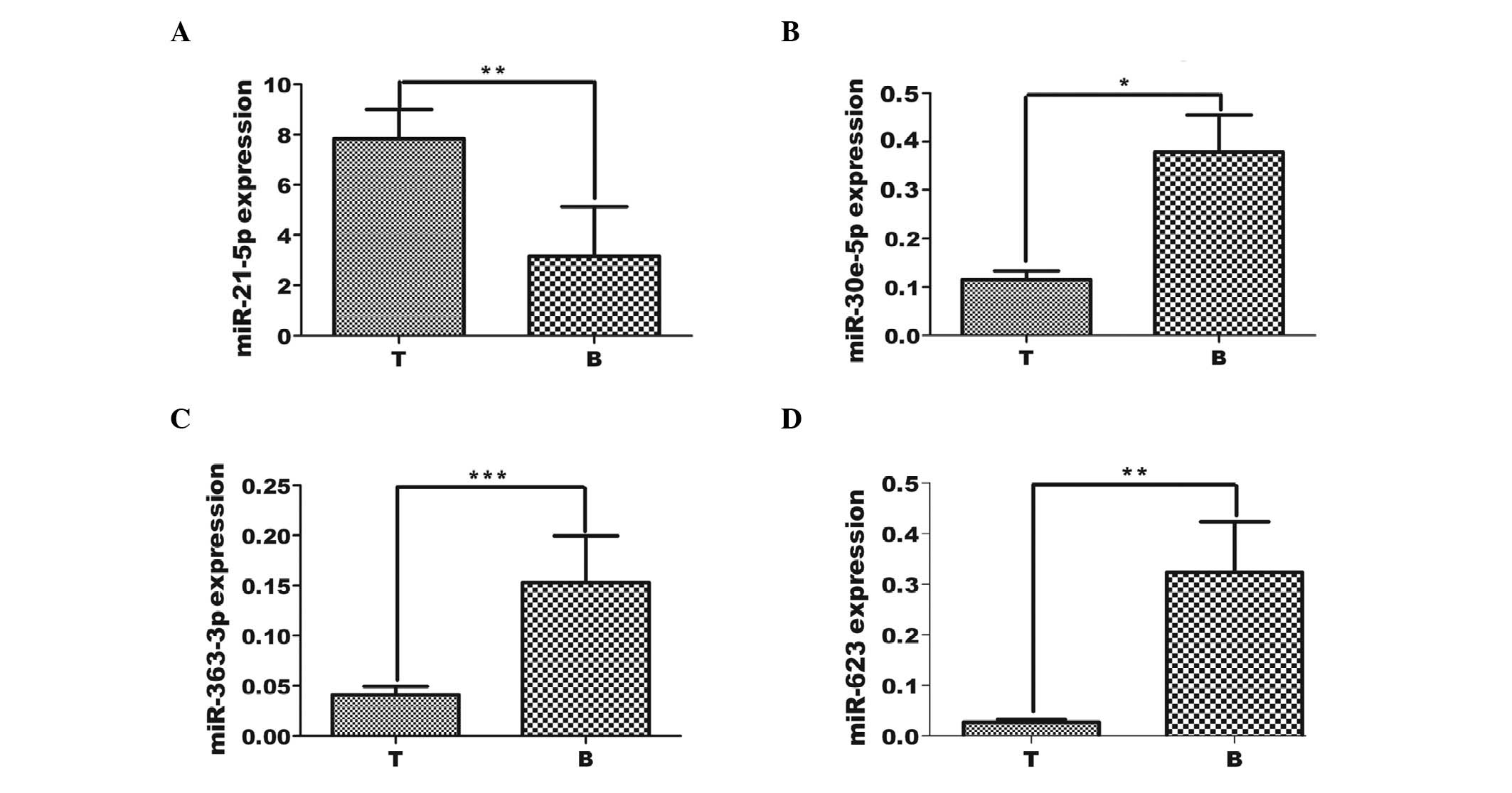
Validation of the selected miRNAs
expression in FFPE tissue
It was hypothesized that FFPE tissue storage may
improve the stability of mRNA (30).
To determine if the identified miRNAs could be reliably detected in
the specimens, and whether the previously identified miRNAs
(miR-21-5p, −10a-5p, −26b-5p, −363-3p, −5100, −650, −145-5p,
−30e-5p and miR-623) were differentially expressed in FFPE samples
from NSCLC compared with those from pulmonary bullae, their
expression levels were quantified by RT-qPCR. Each of the 15 miRNAs
presented Ct values ≤40, indicating that the miRNAs could easily be
measured in these FFPE samples (data not shown). In addition, the
median 2−ΔΔCq levels of miR-623, −21-5p, −30e-5p and
−363-3p were significantly different between NSCLC and pulmonary
bulla patient samples (Fig. 2),
whereas the observed level of miR-26b-5p, −5100, −650 and −145-5p
did not differ significantly between NSCLC and pulmonary bulla
patients (Fig. 2).
Association between
clinicopathological characteristics and differentially expressed
miRNAs in NSCLC
The association between the differentially expressed
miRNAs and the clinicopathological features (such as clinical
stage, lymph node status, tumor size, histological classification
and differentiation) of the associated tumors, was analyzed.
miR-363, −10a and −145 were associated with lymph node status
(P=0.002, 0.005 and 0.007, respectively) and miR-650 and −145 were
associated with differentiation (P=0.01 and 0.05, respectively;
Table I). No association was
identified for the other miRNAs examined. In the FFPE NSCLC
samples, miR-30e-5p correlated with the differentiation of the
tissue (P=0.011) in these patient samples (Table II).
Discussion
The critical role of miRNAs in cancer has become
increasingly apparent. Previous studies have demonstrated that
these small regulatory RNA molecules participate in a diverse set
of cell signaling processes, including apoptosis, cell
proliferation and epithelial-to-mesenchymal transition (12,21,31,32).
In the present study, miRNA expression levels in NSCLC tissues and
their corresponding normal lung tissues were quantified using
RT-qPCR analysis. The present study identified 8 miRNAs that were
significantly differentially expressed in the NSCLC tissues
compared with the corresponding normal specimens, of which 4 miRNAs
were upregulated and 4 miRNAs were downregulated. The data
presented in the current study are in agreement with the results of
previously published miRNA profiling results (18). To the best of our knowledge, the
present study is the first to report the differential expression of
miR-5100 in cancerous tissues and is the first to report altered
miR-363-3p expression in lung cancer. The expression level of
miR-145 was associated with the differentiation and lymph node
status (P=0.05 and 0.007, respectively) and mir-650 was associated
with the differentiation status (P=0.01).
A previous study reported that hsa-miR-145
expression levels are reduced in colon cancer (33), which was also reflected in NSCLC
tissues in the present study. In addition, the present study also
demonstrated that hsa-miR-145 expression levels were associated
with lymph node status (P=0.007). Upregulation of hsa-miR-21
expression levels has been observed in a number of human cancer
specimens and it has been demonstrated that hsa-miR-21 targets and
downregulates the tumor suppressor gene, tumor suppressor
tropomyosin 1 (34). In the present
study, compared with normal lung tissue, the expression level of
miR-21-5p was observed to be increased in the paratumor tissues,
and further increased in the NSCLC tissues (Fig. 1A), suggesting that increased miR-21-5p
expression may also be involved in the development and progression
of lung cancer.
A previous study identified a 5-miRNA signature
(hsa-miR-155, hsa-miR-17-3p, hsa-let-7a-2, hsa-miR-145 and
hsa-miR-21) whose expression was significantly increased in lung
squamous cell carcinoma tissue samples compared with normal lung
tissues, and it correlated with the prognosis and survival outcome
of the patients (35). However, this
5-miRNA signature did not significantly predict patient outcome in
the data set from the present study, in which miR-17 and miR-155
expression levels were not significantly altered in the NSCLC
tissue.
A previous study reported that miR-181a expression
is upregulated in papillary thyroid carcinoma (36), while others have reported it to be
downregulated (37,38). No changes in miR-181a expression
levels were observed in the NSCLC tissues in the present study, in
contrast to Gao et al (38),
who observed low expression levels of miR-181a in tissues from
NSCLC patients. This difference may be due to the different tissue
processing methods and the sample origin.
The present study also identified that miR-30e-5p is
significantly downregulated in NSCLC tissues, by comparing FFPE
non-cancerous adjacent tissues and NSCLC tissues. This result is
consistent with Markou et al (24) who also observed downregulation of
miR-30e-5p in an independent group of 40 matched fresh-frozen
tissues, 37 plasma samples from NSCLC patients and 28 healthy
donors. Consistent with a previous study (39), the present study demonstrated that
miR-30e-5p expression was significantly upregulated in paratumor
tissue compared with adjacent lung tissues or tumor tissues,
however, no significant differences were identified between
miR-30e-5p expression in tumor and adjacent normal lung tissue.
Furthermore, miR-30e-5p expression was significantly associated
with the tissue differentiation in FFPE samples (Fig. 1D).
In the present study, miR-650 expression levels were
significantly higher in NSCLC tissues than in adjacent
non-cancerous tissues (Fig. 1E) and
associated with differentiation (Table
I). Similarly, Huang et al (40) previously demonstrated that miR-650
expression levels were upregulated in human lung adenocarcinoma
tissues and correlated with advanced clinical stage and a higher
incidence of lymph node metastasis (40). However, in the FFPE samples examined
in the present study, no significant change in miR-650 expression
level was observed, which may indicate that the stability of mRNA
is an issue with regard to extraction from FFPE tissues is
necessary (data not shown).
The downregulation of miR-623 expression levels in
malignantly transformed oral leukoplakia has previously been
observed (41). However, in the
present study, miR-623 was significantly upregulated in tumor
tissues compared with adjacent normal tissue (P<0.01; Fig. 1F). Overexpression of miR-623 has also
been observed in esophageal cancer tissues compared with tissues
adjacent to the tumors (24).
There have been few studies investigating miR-363
expression levels in different types of human cancer. To the best
of our knowledge, the present study reports for the first time that
miR-363 is downregulated in NSCLC tissues compared with the
non-cancerous adjacent tissues (Fig.
1H). miR-363 has been proposed as a novel biomarker for the
diagnosis of colorectal cancer (42,43), and
it is downregulated in head and neck squamous cell carcinoma
tissues with lymph node metastasis (44).
Furthermore, the present study demonstrated for the
first time that miR-5100 is upregulated in NSCLC compared with its
corresponding adjacent normal tissue. Fig. 1G indicates that, compared with normal
lung tissues, the expression level of miR-5100 is increased in
paratumor tissues, and further increased in tumor tissues; a
gradual increase was observed.
A previous study demonstrated that miR-10a-5p
expression levels were significantly different in acute myeloid
leukemia compared with control serum samples (45). The results of the present study
demonstrated that miR-10a-5p expression levels in NSCLC tissues
were downregulated compared with normal tissues (Fig. 1I), and that they were associated with
lymph node status (P=0.005).
Previous studies have indicated that RT-qPCR can be
performed to quantify miRNA expression using archival FFPE tissue
samples (46,47). The present study employed FFPE tissue
samples from lung cancer and normal tissues to identify miRNAs
deregulated in patients with lung cancer: miRNA-21-5p was
upregulated and miR-623, −30e-5p and −363-3p were downregulated in
lung cancer samples compared with control pulmonary bulla
specimens. In addition, the expression level of miRNA-30e-5p was
associated with the differentiation status of the tumor (P=0.011;
Table II). However, no alteration in
the expression levels of miR-145-5p, −26b-5p, −650, −5100 and
−10a-5p was detected in FFPE tissue samples compared with normal
pulmonary bulla tissues. One possible explanation is that the
formalin cross-links that form between proteins and nucleic acids
in the tissue complicates the quantification of the miRNAs
(48). Alternatively, the RNA is
significantly damaged prior to, during and following the FFPE
procedure and is degraded into fragments <200 base pairs in
length. Thus, the efficiency of RNA extraction and RT are reduced
and as a result, the quantification of RNA extracted from FFPE
tissues using PCR-based methods is less reliable than in frozen
tissues (49).
The following limitations of the present study
should be taken into consideration: i) The sample size is
relatively small. A larger scale study to confirm these data is
required; ii) the precise mechanisms by which these miRNAs and
their target mRNAs regulate NSCLC progression remain largely
unclear and further mechanistic and external validation through
in vitro and in vivo studies are required to
determine the clinical significance of miRNA expression and their
role in the development of NSCLC; and iii) the current study is
retrospective, with limited scope for generalizability as all the
patients were from China, and the distribution of clinical
characteristics may be different in patients from other countries
of other ethnicities.
In conclusion, the present study demonstrated
aberrant expression of 8 miRNAs in tissues from patients with
NSCLC. miR-650 and miR-10a were aberrantly expressed, miR-145 was
associated with lymph node metastasis; and miR-650 was associated
with differentiation. The present study demonstrates for the first
time that miR-5100 is upregulated and that miR-363 is downregulated
in NSCLC and associated with lymph node metastasis. The expression
levels of miR-26b-5p, miR-1246, miR-211-3p and miR-125b-5p were
unchanged in the NSCLC tissues, in contrast with previous reports
that observed upregulation or downregulation of these miRNAs in
melanoma, multiple myeloma or hepatitis B virus-related
hepatocellular carcinoma (47,50,51).
The present study provides further experimental data for these
candidate miRNAs in order to aid the identification of reliable
molecular diagnostic and prognostic markers of NSCLC in the
future.
Acknowledgements
The present study was supported by the Science &
Technology Innovation Fund of Guangdong Medical University (grant
no. STIF201109); the National Natural Science Foundation of China
project (grant nos. NSFC81172615 and NSFC81570062) and the
Guangdong Medical University Science Fund (grant nos. M2014046 and
M2014032).
Glossary
Abbreviations
Abbreviations:
|
NSCLC
|
non-small cell lung cancer
|
|
FF
|
fresh frozen
|
|
FFPE
|
formalin-fixed, paraffin-embedded
|
References
|
1
|
Siegel R, Ma J, Zou Z and Jemal A: Cancer
statistics, 2014. CA Cancer J Clin. 64:9–29. 2014. View Article : Google Scholar : PubMed/NCBI
|
|
2
|
Minna JD, Roth JA and Gazdar AF: Focus on
lung cancer. Cancer Cell. 1:49–52. 2002. View Article : Google Scholar : PubMed/NCBI
|
|
3
|
Wistuba II: Genetics of preneoplasia:
lessons from lung cancer. Curr Mol Med. 7:3–14. 2007. View Article : Google Scholar : PubMed/NCBI
|
|
4
|
Miller YE: Pathogenesis of lung cancer:
100 year report. Am J Respir Cell Mol Biol. 33:216–223. 2005.
View Article : Google Scholar : PubMed/NCBI
|
|
5
|
Lagos-Quintana M, Rauhut R, Lendeckel W
and Tuschl T: Identification of novel genes coding for small
expressed RNAs. Science. 294:853–858. 2001. View Article : Google Scholar : PubMed/NCBI
|
|
6
|
Garzon R, Marcucci G and Croce CM:
Targeting microRNAs in cancer: rationale, strategies and
challenges. Nat Rev Drug Discov. 9:775–789. 2010. View Article : Google Scholar : PubMed/NCBI
|
|
7
|
Eder M and Scherr M: MicroRNA and lung
cancer. N Engl J Med. 352:2446–2448. 2005. View Article : Google Scholar : PubMed/NCBI
|
|
8
|
Landi MT, Zhao Y, Rotunno M, et al:
MicroRNA expression differentiates histology and predicts survival
of lung cancer. Clin Cancer Res. 16:430–441. 2010. View Article : Google Scholar : PubMed/NCBI
|
|
9
|
Bishop JA, Benjamin H, Cholakh H, et al:
Accurate classification of non-small cell lung carcinoma using a
novel microRNA-based approach. Clin Cancer Res. 16:610–619. 2010.
View Article : Google Scholar : PubMed/NCBI
|
|
10
|
Fassina A, Cappellesso R and Fassan M:
Classification of non-small cell lung carcinoma in transthoracic
needle specimens using microRNA expression profiling. Chest.
140:1305–1311. 2011. View Article : Google Scholar : PubMed/NCBI
|
|
11
|
Sassen S, Miska EA and Caldas C: MicroRNA:
implications for cancer. Virchows Archiv. 452:1–10. 2008.
View Article : Google Scholar : PubMed/NCBI
|
|
12
|
Zhang JG, Wang JJ, Zhao F, Liu Q, Jiang K
and Yang GH: MicroRNA-21 (miR-21) represses tumor suppressor PTEN
and promotes growth and invasion in non-small cell lung cancer
(NSCLC). Clin Chim Acta. 411:846–852. 2010. View Article : Google Scholar : PubMed/NCBI
|
|
13
|
Lu Z, Liu M, Stribinskis V, et al:
MicroRNA-21 promotes cell transformation by targeting the
programmed cell death 4 gene. Oncogene. 27:4373–4379. 2008.
View Article : Google Scholar : PubMed/NCBI
|
|
14
|
Fassan M, Pizzi M, Giacomelli L, et al:
PDCD4 nuclear loss inversely correlates with miR-21 levels in colon
carcinogenesis. Virchows Arch. 458:413–419. 2011. View Article : Google Scholar : PubMed/NCBI
|
|
15
|
Jiang L, Huang Q, Zhang S, et al:
Hsa-miR-125a-3p and hsa-miR-125a-5p are downregulated in non-small
cell lung cancer and have inverse effects on invasion and migration
of lung cancer cells. BMC cancer. 10:3182010. View Article : Google Scholar : PubMed/NCBI
|
|
16
|
Arora S, Ranade AR, Tran NL, et al:
MicroRNA-328 is associated with (non-small) cell lung cancer
(NSCLC) brain metastasis and mediates NSCLC migration. Int J
Cancer. 129:2621–2631. 2011. View Article : Google Scholar : PubMed/NCBI
|
|
17
|
Wang X, Ling C, Bai Y and Zhao J:
MicroRNA-206 is associated with invasion and metastasis of lung
cancer. Anat Rec (Hoboken). 294:88–92. 2011. View Article : Google Scholar : PubMed/NCBI
|
|
18
|
Yanaihara N, Caplen N, Bowman E, et al:
Unique microRNA molecular profiles in lung cancer diagnosis and
prognosis. Cancer Cell. 9:189–198. 2006. View Article : Google Scholar : PubMed/NCBI
|
|
19
|
Volinia S, Calin GA, Liu CG, et al: A
microRNA expression signature of human solid tumors defines cancer
gene targets. Proc Natl Acad Sci USA. 103:2257–2261. 2006.
View Article : Google Scholar : PubMed/NCBI
|
|
20
|
Jeong HC, Kim EK, Lee JH, et al: Aberrant
expression of let-7a miRNA in the blood of non-small cell lung
cancer patients. Mol Med Rep. 4:383–387. 2011.PubMed/NCBI
|
|
21
|
Boeri M, Verri C, Conte D, et al: MicroRNA
signatures in tissues and plasma predict development and prognosis
of computed tomography detected lung cancer. Proc Natl Acad Sci
USA. 108:3713–3718. 2011. View Article : Google Scholar : PubMed/NCBI
|
|
22
|
Foss KM, Sima C, Ugolini D, et al:
miR-1254 and miR-574-5p: serum-based microRNA biomarkers for
early-stage non-small cell lung cancer. J Thorac Oncol. 6:482–488.
2011. View Article : Google Scholar : PubMed/NCBI
|
|
23
|
Liu X, Sempere LF, Ouyang H, et al:
MicroRNA-31 functions as an oncogenic microRNA in mouse and human
lung cancer cells by repressing specific tumor suppressors. J Clin
Invest. 120:1298–1309. 2010. View
Article : Google Scholar : PubMed/NCBI
|
|
24
|
Markou A, Sourvinou I, Vorkas PA, Yousef
GM and Lianidou E: Clinical evaluation of microRNA expression
profiling in non small cell lung cancer. Lung Cancer. 81:388–396.
2013. View Article : Google Scholar : PubMed/NCBI
|
|
25
|
Paci M, Rapicetta C and Maramotti S: New
biomarkers for lung cancer. Expert Opin Med Diagn. 4:201–224.
2010.PubMed/NCBI
|
|
26
|
Xu C, Zheng Y, Lian D, et al: Analysis of
microRNA expression profile identifies novel biomarkers for
non-small cell lung cancer. Tumori. 101:104–110. 2015. View Article : Google Scholar : PubMed/NCBI
|
|
27
|
Solomides CC, Evans BJ, Navenot JM, et al:
MicroRNA profiling in lung cancer reveals new molecular markers for
diagnosis. Acta Cytol. 56:645–654. 2012. View Article : Google Scholar : PubMed/NCBI
|
|
28
|
Guan P, Yin Z, Li X, Wu W and Zhou B:
Meta-analysis of human lung cancer microRNA expression profiling
studies comparing cancer tissues with normal tissues. J Exp Clin
Cancer Res. 31:542012. View Article : Google Scholar : PubMed/NCBI
|
|
29
|
Ma L, Huang Y, Zhu W, et al: An integrated
analysis of miRNA and mRNA expressions in non-small cell lung
cancers. PloS One. 6:e265022011. View Article : Google Scholar : PubMed/NCBI
|
|
30
|
Abrahamsen HN, Steiniche T, Nexo E, et al:
Towards quantitative mRNA analysis in paraffin-embedded tissues
using real-time reverse transcriptase-polymerase chain reaction: a
methodological study on lymph nodes from melanoma patients. J Mol
Diagn. 5:34–41. 2003. View Article : Google Scholar : PubMed/NCBI
|
|
31
|
Mott JL, Kobayashi S, Bronk SF and Gores
GJ: mir-29 regulates Mcl-1 protein expression and apoptosis.
Oncogene. 26:6133–6140. 2007. View Article : Google Scholar : PubMed/NCBI
|
|
32
|
Osada H and Takahashi T: let-7 and
miR-17-92: small-sized major players in lung cancer development.
Cancer Sci. 102:9–17. 2011. View Article : Google Scholar : PubMed/NCBI
|
|
33
|
Michael MZ, O'Connor SM, van Holst
Pellekaan NG, Young GP and James RJ: Reduced accumulation of
specific microRNAs in colorectal neoplasia. Mol Cancer Res.
1:882–891. 2003.PubMed/NCBI
|
|
34
|
Zhu S, Si ML, Wu H and Mo YY: MicroRNA-21
targets the tumor suppressor gene tropomyosin 1 (TPM1). J Biol
Chem. 282:14328–14336. 2007. View Article : Google Scholar : PubMed/NCBI
|
|
35
|
Tan X, Qin W, Zhang L, et al: A 5-microRNA
signature for lung squamous cell carcinoma diagnosis and hsa-miR-31
for prognosis. Clin Cancer Res. 17:6802–6811. 2011. View Article : Google Scholar : PubMed/NCBI
|
|
36
|
Ciafrè SA, Galardi S, Mangiola A, et al:
Extensive modulation of a set of microRNAs in primary glioblastoma.
Biochem Biophys Res Commun. 334:1351–1358. 2005. View Article : Google Scholar : PubMed/NCBI
|
|
37
|
Cahill S, Smyth P, Finn SP, et al: Effect
of ret/PTC 1 rearrangement on transcription and
post-transcriptional regulation in a papillary thyroid carcinoma
model. Mol cancer. 5:702006. View Article : Google Scholar : PubMed/NCBI
|
|
38
|
Gao W, Yu Y, Cao H, et al: Deregulated
expression of miR-21, miR-143 and miR-181a in non small cell lung
cancer is related to clinicopathologic characteristics or patient
prognosis. Biomed Pharmacother. 64:399–408. 2010. View Article : Google Scholar : PubMed/NCBI
|
|
39
|
Busacca S, Germano S, De Cecco L, et al:
MicroRNA signature of malignant mesothelioma with potential
diagnostic and prognostic implications. Am J Respir Cell Mol Biol.
42:312–319. 2010. View Article : Google Scholar : PubMed/NCBI
|
|
40
|
Huang JY, Cui SY, Chen YT, et al:
MicroRNA-650 was a prognostic factor in human lung adenocarcinoma
and confers the docetaxel chemoresistance of lung adenocarcinoma
cells via regulating Bcl-2/Bax expression. PloS One. 8:e726152013.
View Article : Google Scholar : PubMed/NCBI
|
|
41
|
Xiao W, Bao ZX, Zhang CY, et al:
Upregulation of miR-31* is negatively associated with
recurrent/newly formed oral leukoplakia. PloS One. 7:e386482012.
View Article : Google Scholar : PubMed/NCBI
|
|
42
|
Costa A, Afonso J, Osório C, et al:
miR-363-5p regulates endothelial cell properties and their
communication with hematopoietic precursor cells. J Hematol Oncol.
6:872013. View Article : Google Scholar : PubMed/NCBI
|
|
43
|
Xu X, Wu X, Wu S, et al: Study on
miR-490-5p and miR-363 as novel biomarkers for the diagnosis of
colorectal cancer. Zhonghua Wei Chang Wai Ke Za Zhi. 17:45–50.
2014.(In Chinese). PubMed/NCBI
|
|
44
|
Sun Q, Zhang J, Cao W, et al: Dysregulated
miR-363 affects head and neck cancer invasion and metastasis by
targeting podoplanin. Int J Biochem Cell Biol. 45:513–520. 2013.
View Article : Google Scholar : PubMed/NCBI
|
|
45
|
Zhi F, Cao X, Xie X, et al: Identification
of circulating microRNAs as potential biomarkers for detecting
acute myeloid leukemia. PloS One. 8:e567182013. View Article : Google Scholar : PubMed/NCBI
|
|
46
|
Kelly AD, Hill KE, Correll M, et al:
Next-generation sequencing and microarray-based interrogation of
microRNAs from formalin-fixed, paraffin-embedded tissue:
preliminary assessment of cross-platform concordance. Genomics.
102:8–14. 2013. View Article : Google Scholar : PubMed/NCBI
|
|
47
|
Oue N, Anami K, Schetter AJ, et al: High
miR-21 expression from FFPE tissues is associated with poor
survival and response to adjuvant chemotherapy in colon cancer. Int
J Cancer. 134:1926–1934. 2014. View Article : Google Scholar : PubMed/NCBI
|
|
48
|
Foss RD, Guha-Thakurta N, Conran RM and
Gutman P: Effects of fixative and fixation time on the extraction
and polymerase chain reaction amplification of RNA from
paraffin-embedded tissue. Comparison of two housekeeping gene mRNA
controls. Diagn Mol Pathol. 3:148–155. 1994. View Article : Google Scholar : PubMed/NCBI
|
|
49
|
Doleshal M, Magotra AA, Choudhury B, et
al: Evaluation and validation of total RNA extraction methods for
microRNA expression analyses in formalin-fixed, paraffin-embedded
tissues. J Mol Diagn. 10:203–211. 2008. View Article : Google Scholar : PubMed/NCBI
|
|
50
|
Jones CI, Zabolotskaya MV, King AJ, et al:
Identification of circulating microRNAs as diagnostic biomarkers
for use in multiple myeloma. Br J Cancer. 107:1987–1996. 2012.
View Article : Google Scholar : PubMed/NCBI
|
|
51
|
Giray BG, Emekdas G, Tezcan S, et al:
Profiles of serum microRNAs; miR-125b-5p and miR223-3p serve as
novel biomarkers for HBV-positive hepatocellular carcinoma. Mol
Biol Rep. 41:4513–4519. 2014. View Article : Google Scholar : PubMed/NCBI
|