Introduction
The gastric region is the most frequent site of
extra nodal lymphoma (1). Gastric
lymphoma, which originates from the mucosa-associated lymphoid
tissue (MALT), behaves as an indolent disease and has a favorable
long-term prognosis with a 10-year survival rate of >90%. Most
cases of gastric MALT lymphoma remain localized within the gastric
region for many years (2).
Gastric MALT lymphoma is difficult to diagnose due
to its non-specific symptoms and various endoscopic findings
(3). Helicobacter pylori
(H. pylori) infection is associated with gastric MALT
lymphoma. Therefore, H. pylori eradication (HPE) is a
potentially effective therapeutic option in cases of early-stage,
low-grade gastric MALT lymphoma. This leads to remission in >75%
of patients (4). However, the
management strategy for H. pylori-negative, lymphoma
residuals, or recurrent disease is not well-defined. Treatment
guidelines recommend a stage-dependent approach, favoring
radiotherapy (RT) for localized and chemotherapy (CT) for advanced
stages of the disease (5). However,
clinicians and patients have to balance the risks and benefits of
specific treatments without high-level evidence or long-term
survival data. Different prognostic factors for gastric MALT
lymphoma have been identified, including neoplasia stage, depth of
infiltration in the gastric wall, localization in the stomach, and
patient ethnicity (6–9).
We conducted a retrospective analysis in the
patients with gastric MALT lymphoma to analyze the clinical
characteristics, prognostic factors and long-term outcomes.
Materials and methods
Ethics approval
The Ethics Committee of the Tianjin Medical
University Cancer Institute and Hospital approved the current
study. Patient records/information were anonymized prior to the
analysis. Written informed consent was provided by the participants
in order for their clinical records to be used in the current
study.
The hospital database was searched for gastric MALT
lymphoma diagnosed at the Tianjin Medical University Cancer
Institute and Hospital, China, between August 2001 and August 2013.
Histopathologic diagnosis of the gastric MALT lymphoma was made
according to the World Health Organization classification (10). Histology and immunohistochemistry of
the original histological samples (biopsy or resection specimens)
were investigated, and reviewed by at least three
histopathologists. Patients were excluded from the study subjects
if: i) they had another malignancy at the time of diagnosis; ii)
they were human immunodeficiency virus-positive cases; or iii) the
follow-up period was <12 months. Following exclusion of those
patients, the remaining 103 patients were included in the current
study.
Staging procedures
The patients were restaged according to the Lugano
staging system for gastrointestinal non-Hodgkin's lymphoma
(11). The staging workup included
patient history, physical examination, endoscopy, barium meal
examination, chest X-ray or computed tomography scan, abdominal
ultrasound and computed tomography scan, bone marrow aspiration and
biopsy, gastric mucosal biopsies or gastrectomy as well as
immunohistochemistry. The H. pylori infection status was
determined by histologic examination, urea breath test, or both to
evaluate the status of H. pylori infection following HPE.
Routine laboratory tests, including measurement of hemoglobin,
serum lactate dehydrogenase (LDH) and β2-microglobulin (β2-MG),
were also carried out in all 103 patients. Low hemoglobin was
defined as <120 g/l, high serum LDH as >245 U/l, and high
β2-MG as >2.2 mg/l.
Treatment modalities
Of the patients with localized gastric MALT
lymphoma, 69 patients associated with H. pylori-positive and
4 patients with H. pylori-negative underwent HPE therapy.
Other treatment modalities, such as RT and CT, were also used for
treatment. The HPE regimen included proton pump inhibitors
(omeprazole, lansoprazole, or rabeprazole) and a combination of
antibiotics (amoxicillin, clarithromycin, and metronidazole). CT
included monotherapy or a combination of CT and immunotherapy. RT
was performed at a median total dose ranging from 26 to 46 Gy. The
response was assessed according to the international workshop for
NHL standardized criteria (12).
Statistical analysis
Primary endpoints of the survival analysis were
overall survival (OS) and progression-free survival (PFS). OS was
measured from the date of diagnosis until the date of death due to
any cause, or the date of survivors' final follow-up. The PFS was
calculated from the time of diagnosis to the date of treatment
failure, relapse, evidence of disease progression, or death due to
any cause. The Kaplan-Meier method was used for survival
estimations, and the log-rank test for survival comparisons.
Variables that influenced the prognosis (P<0.05) in the
univariate analysis were assessed by a multivariate analysis using
the Cox regression model to determine independent prognostic
factors for survival. The SPSS 17.0 software (SPSS, Inc., Chicago,
IL, USA) was used for statistical analyses. P<0.05 was
considered to indicate a statistically significant difference.
Results
Patient characteristics
Patient characteristics are shown in Table I. Patients (n=103) with gastric MALT
lymphoma had a median age of 53 years (range: 19–85 years), and
included 54 males (M) and 49 females (F) at a ratio of 1:1. The
onset of the disease was often insidious and without specific
clinical manifestation. None of the patients experienced
perforation prior to treatment. Their serum LDH and β2-MG were
usually within normal limits. In all 103 patients, the diagnosis
was made on the basis of an endoscopic biopsy. Macroscopically, the
most commonly involved site was the antrum (60.7%), followed by the
corpus (51.6%), and fundus (23.5%). Most patients appeared
ulcerative (78.4%). Using the Lugano staging system, 40 patients
(39%) had stage I, and 35 patients (35%) were diagnosed with local
or distant nodal involvement. There were also patients with stage
IIIE (20/103, 19%) and stage IV (8/103, 7%) disease.
 | Table I.Characteristics of 103 patients with
gastric MALT lymphoma. |
Table I.
Characteristics of 103 patients with
gastric MALT lymphoma.
| Feature | No. (%) |
|---|
| Gender |
|
|
Female | 51 (50) |
| Male | 52 (50) |
| Age |
|
| ≤60 | 35 (34) |
|
>60 | 38 (66) |
| Lugano staging |
|
|
I–II2 | 75 (73) |
|
IIE-IV | 28 (27) |
| Serum LDH level |
|
|
Normal | 78 (76) |
|
Abnormal | 25 (24) |
| B symptoms |
|
|
Present | 51 (50) |
|
Absent | 52 (50) |
| β2-microglobulin |
|
|
Normal | 72 (70) |
|
Abnormal | 31 (30) |
| m-IPIa |
|
| 0–1 | 78 (76) |
| ≥2 | 15 (15) |
|
Unknown | 10 (9) |
| Infection with H.
pylori |
|
|
Positive | 97 (94) |
|
Negative | 6 (6) |
| Tumor mass |
|
| <5
cm | 56 (54) |
| ≥5
cm | 34 (33) |
|
Unknown | 13 (13) |
| Performance
status |
|
|
<2 | 72 (70) |
| ≥2 | 15 (15) |
|
Unknown | 16 (15) |
|
Anemia |
|
|
Present | 48 (47) |
|
Absent | 55 (53) |
HPE
The H. pylori infection rate was 94%
(97/103), whereas the infection rate was 100% (28/28) in patients
with advanced stages. A total of 73 patients with localized
disease, including 69 patients associated with H.
pylori-positive and 4 patients with H. pylori-negative,
received HPE as a first-line treatment. H. pylori was
eradicated in all the positive patients (69/69), although in 17 of
these patients, a second-line HPE was required. The symptoms
disappeared or were markedly reduced in the majority of patients.
The CR was achieved in 54 of the 69 patients (78%) with H.
pylori-positive and in 2 of the 4 patients (50%) with H.
pylori-negative. The median time to complete remission (CR)
after HPE was 4 months (range: 3–9 months). HPE had a superior
trend in the H. pylori-positive patients but was not
significantly different in the two groups (p=0.194).
Other treatment modalities
The 16 patients, including 14 patients who failed to
achieve CR by HPE and 2 patients associated with H.
pylori-negative, received RT. The remaining 3 patients who
failed to achieve CR by HPE were followed on watch-and-wait
strategy due to improved endoscopic features and relief symptoms.
The success rate of RT was 100% (16/16). Of note, the 3 patients
treated by watch-and-wait strategy, only 1 had disease recurrence
by gastric H. pylori reinfection and a second remission was
obtained following a second-line HPE.
Of the 28 patients with advanced stages, 12 patients
received CT or RT combined with HPE and H. pylori was
eradicated in the 12 patients. CR was achieved in 8 of the 12
patients (67%). The remaining 16 patients received CT or RT. The CR
was achieved in 10 of the 16 patients (63%). There was no
statistical difference in the two groups (p=0.820).
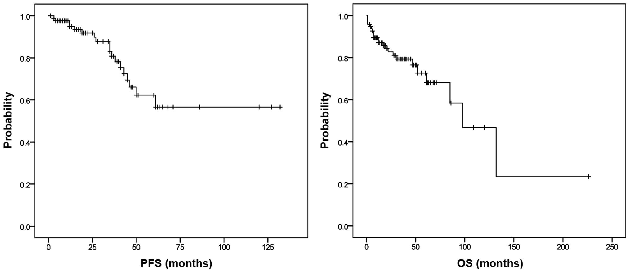
Survival analysis
The median follow-up time was 62.9 months (range:
1–226 months). The median PFS was 41.5 months (range: 1–132
months), and the median OS was 59.5 months (range: 2–226 months).
The 5-year PFS and OS estimates for the 103 patients were 58 and
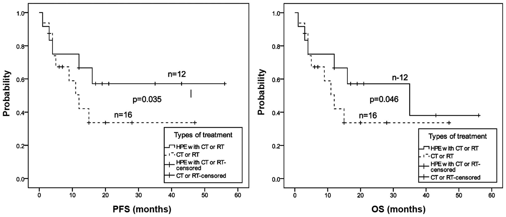
69%, respectively (Fig. 1). The
5-year PFS and OS estimates were compared between patients
receiving CT or RT combined with HPE and those receiving CT or RT.
We found that the five-year PFS and OS estimates were significantly
improved (p<0.05, Fig. 2) for
patients receiving CT or RT combined with HPE than those receiving
CT or RT.
Based on the univariate analysis, the B symptoms,
serum LDH >245 U/l, β2-MG >2.2 mg/l, advanced stages, PS ≥2
and modified-International Prognostic Index (m-IPI) (≥2) adversely
affected PFS and OS (Table II).
Advanced stages were significant only for shorter PFS, and m-IPI
(≥2) retained its prognostic significance for the shorter OS with
multivariate analysis (Table
III).
| Table II.Univariate analysis of prognostic
factors for PFS and OS. |
Table II.
Univariate analysis of prognostic
factors for PFS and OS.
|
| OS | PFS |
|---|
|
|
|
|
|---|
| Clinical
characteristics | χ2
test | p-value | χ2
test | p-value |
|---|
| Age | 3.118 | 0.077 | 0.795 | 0.373 |
| Gender | 0.030 | 0.864 | 0.450 | 0.502 |
| B symptoms | 5.025 | 0.025 | 10.449 | 0.001 |
| Anemia | 1.508 | 0.208 | 0.511 | 0.475 |
| Serum LDH >245
U/l | 6.213 | 0.013 | 7.261 | 0.007 |
| β2-microglobulin
>2.2 mg/l | 9.604 | 0.002 | 8.098 | 0.004 |
| Tumor mass ≥10
cm | 3.873 | 0.275 | 4.215 | 0.239 |
| H.
pylori-positive | 2.747 | 0.253 | 4.238 | 0.120 |
| Advanced
stages | 4.028 | 0.045 | 4.930 | 0.026 |
| PS ≥2 | 5.998 | 0.014 | 74.897 | 0.000 |
| m-IPI ≥2 | 5.784 | 0.017 | 4.389 | 0.036 |
| Table III.Factors retaining prognostic
significance for PFS and OS with multivariate and Cox proportional
hazards analysis. |
Table III.
Factors retaining prognostic
significance for PFS and OS with multivariate and Cox proportional
hazards analysis.
|
| OS | PFS |
|---|
|
|
|
|
|---|
| Prognostic
factors | Relative risk | 95% CI | p-value | Relative risk | 95% CI | p-value |
|---|
| B symptoms | 4.982 | 0.000–0.548 | 0.246 | 0.191 | 0.311–2.172 | 0.514 |
| Serum LDH >245
U/l | 4.266 | 1.216–17.132 | 0.069 | 2.029 | 0.165–3.517 | 0.917 |
| β2-microglobulin
>2.2 mg/l | 1.564 | 0.467–31.345 | 0.211 | 1.273 | 0.417–3.218 | 0.287 |
| Advanced
stages | 2.936 | 0.002–1.524 | 0.087 | 1.157 | 0.924–11.627 | 0.028 |
| PS ≥2 | 0.620 | 0.272–21.203 | 0.431 | 2.137 | 0.056–1.087 | 0.106 |
| m-IPI ≥2 | 6.937 | 3.721–77.677 | 0.008 | 4.682 | 0.826–11.235 | 0.076 |
Discussion
Gastric MALT lymphoma is an indolent disease with a
prolonged clinical course that most often involves the stomach
(3). The clinical presentation is
similar to gastric carcinomas and benign ulcers, and commonly
includes abdominal pain or discomfort, and with less frequent signs
such as bleeding and perforation. The duration of the symptoms
prior to diagnosis is variable, ranging from a few weeks to several
years. B symptoms are exceedingly rare, and adverse biological
prognostic factors such as high serum LDH and β2-MG levels are
infrequently elevated. H. pylori is linked to the
development of gastric MALT lymphoma (12), and they occurred frequently in the
present study. The clinical characteristics of gastric MALT
lymphoma in the present study are similar to those reported in
previous studies (3,13,14).
Identification of the etiologic role of H.
pylori infection in gastric MALT lymphoma has radically changed
the therapeutic approach for such neoplasia. Following successful
HPE, lymphoma remission was achieved in 77.5% of 1,408 patients
with gastric MALT lymphoma at the early stages with a median time
of 5 months (15). In the present
study, we found that the remission of gastric MALT lymphoma was
achieved after HPE in 56 patients (77%) of 73 patients with
localized disease. This figure was comparable to the results of
several reported large cases (16–18). Of
note, in the present study, 2 patients with H.
pylori-negative successfully achieved CR by HPE and there were
no significant differences in the CR rate following HPE between the
H. pylori-positive and -negative groups, which is consistent
with a previous study (19).
Eradication therapy may be useful for H. pylori-negative
gastric MALT lymphoma: first, another infective organism such as
Helicobacter heilmannii, might be involved in the
development of gastric MALT lymphoma (20); second, because of very low bacterial
counts, conventional examination did not detect the organisms
(21). Currently, unlike that for
H. pylori-positive MALT lymphoma, the optimal management of
H. pylori-negative MALT lymphoma remains controversial.
These findings suggest that HPE therapy may be considered as a
first-line treatment regardless of H. pylori infection
status. In addition, there was no difference in 5-year OS between
patients who received CT or RT with HPE and those receiving CT or
RT. Patients who received CT or RT combined with HPE had an
improved 5-year PFS and OS than those who received CT or RT. This
result suggests that HPE may improve the prognosis of advanced
stages of gastric MALT lymphoma. Since advanced gastric MALT
lymphoma is likely a progression from localized to extensive
disease, they may still be completely cured by HPE. According to
the currently published European Society of Medical Oncology
guidelines, HPE should be administered to all gastric MALT
lymphomas, independent of the stages and even in H.
pylori-negative cases (22).
However, large-scale prospective randomized clinical trials are
needed to document any HPE benefits for patients with gastric MALT
lymphoma.
There are no accepted prognostic factors for gastric
MALT lymphoma thus far. A recent study by Wirth et al
(23) showed that there was a trend
to the poorer outcome with increasing age, lymph node involvement
and stomach wall thickening. Previous studies (6,24)
described the grade of malignancy and stage of the disease as the
two major prognostic factors. Translocation t(11;18), the most
common genetic aberration in gastric MALT lymphomas is associated
with poorer outcome as it strongly predicts the response of gastric
MALT lymphoma to HPE (25). The IPI
has been correlated with relapse, while the utility of IPI is
controversial. Previous studies (26,27) have
demonstrated that stage m-IPI is an effective predictive factor for
patients with gastric lymphoma, where m-IPI was more accurate than
the IPI for predicting gastric lymphoma patient survival. In our
analysis, stage m-IPI was identified as an independent prognostic
factor for OS in the multivariate analysis. Patients in the
low-risk group (m-IPI ≤1) had significantly longer survival than
the intermediate/high-risk (m-IPI ≥2) patients. Our multivariate
analysis revealed that advanced stage of lymphoma was an
independent prognostic factor for PFS. Previous findings (27,28) have
also shown that the prognosis for gastric lymphoma is influenced by
the depth of infiltration and is exacerbated with metastasis to
other organs. Ranaldi et al (29) showed there was no difference in the
survival rate between stages I and II gastric lymphoma. However,
there was a trend for increasing hazard as the neoplasia progresses
from stages II to IIE (p=0.001). We believe that advanced lymphoma
stages are crucial for prognostic evaluation, although the exact
relationship between disease stage and outcomes remains to be
established. Studies identifying other prognostic markers remain to
be undertaken.
In conclusion, the results of the present study
confirm the indolent course of gastric MALT lymphoma with late
relapse and prolonged survival. The patients with advanced stages
and m-IPI (≥2), however, tend to have a worse prognosis. As an
effective treatment for gastric MALT lymphoma, HPE therapy should
be administered to all gastric MALT lymphomas, independently of
stage and even in H. pylori-negative cases.
Acknowledgements
The authors would like to thank the medical staff
who contributed to the success of the present study.
References
|
1
|
Zucca E and Cavalli F: Extranodal
lymphomas. Ann Oncol. 11(Suppl 3): 219–222. 2000. View Article : Google Scholar : PubMed/NCBI
|
|
2
|
Du MQ and Atherton JC: Molecular subtyping
of gastric MALT lymphomas: Implications for prognosis and
management. Gut. 55:886–893. 2006. View Article : Google Scholar : PubMed/NCBI
|
|
3
|
Psyrri A, Papageorgiou S and Economopoulos
T: Primary extranodal lymphomas of stomach: clinical presentation,
diagnostic pitfalls and management. Ann Oncol. 19:1992–1999. 2008.
View Article : Google Scholar : PubMed/NCBI
|
|
4
|
Zullo A, Hassan C, Andriani A, Cristofari
F, De Francesco V, Ierardi E, Tomao S, Morini S and Vaira D:
Eradication therapy for Helicobacter pylori in patients with
gastric MALT lymphoma: a pooled data analysis. Am J Gastroenterol.
104:1932–1937, quiz 1938. 2009. View Article : Google Scholar : PubMed/NCBI
|
|
5
|
Ruskoné-Fourmestraux A, Fischbach W,
Aleman BM, Boot H, Du MQ, Megraud F, Montalban C, Raderer M, Savio
A and Wotherspoon A: EGILS group: EGILS consensus report. Gastric
extranodal marginal zone B-cell lymphoma of MALT. Gut. 60:747–758.
2011. View Article : Google Scholar : PubMed/NCBI
|
|
6
|
Radaszkiewicz T, Dragosics B and Bauer P:
Gastrointestinal malignant lymphomas of the mucosa-associated
lymphoid tissue: Factors relevant to prognosis. Gastroenterology.
102:1628–1638. 1992.PubMed/NCBI
|
|
7
|
Ruskoné-Fourmestraux A, Aegerter P, Delmer
A, Brousse N, Galian A and Rambaud JC: Primary digestive tract
lymphoma: a prospective multicentric study of 91 patients. Groupe
d'Etude des Lymphomes Digestifs. Gastroenterology. 105:1662–1671.
1993.PubMed/NCBI
|
|
8
|
Liu H, Ye H, Ruskone-Fourmestraux A, De
Jong D, Pileri S, Thiede C, Lavergne A, Boot H, Caletti G, Wündisch
T, et al: T(11;18) is a marker for all stage gastric MALT lymphomas
that will not respond to H. pylori eradication.
Gastroenterology. 122:1286–1294. 2002. View Article : Google Scholar : PubMed/NCBI
|
|
9
|
Yeh KH, Kuo SH, Chen LT, Mao TL, Doong SL,
Wu MS, Hsu HC, Tzeng YS, Chen CL, Lin JT, et al: Nuclear expression
of BCL10 or nuclear factor kappa B helps predict Helicobacter
pylori-independent status of low-grade gastric
mucosa-associated lymphoid tissue lymphomas with or without
t(11;18)(q21;q21). Blood. 106:1037–1041. 2005. View Article : Google Scholar : PubMed/NCBI
|
|
10
|
Tomonaga M: Outline and direction of
revised WHO classification of Tumors of Haematopoietic and Lymphoid
Tissues. Rinsho Ketsueki. 50:1401–1406. 2009.(In Japanese).
PubMed/NCBI
|
|
11
|
Rohatiner A, d'Amore F, Coiffier B,
Crowther D, Gospodarowicz M, Isaacson P, Lister TA, Norton A, Salem
P, Shipp M, et al: Report on a workshop convened to discuss the
pathological and staging classifications of gastrointestinal tract
lymphoma. Ann Oncol. 5:397–400. 1994.PubMed/NCBI
|
|
12
|
Wotherspoon AC, Doglioni C, Diss TC, Pan
L, Moschini A, de Boni M and Isaacson PG: Regression of primary
low-grade B-cell gastric lymphoma of mucosa-associated lymphoid
tissue type after eradication of Helicobacter pylori.
Lancet. 342:575–577. 1993. View Article : Google Scholar : PubMed/NCBI
|
|
13
|
Nakamura S, Matsumoto T, Iida M, Yao T and
Tsuneyoshi M: Primary gastrointestinal lymphoma in Japan: a
clinicopathologic analysis of 455 patients with special reference
to its time trends. Cancer. 97:2462–2473. 2003. View Article : Google Scholar : PubMed/NCBI
|
|
14
|
Kolve M, Fischbach W, Greiner A and Wilms
K: German Gastrointestinal Lymphoma Study Group: Differences in
endoscopic and clinicopathological features of primary and
secondary gastric non-Hodgkin's lymphoma. Gastrointest Endosc.
49:307–315. 1999. View Article : Google Scholar : PubMed/NCBI
|
|
15
|
Zullo A, Hassan C, Cristofari F, Andriani
A, De Francesco V, Ierardi E, Tomao S, Stolte M, Morini S and Vaira
D: Effects of Helicobacter pylori eradication on early stage
gastric mucosa-associated lymphoid tissue lymphoma. Clin
Gastroenterol Hepatol. 8:105–110. 2010. View Article : Google Scholar : PubMed/NCBI
|
|
16
|
Wündisch T, Thiede C, Morgner A, Dempfle
A, Günther A, Liu H, Ye H, Du MQ, Kim TD, Bayerdörffer E, et al:
Long-term follow-up of gastric MALT lymphoma after Helicobacter
pylori eradication. J Clin Oncol. 23:8018–8024. 2005.
View Article : Google Scholar : PubMed/NCBI
|
|
17
|
Fischbach W, Goebeler ME,
Ruskone-Fourmestraux A, Wündisch T, Neubauer A, Raderer M and Savio
A: EGILS (European Gastro-Intestinal Lymphoma Study) Group: Most
patients with minimal histological residuals of gastric MALT
lymphoma after successful eradication of Helicobacter pylori
can be managed safely by a watch and wait strategy: experience from
a large international series. Gut. 56:1685–1687. 2007. View Article : Google Scholar : PubMed/NCBI
|
|
18
|
Vivas S, de Morales Ruiz JM, Fernandez M,
Hernando M, Herrero B, Casqueiro J and Gutierrez S: Age-related
clinical, serological, and histopathological features of celiac
disease. Am J Gastroenterol. 103:2360–2365, quiz 2366. 2008.
View Article : Google Scholar : PubMed/NCBI
|
|
19
|
Raderer M, Streubel B, Wöhrer S, Häfner M
and Chott A: Successful antibiotic treatment of Helicobacter
pylori negative gastric mucosa associated lymphoid tissue
lymphomas. Gut. 55:616–618. 2006. View Article : Google Scholar : PubMed/NCBI
|
|
20
|
Morgner A, Lehn N, Andersen LP, Thiede C,
Bennedsen M, Trebesius K, Neubauer B, Neubauer A, Stolte M and
Bayerdörffer E: Helicobacter heilmannii-associated primary gastric
low-grade MALT lymphoma: Complete remission after curing the
infection. Gastroenterology. 118:821–828. 2000. View Article : Google Scholar : PubMed/NCBI
|
|
21
|
Steinbach G, Ford R, Glober G, Sample D,
Hagemeister FB, Lynch PM, McLaughlin PW, Rodriguez MA, Romaguera
JE, Sarris AH, et al: Antibiotic treatment of gastric lymphoma of
mucosa-associated lymphoid tissue. An uncontrolled trial. Ann
Intern Med. 131:88–95. 1999. View Article : Google Scholar : PubMed/NCBI
|
|
22
|
Zucca E, Copie-Bergman C, Ricardi U,
Thieblemont C, Raderer M and Ladetto M: ESMO Guidelines Working
Group: Gastric marginal zone lymphoma of MALT type: ESMO Clinical
Practice Guidelines for diagnosis, treatment and follow-up. Ann
Oncol. 24(Suppl 6): vi144–vi148. 2013. View Article : Google Scholar : PubMed/NCBI
|
|
23
|
Wirth A, Gospodarowicz M, Aleman BM,
Bressel M, Ng A, Chao M, Hoppe RT, Thieblemont C, Tsang R, Moser L,
et al: Long-term outcome for gastric marginal zone lymphoma treated
with radiotherapy: a retrospective, multi-centre, International
Extranodal Lymphoma Study Group study. Ann Oncol. 24:1344–1351.
2013. View Article : Google Scholar : PubMed/NCBI
|
|
24
|
Cogliatti SB, Schmid U, Schumacher U,
Eckert F, Hansmann ML, Hedderich J, Takahashi H and Lennert K:
Primary B-cell gastric lymphoma: a clinicopathological study of 145
patients. Gastroenterology. 101:1159–1170. 1991.PubMed/NCBI
|
|
25
|
Lima KS, Albuquerque W, Arantes VN,
Drummond-Lage AP and Coelho LG: Helicobacter pylori and
t(11;18)(q21;q21) translocation in gastric malt lymphoma. Arq
Gastroenterol. 51:84–89. 2014. View Article : Google Scholar : PubMed/NCBI
|
|
26
|
Cortelazzo S, Rossi A, Roggero F, Oldani
E, Zucca E, Tondini C, Ambrosetti A, Pasini F, Pinotti G, Bertini
M, et al: International Extranodal Lymphoma Study Group (IELSG):
Stage-modified international prognostic index effectively predicts
clinical outcome of localized primary gastric diffuse large B-cell
lymphoma. Ann Oncol. 10:1433–1440. 1999. View Article : Google Scholar : PubMed/NCBI
|
|
27
|
Leopardo D, Di Lorenzo G, De Renzo A,
Federico P, Luponio S, Buonerba C, Matano E, Merola G, Imbimbo M,
Montesarchio E, et al: Efficacy of rituximab in gastric diffuse
large B cell lymphoma patients. World J Gastroenterol.
16:2526–2530. 2010. View Article : Google Scholar : PubMed/NCBI
|
|
28
|
Medina-Franco H, Germes SS and Maldonado
CL: Prognostic factors in primary gastric lymphoma. Ann Surg Oncol.
14:2239–2245. 2007. View Article : Google Scholar : PubMed/NCBI
|
|
29
|
Ranaldi R, Goteri G, Baccarini MG,
Mannello B and Bearzi I: A clinicopathological study of 152
surgically treated primary gastric lymphomas with survival analysis
of 109 high grade tumours. J Clin Pathol. 55:346–351. 2002.
View Article : Google Scholar : PubMed/NCBI
|